JBRA Assist. Reprod. 2010;14(3):28-31
ARTIGO ORIGINAL
doi: 10.5935/1518-0557.2010.14.3.05
Predictive factors of intrauterine insemination success: sperm morphology according to the new World Health Organization laboratory manual
Fatores preditivos do sucesso da inseminação intrauterina: a morfologia espermática de acordo com o novo manual de laboratório da Organização Mundial de Saúde
aFertility – Centro de Fertilização Assistida Av. Brigadeiro Luis Antônio, 4545. São Paulo – SP, Brasil. CEP: 01401-002
bInstituto Sapientiae – Centro de Ensino e Pesquisa em Reprodução Assistida Rua Vieira Maciel, 62. São Paulo – SP, Brasil. CEP: 04503-040
cDepartmento de Ginecologia e Obstetrícia, Faculdade de Medicina de Botucatu UNESP Botucatu – SP – Brasil Distrito de Rubião Junior – CEP: 18618-970 Botucatu – SP
*Os dois autores contribuíram igualmente para esse trabalho.
ABSTRACT
Objective: The aim of our study was to assess the likelihood of IUI success as a function of the previously described predictive factors, including sperm morphology according to the new reference values defined by WHO.
Material and Methods: This retrospective study enrolled 300 couples which underwent IUI. Regression analyses were used to correlate maternal age, number of preovulatory follicles on the day of hCG administration, number of inseminated motile sperm, and normal sperm morphology with clinical pregnancy. Results are expressed as odds ratio (OR) with 95% of confidence intervals (CI).
Results: Women older than 35 years showed a lower pregnancy rate (6.5% vs 18.2%, p=0.017). Logistic regression models confirmed the lower chance of pregnancy occurrence for older women (OR: 0.39; CI: 0.16–0.96; p=0.040). The presence of two or more preovulatory follicles on the day of hCG administration resulted in higher pregnancy rate when compared to cases in which only one preovulatory follicle was present (18.6% vs 8.2%, p=0.011). The regression model showed a more than two fold increase on probability of pregnancy when two or more preovulatory follicles were detected (OR: 2.58; CI: 1.22–5.46, p=0.013). The number of inseminated motile sperm positively influenced pregnancy occurrence (OR: 1.47; CI: 0.88–3.14, p=0.027). Similar pregnancy rates were observed when semen samples were classified as having normal or abnormal morphology (10.6% vs 10.2%, p=0.936).
Conclusion: Our results demonstrate that sperm morphological normalcy, according to the new reference value, has no predictive value on IUI outcomes.
Key words: intrauterine insemination, pregnancy rate, sperm morphology
RESUMO
Objetivo: Avaliar a probabilidade de sucesso da IIU em função dos fatores preditivos descritos previamente, incluindo a morfologia espermática de acordo com os novos valores definidos pela OMS.
Métodos: Este estudo retrospectivo envolveu 300 casais que foram submetidos à IIU. A idade materna, o número de folículos pré-ovulatórios no dia da administração do hCG, número de espermatozóides móveis inseminados e morfologia espermática normal foram correlacionados com a gravidez clínica. Os resultados foram expressos como odds ratio (OR) com 95% de intervalos de confiança (IC).
Resultados: Mulheres com mais de 35 anos mostraram uma menor taxa de gravidez (6.5% vs 18.2%, p = 0.017). Modelos de regressão logística confirmaram a menor chance de gravidez para as mulheres mais velhas (OR: 0.39, IC: 0.16-0.96, p = 0.040). A presença de dois ou mais folículos pré-ovulatórios no dia da administração de hCG resultou em maior taxa de gravidez quando comparada aos casos em que apenas um folículo pré-ovulatório estava presente (18.6% vs 8.2%, p = 0.011). Um aumento de mais de duas vezes na probabilidade de gravidez foi observado quando dois ou mais folículos pré-ovulatórios foram detectados (OR: 2.58, IC: 1.22-5.46, p = 0.013). O número de espermatozóides móveis inseminados influenciou positivamente a ocorrência de gravidez (OR: 1.47, IC: 0.88-3.14, p = 0.027). Taxas de gestação semelhantes foram observadas quando as amostras possuíam morfologia normal ou anormal (10.6% vs 10.2%, p = 0.936).
Conclusão: A morfologia espermática de acordo com o novo valor de referência, não tem valor preditivo nos resultados da IIU.
Palavras-chave: inseminação intra-uterina, morfologia espermática, taxa de gestação
INTRODUCTION
Currently, medical advice for infertility is requested for 16% of prospective parents (Merviel et al. 2008), therefore, identifying which couples are likely to benefit from artificial insemination or in vitro fertilization (IVF) techniques is of pivotal importance.
Intrauterine insemination (IUI), which has been used for over 200 years, relies on the increase of gamete density in the site of fertilization (Ombelet et al. 2003). This method is widely used for treating infertility in couples because it is a simple, minimally invasive, relatively inexpensive, and acceptable intervention in reproductive medicine (Karabinus and Gelety 1997; Badawy et al. 2009). There are a variety of indications for IUI such as cervical infertility, relative male factor infertility, anovulation, endometriosis with a healthy fallopian tube, and, lastly, unexplained infertility (Merviel et al., 2008).
Since the introduction of the technique, IUI has progressed through advances in reproductive medicine techniques such as semen preparation, monitoring for preovulatory timing and induction of ovulation with human chorionic gonadotrophin (hCG) (Merviel et al., 2008). Intrauterine insemination can be performed with or without controlled ovarian stimulation (COS). The intention of ovarian stimulation is to induce growth of the two or three most gonadotrophin-sensitive follicles (Freiesleben et al. 2008). The rationale for addition of COS with IUI is to increase the pregnancy rate by multifollicular growth (Cohlen et al. 1998), nonetheless, studies reporting on the number of preovulatory follicles in relation to pregnancy rates in ovarian stimulation with IUI show contradictory results (Tomlinson et al. 1996; NuojuaHuttunen et al. 1999; Stone et al. 1999; Dickey et al. 2002; Iberico et al. 2004; Steures et al. 2004; van Rumste et al. 2006).
Although IUI is a commonly used treatment, a reliable prediction of a successful ongoing pregnancy cannot be given for a specific couple. Several studies have demonstrated that the outcome of IUI is dependent on maternal age, duration of subfertility, the sperm count in the initial analysis or in the catheter, the number of mature follicles, the E2 concentration on the day of hCG administration, and the type of catheter used (Steures et al., 2004; Merviel et al., 2008). Moreover, pregnancy rates after IUI differ from one study to another according to patient inclusion criteria, wide range of infertility factors, different ovarian stimulation protocols, number of cycles performed, variable sperm parameters, and technique of preparation (Badawy et al., 2009).
Despite IUI often has been used as a treatment for male factor infertility, seminal characteristics indicative of IUI success have not been defined clearly (Karabinus and Gelety 1997). Recently, the 5th World Health Organization (WHO) (WHO 2010) data described lower reference limits for the number of morphologically normal spermatozoa. The aim of our study was to assess the likelihood of IUI success as a function of the previously described predictive factors, including sperm morphology according to the new reference values defined by the recently published WHO guideline.
MATERIALS & METHODS
Experimental design
We performed a retrospective study among 300 consecutive couples (women younger than 40 years of age) that had been treated with IUI. The data were collected between June 2002 and April 2009. Institutional Review Board approval was obtained for this study. All couples completed their first IUI cycle, using father’s fresh sperm as a result of relative male factor infertility, ovarian factor or unexplained infertility. Enrolled couples had to have been trying unsuccessfully to conceive for at least 12 months.Pregnancy rates and percentage of patients which presented one and more than one preovulatory follicle on the day of hCG administration (≥16mm) were compared between cycles from female patients with age ≤ 35 years and > 35 years. In addition, pregnancy rates were compared between cycles with number of preovulatory follicles on the day of hCG administration = 1 and ≥ 2. The influences of maternal age and number of preovulatory follicles on the day of hCG administration (≥16mm) on clinical pregnancy were evaluated.Normal sperm morphology was adjusted to the recent WHO guideline (WHO 2010). All semen samples were considered to be normal with ≥4% of morphologically normal spermatozoa. Pregnancy rates were compared between the patients characterized as having normal and abnormal sperm morphology values according to the previous and the recent WHO reference values. The influence of normal sperm morphology on clinical pregnancy was evaluated.The number of inseminated motile sperm was compared between the patients who did and did not achieve a pregnancy and the influence of the number of inseminated motile sperm on clinical outcome was evaluated.In a further analysis the cycles were divided in four groups according to IIU indication (Group UI, unexplained infertility, n=124; Group MF, male factor, n=100; and Group OF, ovarian factor, n = 76). The maternal and paternal age, number of preovulatory follicles on the day of hCG administration (≥16mm), total dose of follicle stimulating hormone administrated (FSH), endometrial length, female body mass index (BMI), sperm morphology, total number of inseminated sperm, number of inseminated motile sperm and clinical pregnancy rates were compared among the groups.
IUI protocol
All IUI cycles were preceded by ovarian stimulation with human recombinant follicle stimulating hormone (rFSH, Gonal-F®, Serono, Geneve, Switzerland). Cycles were monitored by transvaginal ultrasound for the mean folli-cular volume and thickness of the endometrium. Human chorionic gonadotrophin (hCG, Ovidrel®, Serono, Geneve, Switzerland) was administered to induce ovulation when a follicle had a diameter of at least 16mm. Patients were inseminated 36–40 hours thereafter. The IUI was performed with 0.5mL of a suspension of processed spermatozoa introduced into the uterine cavity with an artificial insemination catheter. The luteal phase was routinely supported with micronized progesterone. Serum hCG was determined 2 weeks after hCG injection and clinical pregnancy was defined as the presence of fetal cardiac activity at transvaginal ultrasound at a gestational age of at least five weeks.
Semen analysis and preparation
All semen samples were collected in the laboratory after 3 to 5 days of ejaculatory abstinence. After liquefaction for 30 minutes at room temperature, the semen samples were evaluated according to the threshold values established by the WHO in 1999 (WHO 1999) (concentration ≥20x106/mL, total count ≥40x106 and progressive motility >50%). Typical morphology was evaluated according to Kruger criteria (Kruger et al. 1987).Density gradient centrifugation technique was used for sperm preparation. All procedures were conducted under sterile conditions. Using a sterile pipette 1.0mL of the “lower layer” (90% Isolate, Irvine Scientific, Santa Ana, CA, USA) was transferred into a conical centrifuge tube. Using a new sterile pipette 1.0mL of the “upper layer” (50% Isolate, Irvine Scientific, Santa Ana, CA, USA) was gently dispensed on top of the lower layer. A liquefied 2.0mL semen sample was then placed on top of the upper layer and the tube was centrifuged for 20 minutes at 330 ×g and this process was repeated using additional tubes until the whole ejaculated sample was processed. The upper and lower layers were carefully aspirated without disturbing the pellet. Using a transfer pipette, 1.0mL of HEPES-buffered human tubal fluid medium (mHTF, Irvine Scientific, Santa Ana, CA, USA) was added and the re-suspended pellet was centrifuged for 7 minutes at 330 ×g. The washing procedure was repeated. The supernatant was then removed and the pellet suspended in a volume of 0.5 mL of mHTF. Sperm count and motility were estimated in the recovered fractions.
Data analysis
Proportions (%) were used for categorical variables and compared by the Chi-squared or Fisher exact test, only when expected frequency was five or less. Results were considered to be significant at the 5% critical level (p<0.05). Mean values were used for continuous variables and compared by Mann-Whitney non-parametric test. Regression analyses models were used to correlate maternal age, number of preovulatory follicles on the day of hCG administration, number of inseminated motile sperm and normal sperm morphology with clinical pregnancy. Results are expressed as odds ratio (OR) with 95% of confidence intervals (CI) and considered to be significant at the 5% critical level (p<0.05). Data analysis was carried out using SAS System for Windows.
RESULTS
The overall pregnancy rate was 12.3%. Women older than 35 years showed a lower pregnancy rate as compared to women younger than 35 years (6.5% vs 18.2%, p=0.017). Binary logistic regression models confirmed the significant lower chance of pregnancy occurrence for older women (OD: 0.39; CI: 0.16 – 0.96; p=0.040). The total multiple pregnancy rate was 21.9%.
The presence of two or more preovulatory follicles on the day of hCG administration resulted in higher pregnancy rate when compared to cases in which only one preovulatory follicle was present (18.6% vs 8.2%, p=0.011). The regression model showed a more than two fold increase on probability of pregnancy when two or more preovulatory follicles were detected (OD: 2.58; CI: 1.22 – 5.46, p=0.013). There were no significant differences in the percentage of patients who produced only one follicle ≥16mm (41.3% vs 51.6%, p= 0.0899) or more than one follicle ≥16mm (37.5% vs 29.5%, p= 0.1637) between women up to 35 years old and women older than 35 years. There were no significant differences between the mean total number of inseminated sperm (26.4 ± 21.7 vs 34.86 ± 33.12, p= 0.1237) and the mean number of inseminated motile sperm (22.3 ± 18.6 vs 27.0 ± 24.5, p= 0.3156) between the patients who did and did not achieve a pregnancy. Nonetheless, in order to detect whether there is a minimum number of inseminated motile sperm in which the pregnancy would be impaired, the samples were evaluated and the result shows that the number of inseminated motile sperm ≥ 1x10⁶ M/mL positively influenced pregnancy occurrence (OD: 1.47; CI: 0.88 – 3.14, p=0.027).
Pregnancy rates did not differed neither when the male partners were characterized as having normal or abnormal sperm morphology according to the previous WHO reference values (9.8% vs 11.6%, p= 0.6584), nor when the male partners were characterized as having normal or abnormal sperm morphology according to the recent WHO reference values (9.4% vs 11.3%, p= 0.7469). Finally, normal sperm morphology had no influence on pregnancy rate (OD: 1.05; CI: 0.56 – 3.02, p=0.936).
Regarding the relationship between IUI indication and cycles’ characteristics and outcomes, endometrial length was significantly higher in Group MF as compared to Group OF. Sperm morphology was significantly higher in Group OF as compared to all the other groups and significantly lower in MF group as compared to all the other groups. Nevertheless, no differences were observed in pregnancy rates. These results are shown in Table 1.
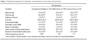
Table 1. Relationship between IUI indication, characteristics of the cycles and outcomes
DISCUSSION
Treatment of subfertile couples with IUI appears to be a valuable first line treatment before introducing more invasive and more expensive techniques of assisted fertilization in couples with various subfertility causes. Although IUI is the oldest technique in the spectrum of assisted reproduction, the pregnancy rates have not progressed as compared to IVF remarkable pregnancy rate improvement.
This study sought to determine the significance of different variables in the prediction of pregnancy rates in IUI patients. Our data suggests that female age, number of preovulatory follicles and number of inseminated motile spermatozoa are the best predictors of pregnancy rates in IUI cycles. No influence of the sperm morphology according to the previous or the recent WHO reference values on pregnancy rate was observed.
Female age has previously been reported as predictor of pregnancy rate in IUI cycles (Steures et al., 2004). Accordingly, Badawy et al (2009) also observed that women older than 35 years of age have lower chances of success in IUI cycles as compared to younger patients. In another study, the woman’s age was the strongest predictor of success in all indications (Merviel et al., 2008).
Our data suggests that the number of preovulatory follicles on the day of hCG administration predicts pregnancy rates in IUI cycles. Park et al (2007) addressed this correlation and reported that multiple follicular response increases pregnancy rate in IUI cycles. In a meta-analysis, multifollicular growth was associated with increased pregnancy rates in IUI with controlled ovarian hyperstimulation (van Rumste et al., 2006).
In our study, the number of inseminated motile spermatozoa was determinant to the likelihood of pregnancy. This finding is in accordance with Tomlinson et al. (1996). We observed a significant different pregnancy rates when the number of inseminated motile spermatozoa was bellow and superior to 1 million. Accordingly, Belaisch-Allart et al. (1999) reported similar results. Nonetheless, one study observed that the pregnancy rates per cycle were not statistically significant when the number of inseminated motile spermatozoa was below and superior to 1 million (Merviel et al., 2008). Contrarily, Campan et al. (Campana et al. 1996) did not obtain any pregnancies when the number of inseminated motile spermatozoa was below 1million.
Universally, the most common accepted classification system used for sperm morphology is the WHO criteria (Badawy et al., 2009). The threshold of spermatozoa with normal morphology below which IVF is recommended varies widely in the literature (Francavilla et al. 1990; Burr et al. 1996; Hauser et al. 2001). However, the implication of sperm morphology for choice and outcome of IUI is controversial. Our study failed to correlate the minimal number of spermatozoa with normal morphology (≥4%), according to the WHO (WHO 2010), with IUI outcome.
Burr et al. (1996) observed a decrease in pregnancy rate when teratospermia reached 90%. It has been suggested that a teratospermia rate over 80% implicates in the impairment of IUI success (Belaisch-Allart et al., 1999). Another study reported that the pregnancy rates were not significantly influenced by teratospermia as long as more than 5 million motile spermatozoa were available for insemination (Wainer et al. 2004). Contrarily, Merviel et al (2008) observed a significant decrease in pregnancy rate with a degree of teratospermia > 70%. Moreover, many previous studies also demonstrated that sperm morphology, before or after preparation, did not predict IUI pregnancy rates (Matorras et al. 1995; Karabinus and Gelety 1997; Dickey et al., 2002).
As an attempt to identify which infertility causes should benefit the most from IUI techniques, the cycles were divided into three groups according to IUI indication. Despite the observed differences regarding endometrial length and sperm morphology, pregnancy rates did not differed among the groups.
CONCLUSION
Our results suggest that couples with the best probability of pregnancy are those in which the woman is under 35 years old and the inseminated motile sperm count was at least 1 million. In addition, the ideal stimulation cycle allows the recruitment of at least two follicles measuring more than 16mm on the day of hCG administration. Our results demonstrate that even after the establishment of new reference values for semen parameters, sperm morphological normalcy still does not have any predictive value on IUI outcomes.
Referências Bibliográficas
1. Badawy A, Elnashar A and Eltotongy M. Effect of sperm morphology and number on success of intrauterine insemination. Fertil Steril. 2009;91:777-81.