JBRA Assist. Reprod. 2010;14(3):43-48
ARTIGO DE REVISÃO
doi: 10.5935/1518-0557.2010.14.3.09
Sperm selection for intracytoplasmic sperm injection
Seleção de espermatozoides para ICSI
1Center for Human Reproduction Prof. Franco Junior, Ribeirão Preto, Brazil
2Paulista Center for Diagnosis, Research and Training, Ribeirão Preto, Brazil
3Department of Gynecology and Obstetrics, Botucatu Medical School São Paulo State University, Botucatu, Brazil
ABSTRACT
The application of assisted reproduction techniques has provided help to many men seeking to father a child, although the current success of these procedures remains suboptimal. Today some protocols allow sperm to be selected according to their ultrastructural morphology or surface molecular characteristics. On the other hand, successful human reproduction relies partly on the inherent integrity of sperm DNA. Therefore, it is now necessary to improve the safety of the sperm selection method. It is urgent to optimize procedures to isolate spermatozoa for ICSI with low risk of DNA damage. In recent years, two technologies have attracted the attention of specialists as methods capable of identifying a spermatozoon with low risk of DNA damage: Ultrastructural morphology sperm selection at high magnification and sperm head birefringence selection. This review analyses these two technologies
Key words: sperm selection, high magnification sperm morphology, birefringence
RESUMO
A aplicação de técnicas de reprodução assistida tem ajudado muitos homens, embora o sucesso desses procedimentos continue abaixo do ideal. Hoje alguns protocolos permitem a seleção do esperma de acordo com sua morfologia ultra-estrutural ou características moleculares de superfície. Por outro lado, a reprodução humana bem sucedida depende, em parte, da integridade inerente do DNA do espermatozoide. Portanto, é necessário melhorar a segurança do método de seleção, sendo urgente a otimização de procedimentos para isolar espermatozoides para ICSI com baixo risco de danos no DNA. Nos últimos anos, duas tecnologias têm atraído a atenção de especialistas como métodos capazes de identificar espermatozoides com baixo risco de danos no DNA: a seleção de esperma pela ultra-morfologia em alta magnificação e pela birrefringência da cabeça. Esta revisão analisa essas duas tecnologias
Palavras-chave: seleção do esoerma, alta magnificação, mofologia do esperma, birrefringencia
The application of assisted reproduction techniques has provided help to many men seeking to father a child, although the current success of these procedures remains suboptimal. For many years the sperm selection methods were based on washing procedures with subsequent resuspension of the male germ cells. Double density gradient centrifugation and the swim-up procedure were used as standard preparations. Today some protocols allow sperm to be selected according to their ultrastructural morphology or surface molecular characteristics.
On the other hand, successful human reproduction relies partly on the inherent integrity of sperm DNA. Clinical evidence has shown that sperm nuclear DNA damage is closely related to male-derived repeated failure of ICSI attempts (Tesarik et al, 2004; Tesarik et al, 2005). It was also noted that the late paternal effect, but not the early one, is associated with increased sperm DNA fragmentation (Tesarik et al, 2004). Sperm DNA damage is associated with a significantly increased risk of pregnancy loss after IVF and ICSI (Zini et al., 2008).
Therefore, it is now necessary to improve the safety of the sperm selection method. It is urgent to optimize procedures to isolate spermatozoa for ICSI with low risk of DNA damage. In recent years, two technologies have attracted the attention of specialists as methods capable of identifying a spermatozoon with low risk of DNA damage: I - Ultrastructural morphology sperm selection at high magnification (Bartoov et al., 2001; Bartoov et al., 2002); II- Sperm head birefringence selection (Gianaroli et al., 2008).
I- Ultrastructural morphology sperm selection at high magnification
The accuracy with which morphological normality of spermatozoa for ICSI can be assessed depends on the resolution power of the optical magnification system. Conventionally, ICSI is performed with a x20/x40 objective, resulting in an overall optical magnification of x200 to x400 (Bartoov et al., 2003; De Vos et al., 2003). At this magnification, only major sperm morphological defects can be observed, whereas it is more difficult to identify subtle sperm organelle malformations that seem to be related to the ICSI outcome. To test this latter hypothesis, in 2001, Bartoov’s group developed a new method of unstained, real-time, high magnification motile sperm organellar morphology examination called MSOME. High magnification is made possible by the use of an inverted light microscope equipped with high-power Nomarski differential interference contrast optics enhanced by digital imaging to achieve a magnification of up to x6600 (Bartoov et al., 2001; Bartoov et al., 2002). Inclusion of MSOME, together with a micromanipulation system, enables the retrieval of a single motile spermatozoon with strictly defined morphologically normal nucleus to be injected into the retrieved oocytes. Bartoov and his group named this modified IVF procedure intracytoplasmic morphologically selected sperm injection (IMSI) (Bartoov et al., 2001).
Furthermore, spermatozoa appearing morphologically normal at this magnification (x400) may in fact carry various structural abnormalities that can only be detected at higher optical magnification. The spermatozoa with vacuoles would not be detected in conventional ICSI (Bartoov et al., 2003). This is a serious disadvantage, because microinjection of spermatozoa with vacuolated nuclei has been shown to be associated with low implantation and pregnancy rates, and with early miscarriage (Berkovitz et al. 2006; Hazout et al. 2006).
Sperm preparation and microscopy equipment for MSOME In our laboratory, a 1µl aliquot of sperm cell suspension was transferred to a 5µl microdroplet of modified HTF medium containing 7% polyvinylpyrrolidone solution (PVP medium; Irvine Scientific). This microdroplet was placed in a sterile glass dish (FluoroDish; Word Precision Instrument, USA) under sterile paraffin oil (Ovoil-100; VitroLife, Goteborg, Sweden). The sperm cells, suspended in the microdroplet, were placed on a microscope stage above an Uplan Apo x100 oil/1.35 objective lens previously covered by a droplet of immersion oil. In this manner, suspended motile sperm cells in the observation droplet could be examined at high magnification by an inverted microscope (Eclipse TE 2000 U; Nikon, Japan) equipped with high-power differential interference contrast optics (DIC/Nomarski). The images were captured by a color video camera containing effective picture elements (pixel) for high quality image production, and a color video monitor (Fig.1). Morphological evaluation was accomplished on a monitor screen and the total calculated magnification was = x8400 (total magnification: objective magnification X magnification selector X video coupler magnification X calculated video magnification) (Franco et al., 2008).
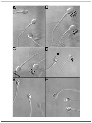
Figure 1. Microscope equipment for MSOME: (A) DIC system; (B) – Plan Fluor Apo x100 oil/1.35 lens + Prism; (C) Color video camera; (D) Immersion oil on objective; (E) Video monitor 21’.
MSOME criteria for IMSI selected sperm cells
At least 200 motile spermatozoa per patient were evaluated and the percentage of normal spermatozoa was determined.
Normal spermatozoa
A spermatozoon was classified as morphologically normal (Fig. 2A) when it exhibited a normal nucleus as well as acrosome, post-acrosomal lamina, neck and tail, besides not presenting a cytoplasmic droplet or cytoplasm around the head (Bartoov et al., 2001; Bartoov et al., 2002). For the nucleus, the morphological state was defined by the form and content of the chromatin. The criterion for normality of nuclear form was a smooth, symmetric and oval configuration. Normal means for length and width were estimated as 4.75±2.8 and 3.28±0.20µm, respectively, whereas the form classified as abnormal presented variation of 2SD in at least one of the axes (length: ≥5.31 or ≤4.19µm, width: >3.7 or <2.9mm). For rapid evaluation of nuclear form, a fixed, transparent, celluloid form of sperm nucleus fitting the criteria was superimposed on the examined cell (chablon construction based on ASTM E 1951-2). In the same manner, the form of the nucleus was considered normal if no extrusion or invagination of the nuclear chromatin mass had been detected (regional abnormality of nuclear form). Chromatin content was considered normal if the total vacuole area was found to occupy less than 4% of the nuclear area. A nucleus was considered normal if both nuclear form and chromatin content were normal.
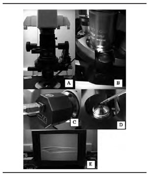
Figure 2. (A) Normal spermatozoa, (B-C); Abnormality of nuclear form: spermatozoa with small or large oval nuclear forms (B); spermatozoa with wide or narrow nuclear forms (C); (D-F) Abnormalities in nuclear chromatin: spermatozoa with regional shape abnormality of nuclear form (D), spermatozoa with vacuoles occupying 5-50% of the nuclear area (E) spermatozoa with large nuclear vacuoles (>50% of the nuclear area) (F).
Abnormalities of nuclear form
a- Spermatozoa with small or large oval nuclear forms (Fig.2B). Sperm cells exhibiting an abnormal but oval nuclear shape and a morphologically normal nucleus, content length ≤4.19µm or ≥5.31µm.b-Spermatozoa with wide or narrow nuclear forms (Fig.2C) - Sperm cells with non-oval, abnormal nuclear shapes, but with normal nuclear content, width: >3.7 or <2.9mm.c-Spermatozoa with regional shape abnormality of nuclear form (Fig.2D) - sperm cells with an extrusion or invagination of the nuclear mass.
Abnormalities of nuclear chromatin content
a. Spermatozoa with vacuoles occupying >4-50% of the nuclear area (Fig.2E)b. Spermatozoa with large nuclear vacuoles (Fig.2F) - sperm cells with vacuoles occupying >50% of the nuclear area.Sperm cells with a severe abnormality (such as pin, amorphous, tapered, round, multinucleated head, double tail) easily identified at low magnification (200x–400x) were not assessed. The abnormalities observed at high magnification, in both form and nuclear content, also presented normal acrosome, post-acrosomal lamina, neck and tail, and did not show a cytoplasmic droplet or cytoplasm around the head. Spermatozoids that presented more than one alteration were classified as having the most severe alteration (small/large < wide/narrow < regional shape abnormality < with vacuoles occupying >4%-50% < with vacuoles occupying >50% of the nuclear area).
IMSI and sperm nuclear vacuoles
One specific sperm malformation, which has been negatively associated with natural male fertility potential, is the presence of large nuclear vacuoles. In 2006, Berkovitz et al. carried out a more specific analysis on the impact of sperm cells with normal nuclear shape but large vacuoles, identified by MSOME, on ICSI pregnancy outcome. They performed a comparative study testing the outcomes of two matched IMSI groups: an experimental group (n=28), where spermatozoa with strictly defined normal nuclear shape but large vacuoles were available for oocyte microinjection, and a control group (n=28), where strictly defined morphologically normal spermatozoa (including nuclear shape and content) were retrieved for microinjection into the oocytes. Both groups satisfied the following selection criteria: maternal age <40 years and three or more retrieved metaphase II oocytes in the present cycle. As a result, the groups showed no differences as to fertilization, implantation rates or development of top quality embryos, whereas the pregnancy rate per cycle in the experimental IMSI groups was significantly lower, and the early miscarriage rate per pregnancy was significantly higher than that of the control group (18% versus 50%, p<0.01, and 80% versus 7%, respectively, p < 0.01). In this work, therefore, retrieval of spermatozoa with strictly normal nuclear shape but large vacuoles appeared to reduce ICSI pregnancy outcome and to be associated with early miscarriage. In fact, embryo development seemed normal at the early stages (no differences in top quality embryos, normal fertilization or implantation), whereas it seemed impaired at the later ones (low pregnancy and high miscarriage rates). In addition, according to Berkovitz et al. (2007) sperm nucleus morphological normality, assessed at high magnification, could decrease the prevalence of major fetal malformations in ICSI offspring.
IMSI and embryo development
Recently, it has been demonstrated that IMSI has no significant effect on embryo quality at day 2 in relation to the conventional ICSI procedure (Mauri et al., 2010). In addition, a relationship has been shown between defective spermatozoa and higher early miscarriage rates, despite the apparent lack of decrease in embryo quality on day 3 (Hazout et al. 2006). Based on the hypothesis that the employment of spermatozoa with large nuclear vacuoles would not produce any early paternal effects on embryo development (up to day 3), Vanderzwalmen and his group (Vanderzwalmen et al., 2008) investigated the possible influence of such nuclear vacuoles on the embryo’s competence to develop to the blastocyst stage; according to the researchers this may suggest a late paternal influence on embryo development after paternal DNA content begins to contribute to such advancement at around day 3 after fertilization. The outcome of embryo development (until day 5) was assessed in a group of 25 patients who underwent sibling oocyte microinjection with four different grades of sperm cells: (i) grade I (absence of vacuoles); grade II (≤2 small vacuoles); (iii) grade III (>2 small vacuoles or ≥1 large vacuole); and grade IV (large vacuoles with abnormal head shape or other abnormalities). Small (<4% of the head volume) and large vacuoles were defined according the classification of Bartoov (Bartoov et al., 2001; Bartoov et al., 2002), while grading was performed between x6000 and x12000 high magnification. To reduce the influence of female factor infertility, the inclusion criteria were female age less than 40 years and availability of at least eight oocytes at retrieval. As a whole, the four groups did not differ significantly as to the number of zygotes and embryo development up to day 3, including the subgroup analysis; on the contrary, the data showed highly significant differences in the development to blastocysts and the blastocyst quality among the four grades (p<0.001). On the other hand, comparing the groups one by one with regard to development to blastocysts, statistically signi-ficant differences were found between groups I (56.3%) and II (61.4%) and between groups III (5.1%) and IV (0%). Even when combining the groups into pairs, no significant difference in terms of embryo development to day 3 was seen (group I/II: 87.1% versus group III/IV: 66.7%), whereas the incidence of blastocyst and good quality blastocyst formation was significantly different between combined grades I/II and grades III/IV spermatozoa (43.5 and 19.1% versus 10.1 and 2.9% respectively, P < 0.001). Based on these results, the researchers postulated that the size and number of sperm nuclear vacuoles, identified accurately at high magnification, negatively affected blastocyst development, especially after a day-3 embryo transfer, and reinforced previous studies suggesting the paternal effects on initial embryonic development (Nadalini et al., 2009; Vanderzwalmen et al. 2008).
MSOME for routine laboratory semen analysis
Although MSOME was initially developed by its creator only as a selection criterion (Bartoov et al., 2001), its application as a morphological semen classification method could represent an improvement in routine laboratory sperm analysis, with potential clinical implications, especially in the field of medically assisted reproduction. In the past, only one study (Bartoov et al., 2002) has examined the relationship between normal spermatozoa obtained by the WHO routine method (World Health Organization, 1999) and by MSOME in 20 patients. As a result, no significant correlation was found between the incidence of morphologically normal spermatozoa as defined by the WHO and by MSOME. Nevertheless, routine analysis reported a significantly higher percentage of sperm normality in relation to MSOME. In a very recent paper, Oliveira et al. (2009) adopted a similar approach, evaluating the correlation between MSOME classification and a highly diffuse sperm morphology classification (Tygerberg criteria) (Menkveld et al., 1990), in order to better understand the potential diagnostic/prognostic value of the MSOME method. The study design included 97 randomly selected semen samples. Regression analysis showed a significant positive correlation between the incidence of normal sperm forms by Tygerberg criteria and that obtained by MSOME (r=0.8395%; CI: 0.75 to 0.88; P<0.0001). Similarly to the work presented by Bartoov et al. (2002), the MSOME criteria appear to be much more restrictive, presenting significantly lower sperm normality percentages for the semen samples in comparison to those found after routine analysis by the Tygerberg classification (3.3±3.2%, range 0-18%; and 9.4 ± 4.8%, range 2-23% respectively, P<0.0001). Based on these results, the researchers postulated that despite the high positive correlation, MSOME represented a much stricter evaluation criterion for sperm morphology, since its resolution power (≥x6000) enabled the identification of vacuoles and chromatin abnormalities that could not be described with the same accuracy by a Tygerberg method analysis. In addition, its focus on motile sperm fractions could only represent an additional advantage for MSOME by providing information on the fertilization and development potential of the sample fraction referred for assisted reproduction treatment. Therefore, our group stressed the importance of not only including MSOME among the criteria for routine laboratory semen analysis, but of performing this step prior to the conventional ICSI procedure (Oliveira et al., 2009).In 2006, Hazout et al., reported a positive association between high-magnification selection of sperm cells with normal nuclear shape and pregnancy outcome in patients with repeated conventional ICSI failures; in a subgroup of patients (n=72) involved in the study, the level of sperm DNA integrity (by TUNEL assay) was assessed, and the outcomes of IMSI could be compared in patients with several degrees of sperm DNA damage. The improvement of clinical ICSI outcomes was evident both in patients with an elevated degree of sperm DNA fragmentation and in those with normal sperm DNA status.In 2008, Franco et al. (2008) compared the amount of DNA fragmentation (by TUNEL assay) and the incidence of denatured single-stranded or normal double-stranded DNA (by acridine orange fluorescence method) in sperm cells characterized by the presence of large nuclear vacuoles (LNV group) and in strictly morphologically normal spermatozoa (NN group), both selected at high magnification (x8400). The analyses were carried out on fresh semen samples of 30 unselected patients. The authors reported a significantly higher level of DNA fragmentation in LNV sperm cells than in NN spermatozoa (29.1% versus 15.9%, P<0.0001). In addition, the LNV group also showed a significantly increased amount of single-stranded denatured DNA with respect to the NN sperm cells (67.9% versus 33.1%, P < 0.0001). Thus, we postulated that the high levels of denatured DNA in LNV sperm cells pointed to early decondensation and disaggregation of sperm chromatin fibers: an unwanted high degree of sperm decondensation could result in asynchronous chromosome decondensation, and may lead to cytoplasmic fragments in the embryo (Menezo et al., 2007). Therefore, the data presented stressed the link between the presence of large nuclear vacuoles and increased DNA damage in sperm cells and supported the routine selection of morphologically motile spermatozoa at high magnification (by MSOME) before conventional ICSI. A similar approach was adopted by Garolla et al. (2008) in particular, after observation via a high magnification system (x13000), 20 strictly morphologically normal sperm cells (for acrosome, head, neck and tail, including those cells which had large nuclear vacuoles, but were otherwise normal) were selected from each semen sample of 10 patients with severe testicular alteration and absent sperm motility: 10 with normal morphology and no vacuoles (group A) and 10 with normal morphology and at least one large head vacuole (group B), for a total of 200 sperm cells. Each spermatozoon was studied for mitochondrial membrane potential, DNA integrity (by acridine orange staining), DNA fragmentation (by TUNEL assay) and sperm aneuploidies (by FISH analysis). The data showed that single cells from group A exhibited a significantly better physiological status than cells from group B with regard to mitochondrial function, DNA integrity and DNA fragmentation (13.3 ± 4.9% versus 52.2 ± 14.7%, 5.3 ± 3.0% versus 71.9 ± 11.1%, and 9.3 ± 4.8% versus 40.1 ± 11.6% respectively, all P<0.001). No chromosomal alteration was present in cells from group A. Therefore, although the study was conducted on a highly restricted number of cells, the results strengthened the concept of an association between the incidence of sperm DNA damage and the presence of nuclear vacuoles and stressed the importance of a morphological selection by high-magnification microscopy (Nadalini et al., 2009).In conclusion, all the publications (Antinori et al., 2008; Bartoov et al., 2003; Berkivitz et al., 2006; Hazout et al., 2006) about IMSI reported not only better results as to implantation and clinical pregnancy rates but also a reduction in the miscarriage rates in couples whose sperm cells were strictly morphologically selected at high magnification (MSOME). In addition, the IMSI procedure improved the clinical ICSI outcome in patients with several degrees of sperm DNA damage.It has been reported that the presence of vacuoles could reflect molecular anomalies responsible for abnormal chromatin remodeling during the sperm maturation process and may contribute to making the spermatozoa more susceptible to sperm DNA impairment. The current results establish a crucial association between normal blastocyst development and both the number and size of vacuoles, indicating that routine morphological analysis of sperm cells, performed at high magnification (x6000), is of fundamental importance to improving the embryo implantation potential. In addition, the presence of vacuoles in the nuclei of spermatozoa is also associated with reduced pregnancy and with a high level of DNA damage.Therefore, in light of these findings, the MSOME method seems to be a powerful tool for selecting strictly morphologically normal spermatozoa, with a lower incidence of DNA defects that cannot be detected at routine ICSI magnification.
II. Sperm head birefringence (SHBF) selection
It is clear that in cases of intracytoplamic sperm injection (ICSI), although the “best” spermatozoon is selected, cells with DNA damage are routinely injected into the oocyte. Baccetti et al. (2004) demonstrated that it is possible to combine a polarization apparatus with an inverted light microscope used by ICSI to analyze the human motile sperm head birefringence (SHBF) to indicate sperm structural normality. Gianaroli et al. (2008) were the first to use SHBF characteristics and apply polarization inverted microscopy to select sperm for ICSI and demonstrated better embryo development and clinical outcome results after injection of SHBF spermatozoa when compared with controls (no SHBF spermatozoa). Recently, the same group carefully evaluated two types of SHBF on the basis of their acrosomal integrity (acro-some-reacted and acrosome-non-reacted spermatozoa) and demonstrated better clinical results when acrosomereacted spermatozoa where used for ICSI (Gianaroli et al., 2010). The data from this study point to the injection of reacted spermatozoa as the best option to achieve pregnancy in patients with male factor infertility.
Semen evaluation and processing
Semen samples were collected in sterile containers by masturbation after a period of 2 to 5 days of sexual abstinence. The semen was analyzed for sperm concentration, volume and quality of motility. The liquefied fresh semen samples were prepared by using a layering method that consisted of permitting the ejaculated sperm sample to migrate up to the human tubal fluid (HTF) medium with 10% human serum albumin (10% HAS) deposited on top of the fresh semen sample, at 2:1 proportion, for 30 minutes at the temperature of 37ºC. The portion of motile sperm was resuspended in HTF/10%HSA medium and the concentration was adjusted to 3x10⁶sperm/ml.
Birefringence evaluation
The SHBF was assessed and selected by using an inverted microscope (TE 300 Nikon, Japan) equipped with Hoffman contrast and polarizing lenses. The total calculated magnification was 2,500X obtained by using x20 Hoffman objective, camera, C-mount and 21-inch monitor (Fig. 3). SHBF selection was performed after a half hour of the deposition of 1µl of prepared sperm into a 10µl microdrop of 7% polyvinylpyrrolidone solution (PVP 7%) (Irvine, USA) covered by oil (Vitrolife, Sweden) and presented in a plastic Petri dish (Corning ref: 430166) at room temperature. Two types of SHBF can be selected: 1. SHBF total (SHBF-T) consists of motile spermatozoa that presented BF throughout their head (Fig. 4A); SHBF partial (SHBF-P) is comprised of those motile spermatozoa with BF present in 50% of their head, with this birefringence being localized in the post-acrosomal region (Fig. 4B).
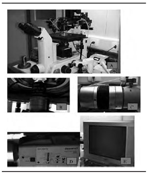
Figure 3. (A) The polarization inverted microscope (TE 300 Nikon, Japan) equipped with Hoffman contrast and polarizing lens; (B) Hoffman objective of 20x; (C) C-mount; (D) Camera and 1.E-21-inch monitor.
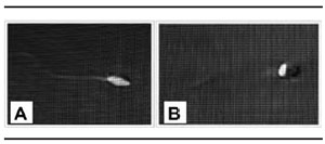
Figure 4. (A) Spermatozoa with total sperm head birefringence (SHBF-T) observed at 2,500 x magnification; (B) Spermatozoa with partial sperm head birefringence (SHBFP) observed at 2,500X magnification
The analysis of birefringence in sperm cells by using polarization microscopy has been reported to be an indicator of nuclear structural normality. The presence of SHBF is expressed by an organized and highly compacted texture that characterizes normal sperm nuclei, acrosomes, and motile tails (Baccettti, 2004).Gianaroli et al. (2008) have proposed the injection of spermatozoa with birefringence in their head as a diagnostic tool to improve clinical outcome in treatment of the most severe male factor cases. Along this line of thought, the same group recently defended the hypothesis that human spermatozoa possess birefringence characteristics that reflect the state of the inner protoplasmic structures (Gianaroli et al., 2010). The authors argue the hypothesis that abnormalities in the protoplasmic compartment of the sperm head may be related to anomalies of sperm chromatin packaging and incomplete nuclear remodeling occurring in the final phase of spermatogenesis. However, there are no studies in the literature that define the precise correlation between birefringence and protoplasmic structure.In the literature, only one study analyzed the relationship between sperm head birefringence and DNA damage (Crippa et al., 2009). The authors found a negative correlation between SHBF and DNA fragmentation, i.e. they showed that the proportion of sperm birefringence was inversely correlated with the incidence of fragmented DNA. In their study, they evaluated the total semen sample both by the presence of SHBF in their head and the DNA fragmentation, but did not describe the types of SHBF (partial and total).Recently, our group evaluated two types of SHBF (total and partial) and their relationship with DNA fragmentation, directly from those spermatozoa selected according to their birefringence. The data show that DNA fragmentation values are significantly higher in spermatozoa with total SHBF, compared to those with partial SHBF (Petersen et al., 2010). This fact could explain the results obtained by Gianaroli et al. (2010), who reported higher embryo implantation rate (39% vs. 8.6%) when oocytes were injected with reacted spermatozoa (SHBF-P) than when injected with non-reacted (SHBF-T)) spermatozoa. Gianaroli et al. (2010) also suggest that birefringence in the absence (SHBF-T) or presence (SHBF-P) of the acrosomic reaction would be their selection criterion for identifying sperm health. But this hypothesis remains to be proven.However, DNA damage should alter the special cellular functions of human spermatozoa, and lead to diminished acrosome reaction with reduced fertilization rates. Moreover, negative correlation was identified between DNA damage and acrosome reaction and/or viability of human spermatozoa (Ozmen et al., 2007).Recently, our group demonstrated that the specific SHBF pattern (total or partial) was not an efficacious means of selecting sperm chromatin packaging abnormalities, at least when observed by chromomycin A3 staining.The percentage of positive chromomycin A3 (abnormal chromatin packaging) in spermatozoa with SHBF-T was (40.5%) not significantly different (Fisher’s exact test; p=0.28) from the percentage (37.3%) of spermatozoa with SHBF-P (Vagnini et al., 2010).In conclusion, there was significantly more DNA fragmentation in spermatozoa with SHBF-T than in those with SHBF-P. The results support a positive relationship between spermatozoa with SHBF-T and increase in DNA fragmentation and the use of SHBF as a sperm selection method.