JBRA Assist. Reprod. 2011;15(5):34-38
ARTIGO ORIGINAL
doi: 10.5935/1518-0557.2011.15.5.06
The prognostic value of the testicular histopathological pattern for sperm retrieval and intracytoplasmic sperm injection outcomes in non-obstructive azoospermic patients
O valor prognóstico do padrão histopatológico testicular na recuperação de espermatozóides e nos resultados da injeção intracitoplasmática de espermatozóides em pacientes com azoospermia não-obstrutiva
aFertility - Assisted Fertilization Center Av. Brigadeiro Luis Antônio, 4545 - São Paulo - SP, Brazil
bSapientiae Institute - Educational and Research Center in Assisted Reproduction Rua Vieira Maciel, 62 - São Paulo - SP, Brazil
cInstitute of Biotechnology, University of Caxias do Sul Rio Grande do Sul, Brazil
ABSTRACT
Objective: To evaluate the prognostic value of testicular histology on sperm retrieval success and intracytoplasmic sperm injection (ICSI) outcomes.
Methods: Sixty couples undergoing testicular sperm aspiration (TESA) for ICSI were divided into three groups according to their testicular histopathological diagnosis: (i) Hypospermatogenesis (HS, N=24), (ii) Maturation arrest (MA, N=17), and (iii) Sertoli-cell-only syndrome (SCOS, N=19). Sperm retrieval success and ICSI outcomes were compared. The relationship between histological diagnosis and (i) serum FSH value and (ii) testicular volume was also investigated.
Results: Sperm retrieval rate (HS: 91.6%, MA: 35.2% and SCOS: 26.3%, p<0.001) and fertilization rate (HS: 69.9%, MA: 49.3%, and SCOS: 43.3%, p=0.048) were significantly higher in HS group. A trend towards a higher percentage of high-quality embryos was noted in HS group (HS: 55.0%, MA: 31.0% and SCOS: 30.9%, p=0.076). No differences in testicular volume were observed. Nevertheless, FSH value was significantly higher in SCOS patients (HS: 10.2±5.4 mIU/mL, MA: 9.9±6.8 mIU/mL, and SCOS: 26.1±15.3 mIU/mL, p< 0.01).
Conclusions: Histological findings present in testicular biopsy specimens of NOA patients were able to predict sperm retrieval success and ICSI outcomes. An elevated serum FSH level is significantly correlated with SCOS presence in azoospermic patients.
Palavras-chave: Hipoespermatogênese, parada de maturação, síndrome Sertoli-cell-only, azoospermia não-obstrutiva, Injeção Intracitoplasmática de Espermatozóide.
RESUMO
Objetivo: Avaliar o valor prognóstico da histologia testicular no sucesso da recuperação de espermatozóides e nos resultados da injeção intracitoplasmática de espermatozóides (ICSI).
Métodos: Sessenta casais que se submeteram a aspiração de espermatozóides testiculares (TESA) para ICSI foram divididos em três grupos de acordo com o diagnóstico histopatológico testicular: (i) hipoespermatogênese (HS, N = 24), (ii) parada de maturação (MA, N = 17), e (iii) Sertoli-cell-only syndrome (SCOS, N = 19). O sucesso da recuperação de espermatozóides e os resultados de ICSI foram comparados entre os grupos. As relações entre o diagnóstico histológico e (i) o valor FSH sérico e (ii) volume testicular também foram investigadas.
Resultados: A taxa de recuperação de espermatozóides (HS: 91,6%, MA: 35,2% e SCOS: 26,3%, p <0,001) e taxa de fertilização (HS: 69,9%, MA: 49,3%, e SCOS: 43,3%, p = 0,048) foram significativamente maiores no grupo HS. A tendência para um maior percentual de alta qualidade embriões foi observado no grupo HS (HS: 55,0%, MA: 31,0% e SCOS: 30,9%, p = 0,076). Nenhuma diferença no volume testicular foi observada. No entanto, o valor de FSH foi significativamente maior em pacientes SCOS (HS: 10,2 ± 5,4 mIU / mL, MA: 9,9 ± 6,8 mUI / mL, e SCOS: 26,1 ± 15,3 mUI / mL, p <0,01).
Conclusões: Os achados histológicos presentes em amostras de biópsia testicular de pacientes com azoospermia não-obstrutiva foram capazes de prever o sucesso de recuperação de espermatozóides e os resultados ICSI. Um nível elevado de FSH sérico está significativamente correlacionado com a presença de SCOS em casos de azoospermia.
Key words: Hypospermatogenesis, Maturation arrest, Sertoli-cell-only syndrome, non-obstructive azoospermia, Intracytoplasmic Sperm Injection
INTRODUCTION
Azoospermia, the absence of sperm in the ejaculate, is present in about 15% of infertile men and is classified as either obstructive azoospermia (OA), in which patients have normal spermatogenesis, or non-obstructive (NOA), which is characterized by impaired spermatogenesis (Tournaye et al., 1997). Since its introduction in 1992 (Palermo et al., 1992), the Intracytoplasmic Sperm Injection (ICSI) has become the treatment of choice for severe male factor infertility and, in addition to ejaculated sperm, testicular and epididymal sperm can all be used for injection, resulting in high fertilization and good pregnancy rates (Pasqualotto et al., 2002).
Testicular sperm extraction (TESE), testicular sperm aspiration (TESA), and epididymal sperm aspiration (PESA) combined with ICSI offers azoospermic patients the possibility of having their own genetic children (Craft and Shrivastav, 1994). In patients with OA, mature sperm cells can be retrieved in most cases, while in NOA patients, sperm is retrieved in approximately 50% of cases (Tournaye et al., 1997).
In fact, some men with NOA have focal areas of spermatogenesis within the testis, despite the fact that the overall spermatogenic function of the testis is severely impaired (Tash and Schlegel, 2001). In these cases, the ability to find sperm in the testis vary according to the histopathological pattern of the testis (De Croo et al., 2000), which has been classified as follows: (i) hypospermatogenesis (HS), (ii) maturation arrest (MA), (iii) Sertoli-cell-only syndrome (SCOS), and (iv) tubular hyalinization (McLachlan et al., 2007).
Even when testicular sperm are found, ICSI outcomes are reportedly affected by the site of sperm origin. It has been observed that ejaculated and epididymal spermatozoa provide better quality embryos than testicular sperm (Rossi-Ferragut et al., 2003). Furthermore, it has been described that testicular sperm injection results in lower fertilization and pregnancy rates compared to epididymal sperm injection (Pasqualotto et al., 2002; Nicopoullos et al., 2004).
Although it has been suggested that the probability of finding viable sperm in the testis depends on the diagnostic of the testicular biopsy (Seo and Ko, 2001; Tsujimura et al., 2002), whether or not the testicular histophatological pattern influences the ICSI outcomes has not been fully elucidated. Therefore, our goal in this study was to evaluate the prognostic significance of testicular histology on sperm retrieval success and ICSI outcomes in NOA patients. In addition, we examined the relationship between the testicular histopathological diagnosis and the serum FSH value and the testicular volume.
MATERIALS AND METHODS
Experimental design
The present study included 60 couples undergoing TESA for ICSI. The patients were divided into following three groups according to the testicular biopsy diagnosis: (i) HS (N=24), (ii) MA (N=17), and (iii) SCOS (N=19). The sperm retrieval success, fertilization rate, percentage of high-quality embryos, implantation rate, and pregnancy rate were compared among the groups. In addition, the relationships between the histopathological diagnosis and (i) the serum FSH value and (ii) the testicular volume were investigated. In all cases testis volume was manually determined by the same operator. Positive sperm retrieval was defined as the presence of spermatozoa in the biopsy specimen. The implantation rate was defined as the total number of gestational sacs divided by the total number of embryos transferred. Clinical pregnancy was defined as the presence of a gestational sac on ultrasound 4 to 6 weeks after embryo transfer. The study was approved by the local institutional review board and written informed consent was obtained from all patients, in which they agreed to share the outcomes of their ICSI cycles for research purposes.
Controlled ovarian stimulation
Controlled ovarian stimulation was achieved by long pituitary down-regulation using a GnRH agonist (Lupron Kit™, Abbott S.A Societé Française des Laboratoires, Paris, France) followed by ovarian stimulation with recombinant-FSH (Gonal-F®, Merck-Serono, Geneva, Switzerland). The follicular dynamic was followed with ultrasound and when adequate follicular growth and serum estradiol levels were observed, recombinant human chorionic gonadotrophin (rhCG, Ovidrel™, Merck-Serono, Geneva, Switzerland) was administered to trigger the final follicular maturation. Oocytes were collected 34-36 hours after hCG administration using transvaginal ultrasound ovum pick-up.
Preparation of oocytes
After retrieval, oocytes were placed in human tubal cultured medium (HTF, Irvine Scientific, Santa Ana, USA) supplemented with 10% Human Serum Albumin (HSA, Irvine Scientific, Santa Ana, USA) covered with mineral oil (Ovoil™, Vitrolife, Kungsbacka, Sweden) and incubated at 37ºC in 6% CO2 for 5 hours. Cumulus cells were removed with 30s exposure to a HEPES-buffered HTF medium (Irvine Scientific, Santa Ana, USA) containing 80IU/mL hyaluronidase (Irvine Scientific, Santa Ana, USA). Coronal cells were then manually removed using a finely drawn glass Pasteur pipette (Humagen Fertility Diagnostics, Charlottesville, Virginia, USA). Denuded oocytes were then assessed for nuclear status. Oocytes that were observed to have released the first polar body were considered mature and used for ICSI.Sperm samples - Testicular Sperm AspirationAfter administration of cord block anesthesia, TESA was performed by longitudinally inserting a 21-gauge butterfly needle into the superior testicular pole while avoiding the epididymis. Forward and backward movements were made, and the needle direction was slightly changed to sample eight different spots in the testis, thereby increasing the chance of contacting a spermatogenesis focus in a multifocal procedure. In addition, the negative pressure applied during needle aspiration (which was obtained by connecting a 10mL syringe to the end of the butterfly catheter) allowed for the extraction of parts of the seminiferous tubules for use in further dissection to search for germ cells. Morphologically normal and motile sperm were immobilized, aspirated into the microinjection needle and injected into the MII oocytes (Palermo et al., 1992). In all patients, a small piece of testicular tissue was placed in Bouin’s solution and sent for pathological evaluation. Histological findings were classified based on the most advanced pattern observed on biopsy specimens, as previously published classifications (McLachlan et al., 2007; Levin, 1979).
Assessment of fertilization, embryo quality, and embryo transfer
Fertilization was assessed 18 hours after ICSI, and normal fertilization was declared when two distinct pronuclei were present. Embryo transfer was performed on the third day of development. One to three embryos from each couple were transferred. Embryo selection for transfer was performed based on embryo morphological parameters. High-quality embryos were defined as those possessing all of the following characteristics: 8 to 10 cells, less than 15% fragmentation, symmetric blastomeres, an absence of multinucleation, colorless cytoplasm with moderate granulation and no inclusions, an absence of perivitelline space granularity, and an absence of zona pellucida dysmorphism.
Statistical analysis
Results were expressed as mean ± standard deviation for numeric variables, while proportions (%) were used for categorical variables. Proportions were compared by the Chi-squared or Fisher exact test, only when expected frequency was five or less. ANOVA was employed for the analysis of continuous variables, and residual normality was tested for all variables. Transformations were performed whenever necessary. Results were described as untransformed means and standard deviations. Results were considered to be significant at the 5% critical level (p<0.05). Data analysis was carried out using Minitab (version 14) Statistical Software.
RESULTS
Hypospermatogenesis was the most common histopathological diagnosis identified (HS: 40.0%, MA: 28.3% and SCOS: 31.7%). No cases of testicular hyalinization were found. The couples’ general characteristics and the ICSI cycle characteristics were equally distributed among the groups (Table 1).
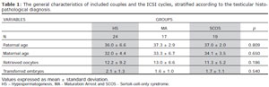
Table 1. The general characteristics of included couples and the ICSI cycles, stratified according to the testicular histopathological diagnosis.
The sperm retrieval rate was significantly higher in the HS group than in the other groups, however it did not differ between the MA and SCOS groups (HS: 91.6%, MA: 35.2% and SCOS: 26.3%, p<0.001). The fertilization rate was also significantly higher when HS was diagnosed as compared to when MA or SCOS was diagnosed (HS: 69.9%, MA: 49.3%, and SCOS: 43.3%, p=0.048).
Moreover, a trend toward a higher percentage of high-quality embryos was noted in the HS group as compared to the other groups (HS: 55.0%, MA: 31.0% and SCOS: 30.9%, p=0.076) (Fig.1).
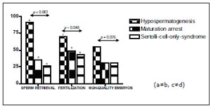
Figure 1. Sperm recuperation rate, fertilization rate and percentage of high quality embryos according to the testicular histology.
However, the pregnancy (HS: 27.8%, MA: 25.0% and SCOS: 16.7%, p=0.854) and implantation rates (HS: 14.0%, MA: 12.2% and SCOS: 4.2%, p=0.684) did not differ among the three groups (Fig.2).
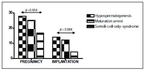
Figure 2. Clinical outcomes according to the testicular histology.
No differences in the testicular volume (HS: 10.9±11.1cm3 MA: 14.2±6.0cm3, and SCOS: 11.8±7.9cm3, p=0.657) was observed when the three testicular histopathological patterns were compared. However, it the FSH value was found to be significantly higher among SCOS patients than among patients with other histopathological diagnoses (HS: 10.2±5.4mIU/mL MA: 9.9±6.8mIU/mL, and SCOS: 26.1±15.3mIU/mL, p<0.01).
DISCUSSION
The advent of ICSI using testicular spermatozoa has significantly improved the treatment options available to NOA patients. However, failure of surgical sperm aspiration may occur in up to 50% of attempts (Schlegel et al., 1997; Rosenlund et al., 1998), and even when sperm is recovered from the testis, the results of the fertilization, embryo development, and pregnancy may be rather disappointing (Nicopoullos et al., 2004; Rossi-Ferragut et al., 2003; Pasqualotto et al., 2002). The present study examined the influence of the testicular histopathological pattern of NOA patients not only in terms of sperm retrieval success, but also in terms of fertilization ability and embryo development.
Since endocrine and genetic tests cannot reliably distinguish OA from NOA, it has been reported that the testicular biopsy has diagnostic importance for assisted reproductive technologies for patients with male factor infertility. The prognostic importance of testicular histology in NOA patients, however, remains under debate. While some reports have found an important correlation between the histopathological pattern of the testis and successful sperm isolation (Seo and Ko, 2001; Tsujimura et al., 2002; Silber, 2000), others have concluded that histology is of limited prognostic significance (Schoor et al., 2002).
In our study, a significant correlation was observed between the testicular histology and successful sperm retrieval. Despite testicular histology limitation to reflect the overall testicular biology an accurate assessment is important for planning infertility treatments because the presence of even small areas of complete spermatogenesis makes successful sperm retrieval very likely. Moreover, we noted that the fertilization rate and percentage of high-quality embryos were significantly higher in HS patients than among patients in the other groups and that the outcomes of the SCOS patients were the worst among the three groups.
Spermatogenesis is an elaborate process of cell differentiation. The first step in spermatogenesis is spermatogonial proliferation, which leads to the maintenance of the stem cells that can enter the spermatogenetic process resulting in the production of spermatozoa (de Kretser et al., 1998; de Rooij, 2001). Throughout adult life, spermatogonial stem cells furnish cells for maturation in a cyclical pattern, while at the same time renewing them to maintain a constant reservoir to allow continuous production of spermatozoa. The second step in spermatogenesis is the differentiation of spermatogonia to spermatozoa through meiosis and spermiogenesis (Chemes, 2001; Griswold, 1998).
The etiology of HS has not been fully elucidated. The histological findings of the testicular biopsy specimens of patients with HS demonstrate various patterns. It has been suspected that some patients have a decrease in the spermatogonial population, as result of a cell proliferative disorder during the first step of spermatogenesis (Takagi et al., 2001). Our results suggest that finding normal spermatogenic foci in patients with HS is common, and that the fertilization and embryo development ability of the retrieved sperm may be comparable to those retrieved from OA patients.
Sertoli-cell-only syndrome is diagnosed when testicular biopsy reveals that seminiferous tubules are lined only by Sertoli cells, with a complete absence of germ cells but with a normal interstitium (Rothman et al., 1982). In this case, sperm retrieval is likely to be successful in about a quarter of patients (Okada et al., 2002; Tsujimura et al., 2002), reflecting focal spermatogenesis that is not apparent on biopsy. Different etiologies of SCOS have been described: idiopathic infertility, Y chromosome microdeletions, previous orchitis, chemoor radiotherapy, or an embryological failure of germ cell migration to the gonadal ridge (McLachlan et al., 2007). Sertoli cells secrete soluble factors and play important roles in spermatogenesis. Impairment of Sertoli cells is a possible cause of several types of spermatogenic impairment, including SCOS (Fujita et al., 2005). The lower fertilization rate that we observed among SCOS patients prompts us to speculate that although some spermatogenic foci may be identified and that sperm may be retrieved from SCOS patients, the fertilization ability of a single injected sperm may be impaired.
In fact, it is well-recognized that the quality of sperm plays a key role during fertilization (Swann et al., 2006). It has also been shown that pre-implantation embryonic development can be compromised by deficiencies in both the nuclear genome of the sperm or by sperm-derived cytoplasmic factors (Tesarik, 2005). In fact, we observed that when sperm retrieved from testes with different histological patterns are injected into oocytes, embryonic development may also be affected.
The present study also examined the relationship between testicular histopathological pattern and the serum FSH value and testicular volume. It has been well-established that the reproductive hormone profile (especially the serum concentrations of inhibin B and FSH) of adult men reflects the functional state of their seminiferous epithelium in testes (Jensen et al., 1997). In addition, Seo and Ko (2001) sought to investigate the factors that were predictive sperm of recovery in NOA patients and found that spermatozoa recovery had no correlation with serum FSH level. Our data showed that there was a significant increase in the FSH level when SCOS was observed. The correlation between serum FSH levels and SCOS has been previously demonstrated and it has been suggested that elevated serum FSH levels make testicular biopsies superfluous for diagnostic purposes in these patients (Bergmann et al., 1994).
In conclusion, our data demonstrated that testicular histopathology is able to predict the sperm retrieval rate, fertilization ability, and embryo developmental competence of the injected spermatozoa of NOA patients used in ICSI. Moreover, the serum FSH level is significantly correlated with the presence of SCOS in azoospermic patients.
Chemes HE, Infancy is not a quiescent period of testicular development. Int J Androl. 2001;24:2-7.
Craft I, Shrivastav, P, Treatment of male infertility. Lancet. 1994;344:191-192.
Rothman CM, Sims, CA, Stotts, CL, Sertoli cell only syndrome 1982. Fertil Steril. 1982;38:388-390.