JBRA Assist. Reprod. 2015;19 (3):119-124
ORIGINAL ARTICLE
doi: 10.5935/1518-0557.20150027
Non-Invasive Prediction of Blastocyst Formation by Day Three Embryo Culture Medium Mass Spectrometry Lipid Fingerprinting
1Disciplina de Urologia, Departamento de Cirurgia – UNIFESP - Brasil
2Instituto Sapientiae – Centro de Estudos e Pesquisa em Reprodução Assistida - Brasil
3Fertility – Medical Group – Sao Paulo - Brasil
4Centro Pluridisciplinar de Pesquisas Químicas, Biológicas e Agrícolas – CPQBA – UNICAMP - Brasil
5Laboratório ThoMSon de Espectrometria de Massas – Instituto de Química – UNICAMP - Brasil
CONFLICT OF INTERESTS
No conflict of interest have been declared.
ABSTRACT
Objective: To identify lipid markers of blastocyst formation by day three culture medium mass spectrometry (MS) fingerprinting.
Methods: For this study, 50 embryo samples from culture media were harvested on day three, from patients undergoing embryo transfers on day five. Samples were split into groups based on their degree of expansion and hatching status on day five (Complete-Blastocyst, n=25 and No-Blastocyst, n=25) and its secretomes were analysed by MS. Mass spectra fingerprinting was acquired using a Q-Tof spectrometer (LC-MS, Agilent 6550 iFunnel Q-TOF) equipped with an automated injector. The data was analysed using the principal component analysis (PCA) followed by a partial least square discrimination analysis (PLS-DA), combined with variable influence in the projection (VIP) scores.
Results: In total, there were 1,657 ions found, in which 165 ions were differently expressed between groups, with a fold chance ≥ 4x and P<0.001, in the t-test. PLS-DA showed a clear separation between the groups and among 15 VIPs selected by the program, 13 of them were highly expressed in the Complete-Blastocyst Group and two were expressed in the No-Blastocyts Group. Besides embryo status on day five, the PLS-DA was also able to classify samples according to patients’ age. Lipids supposedly highly expressed in the Complete-Blastocyst Group included: isoprenoids, diacylglycerols, sterols, fatty esters, secosteroids, phosphosphingolipids, glycerophosphates and diacylglycerophosphates, while fatty amides were suggested to be highly expressed in the No-Blastocysts Group.
Conclusions: Day three culture medium MS is a promising approach for the identification of embryos that should be cultured until day five.
Keywords: Lipid, Fingerprinting, Embryo, Blastocyst, Mass Spectrometry
INTRODUCTION
Although more than two million children have already been born as a consequence of assisted reproductive technologies (ART), around 70-80% of in vitro produced embryos fail to implant, and 66% in vitro fertilization (IVF) cycles fail to result in pregnancy (Assou et al., 2011; Seli et al., 2004; Patrizio and Sakkas, 2009). The identification of factors that may influence the implantation of in vitro-produced embryos is one of the most investigated fields in ART. Although high-quality embryos may be available for transfer, choosing the best embryo for transfer has become a major challenge in IVF.
Serial observation of embryo morphology, which is a common technique for embryo evaluation, has been considered to be a key predictor of implantation and pregnancy (Balaban et al., 2001; Lundin et al., 2001; Lan et al. 2003; Nagy et al., 2003; Payne et al., 2005; Holte et al., 2007; Racowsky et al., 2010; Ahlstrom et al., 2011; Alpha Scientists, 2011; Aydin et al., 2011). However, it has been suggested that embryo morphology is insufficient for predicting successful implantation (Katz-Jaffe et al., 2009; Assou et al., 2011; Mastenbroek et al., 2011). In addition, this method is highly subjective (Paternot et al., 2009;Filho et al., 2010).
Prolonging the embryo culture period allows for a better selection of embryos for transfer because laboratory assessment is performed after the embryonic genome has begun to be expressed (Tesarik et al., 1988). However, because of our continuing inability to predict which cleavage-stage embryos will develop into viable blastocysts (Schoolcraft and Gardner, 2001; Westphal et al., 2003), assisted reproduction centres are reluctant to adopt extended embryo culture to avoid embryo transfer cancellations (Sepulveda et al., 2011).
In the post-genomic era, many “omics” efforts are being focused on understanding the relationships between genome, DNA transcripts, proteins, metabolites and phenotypes in cells and organisms (Katz-Jaffe et al., 2009). Non-invasive approaches for embryonic development potential have the advantage of increasing the knowledge of embryo physiology, therefore enabling the development of methods to predict developmental competence and viability (Hamel et al., 2008). These approaches include genomic and proteomic profiling, embryonic metabolome analytical examination (Botros et al., 2008; Bromer and Seli, 2008; Katz-Jaffe et al., 2009; Aydiner et al., 2010; Ferreira et al., 2010; Seli et al., 2010; Cortezzi et al., 2011), and most recently: lipidomics (Quehenberger et al., 2010).
Mass spectrometry (MS) fingerprinting has been shown to provide a reliable approach to evaluate culture media profiles (Ferreira et al., 2009). MS fingerprinting has been applied to evaluate embryo proteome and metabolome, with promising results (Katz-Jaffe et al., 2006; Cortezzi et al., 2011; D’Alessandro et al., 2012; Lagarrigue et al., 2012; Sanchez-Ribas et al., 2012; Cortezzi et al., 2013).
Therefore, the goal for the present study is to utilise the analytical power of MS with minimal sample preparation and minute analysis to identify possible lipid markers of embryo developmental competence in the surrounding culture medium.
MATERIAL AND METHODS
Experimental Design
For this study, 50 culture media samples were harvest on day three, from patients undergoing day five embryo transfers. Embryos were split into groups based on their degree of expansion and hatching status on day five (Complete-Blastocyst, n=25 and No-Blastocyst, n=25) and their secretomes were analysed by MS.
The patients signed an informed consent form, in which they agreed to share the outcomes of their cycles for research purposes. The local institutional review board approved the study.
Controlled Ovarian Stimulation and Oocyte Retrieval
Controlled ovarian stimulation was achieved by using recombinant FSH (Gonal-F; Serono, Geneva, Switzerland), as a daily dose, starting on day three of the cycle. Pituitary blockage was performed by using a GnRH antagonist (Cetrotide, Serono, Geneva, Switzerland), starting when at least one follicle ≥14 mm was visualised.
Follicular growth was monitored using transvaginal ultrasound examination starting on day four of the gonadotropin administration. When adequate follicular growth and serum 17β estradiol levels were observed, recombinant hCG (Ovidrel; Serono, Geneva, Switzerland) was administered to trigger the final follicular maturation. The oocytes were collected 35 hours after hCG administration through transvaginal ultrasound ovum pick-up.
Preparation of Oocytes and Morphology Assessment
Retrieved oocytes were maintained in culture medium (Global® for Fertilization, LifeGlobal, Connecticut, USA) supplemented with 10% Human Synthetic Albumin (HSA, Irvine Scientific, Santa Ana, USA), covered with mineral oil (Ovoil™ - Vitrolife, Kungsbacka, Sweden) at 37°C and 6% CO2 for 5 hours. Surrounding cumulus cells were removed with exposure to a HEPES buffered-medium containing hyaluronidase (80 IU/mL, Irvine Scientific, Santa Ana, USA). The remaining cumulus cells were then mechanically removed by gentle pipetting with a hand-drawn Pasteur pipette (Humagen Fertility Diagnostics, Charlottesville, Virginia, USA). The oocytes were checked for oocyte maturation and those which had released the first polar body (metaphase II oocytes – MII) were considered mature and used for ICSI.
Intracytoplasmic Sperm Injection
Intracytoplasmic Sperm Injection was performed on all MII oocytes using the technique described by Palermo et al. (1992). The oocytes were individually placed in 4-µL droplets of buffered medium (Global® w/HEPES, LifeGlobal, Connecticut, USA), and sperm was placed in a central 4-µL droplet of polyvinylpyrrolidone solution (PVP, Irvine Scientific, Santa Ana, USA) in a 50 X 40-mm glass culture dish (WillCo-dish®, New Jersey, USA) covered with warm mineral oil (Ovoil™, Vitrolife, Kungsbacka, Sweden), on a heated stage (37.0 ± 0.5°C) of an inverted microscope.
Fertilization, Embryo Quality and Embryo Transfer Assessments
After ICSI, the presumptive embryos were individually maintained in a 50-µL drop of culture medium (Global®, LifeGlobal, Connecticut, USA) supplemented with 10% human serum albumin (HSA) and covered with mineral oil in a humidified atmosphere with 6% CO2 at 37 °C until transferred - which occurred on the fifty day of development.
Approximately 18h after ICSI, fertilization was confirmed by the presence of two pronuclei and the extrusion of the second polar body. Subsequently, embryos were transferred to new drops of culture medium to be individually cultured for 48 hours. On day three, the culture media was refreshed and the embryos were transferred to another dish and cultured until day five.
For the blastocyst stage morphology, the following characteristics were recorded: developmental degree and hatching, status, size and compactness of the ICM, cohesiveness and number of TE cells. Briefly, embryos were given a numerical score from one to six on the basis of their degree of expansion and hatching status, as follows: 1, an early blastocyst with blastocoels that occupied less than half the volume of the embryos; 2, a blastocyst with a blastocoel that was greater than half the volume of the embryo; 3, a full blastocyst with blastocoels completely filling the embryo; 4, an expanded blastocyst; 5, hatching blastocyst; and 6, a hatched blastocyst. Full blastocysts onward were considered for the Complete-Blastocyst Group, the other embryos were considered for the Non-Blastocyst Group.
Sample Preparation, Mass Spectrometry Analysis and Data Analysis
On day three of embryo development, the culture media was individually collected and stored at -20 °C, while embryos were moved to another culture dish with refreshed culture medium until the day of transfer.
The lipids from culture medium were individually extracted using the Bligh and Dyer method (Bligh & Dyer, 1959), dried and diluted in 400 µL of MeOH. Mass spectra were obtained with a direct infusion of both the negative and positive ion modes into a Q-Tof mass spectrometer (LC-MS, Agilent 6550 iFunnel Q-TOF) equipped with an automated injector. The data was analysed using the principal component analysis (PCA), followed by a partial least square discrimination analysis (PLS-DA), combined with variable influence in the projection (VIP) scores. The statistical analysis was performed using the Metabo-Analyst 2.0 (http://www.metaboanalyst.ca).
RESULTS
Overall, 1,657 ions were found. When the univariate analysis was performed, 165 ions were found to be differentially expressed between the groups, with a fold likelihood ≥ 4x and P<0.001, in the t-test (Figure 1).
PLS-DA showed a clear separation between the Complete-Blastocyst and Non-Blastocyst groups (Figure 2). As shown in figure 2, besides the embryo status on day five, the samples were split by the program into two more groups. When these samples were analysed, we noted that the MS lipid fingerprinting also correlated with the patient’s age. The PLS-DA showed a separation between samples obtained from patients >38 years old and those ≤ 37 years old.
Among 15 VIPs selected by the program, 13 of them were highly expressed in the Complete-Blastocyst Group and 2 in the Non-Blastocyst Group (Figure 3).
The lipids supposedly highly-expressed in the Complete-Blastocyst Group included isoprenoids, diacylglycerols, sterols, fatty esters, secosteroids, phosphosphingolipids, glycerophosphates and diacylglycerophosphates, while fatty amides were suggested to be highly expressed in the Non-Blastocysts Group.
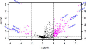
Figure 1. Volcano Plot Graphic showing ions differentially expressed between Complete-Blastocyts and No-Blastocysts groups, with a fold chance ≥ 4x and P<0.001, in the t test.
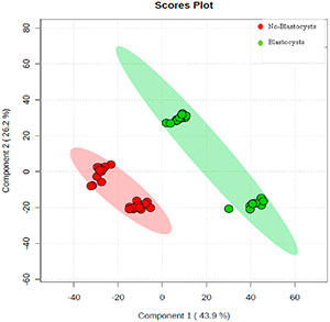
Figure 2. A Pls-Da plot of the scores for samples from the Complete-Blastocysts and No-Blastocyts groups.
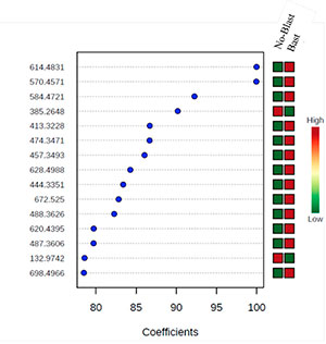
Figure 3. The most important VIP ions for separating the groups Complete-Blastocyst and No-Blastocyst groups.
DISCUSSION
Modern approaches for lipidomics are dominated by MS, commonly preceded by separation by liquid chromatography or gas chromatography. The novel MS-based lipidomics afford the study of intact lipid molecular species from very small amounts of samples, and such methods, due to their wide dynamic range, enable quantitative or relative determination of compounds across a broad range of concentrations. Therefore, as an emerging ‘‘omics’’ field, lipidomics provides a powerful approach to understanding cellular biology (Quehenberger et al., 2010) and integrate the investigation of the role of genomics, transcriptomics, proteomics, and metabolomics in cell function.
To the best of our knowledge, in the present study, possible lipid biomarkers for blastocyst formation competence were suggested. By means of the PLS-DA loading analysis, which identifies the most important ions for separating the two groups based on their m/z values, we were able to observe several ions with higher potential for acting as biomarkers. Not only the presence or absence of specific ions but also their relative abundance in the spectra allowed separation between the complete and no-blastocyst groups.
Recently, the ‘‘freeze-all’ policy has emerged as an alternative to fresh embryo transfer, to improve IVF outcomes (Shapiro et al., 2011a; Shapiro et al., 2011b; Roque et al., 2013). In this method, the entire cohort of embryos is cryopreserved, and embryo transfer is performed later in a natural cycle, or in a cycle with hormonal replacement for endometrial priming (Roque et al., 2015). The potential advantage of this method is that it provides a more physiologic environment in which embryo transfer occurs; this approach could lead to better pregnancy rates and decrease maternal and perinatal morbidity (Barnhart, 2014). However, the patient’s selection and the cost for cryopreservation are potential downsides of this technique. Alternatively, the best embryos can be cryopreserved and transferred into a more receptive endometrium while the other embryo may be transferred in the fresh cycle. Therefore, the identification of possible biomarkers of blastocyst formation would be crucial for the implementation of embryos cryopreservation as a routine procedure, avoiding the deleterious effects of controlled ovarian stimulation on endometrium receptivity.
Moreover, the identification of patients able to benefit from extended embryo culture programs would be imperative for assisted reproduction success. In fact, although it is known that prolonging the embryo culture period enables a better selection of embryos for transfer, leading to increased implantation rates and reduced risk of twins and higher order pregnancies (Forman et al., 2013), there is concern that a strategy of blastocyst culture may result in higher cycle cancellation rates (Glujovsky et al., 2012).
Many metabolic parameters of developing embryos have been studied using a variety of non-invasive methods (Sakkas & Gardner, 2005). These studies demonstrate an underlying molecule expression difference between embryos that converts into blastocysts and those that do not, and this lays the groundworks of a metabolomic approach in the assessment of embryo viability.
Gardner et al. (2001) reported that glucose uptake was larger in human blastocysts of higher grade, whereas Brison et al. (2004) found that elevated asparagine, and decreased glycine and leucine levels in embryo culture media correlate with pregnancy. These and other studies suggest that embryos with positive and negative reproductive potential alter their environment differently and this is reflected in the surrounding metabolites (Scott et al., 2008; Seli et al., 2008; Vergouw et al., 2008; Seli et al., 2010; Ahlstrom et al., 2011b; Sfontouris et al., 2013).
On these previous studies, metabolomic profiling of spent embryo culture media was mainly performed using spectroscopy. In the present study, a different method for embryonic evaluation, the MS, was applied. Mass Spectrometry allied with PLS-DA model has been successfully employed in biomarker identification in other areas than reproduction (Duan et al., 2011; Wang et al., 2012a; Wang et al., 2012b; Yang et al., 2012). Recently, our group also applied an embryo viability prediction (Cortezzi et al., 2013).
In conclusion, our findings demonstrated that the day three culture medium MS may identify possible lipid biomarkers of embryos which are able to convert into blastocysts; therefore, this may be a promising approach for the identification of embryos that should be cultured until day five or even cryopreserved and transferred latter into a more receptive endometrium.
REFERENCES
Ahlstrom A, Westin C, Reismer E, Wikland M, Hardarson T. Trophectoderm morphology: an important parameter for predicting live birth after single blastocyst transfer. Hum Reprod. 2011a; 26:3289-96.
Medline Crossref
Ahlstrom A, Wikland M, Rogberg L, Barnett JS, Tucker M, Hardarson T. Cross-validation and predictive value of near-infrared spectroscopy algorithms for day-5 blastocyst transfer. Reprod Biomed Online. 2011b; 22:477-84.
Medline Crossref
Alpha Scientists in Reproductive Medicine, ESHRE Special Interest Group of Embryology. The Istanbul consensus workshop on embryo assessment: proceedings of an expert meeting. Hum Reprod. 2011; 26:1270-83.
Medline Crossref
Assou S, Boumela I, Haouzi D, Anahory T, Dechaud H, De Vos J, Hamamah S. Dynamic changes in gene expression during human early embryo development: from fundamental aspects to clinical applications. Hum Reprod Update. 2011; 17: 272-90.
Medline Crossref
Aydin S, Cinar O, Demir B, Korkmaz C, Ozdegirmenci O, Dilbaz S, Goktolga U. Is pronuclear scoring a really good predictor for ICSI cycles? Gynecol Endocrinol. 2011; 27: 742-7.
Medline Crossref
Balaban B, Urman B, Isiklar A, Alatas C, Aksoy S, Mercan R, Mumcu A, Nuhoglu A. The effect of pronuclear morphology on embryo quality parameters and blastocyst transfer outcome. Hum Reprod. 2001; 16:2357-61.
Medline Crossref
Barnhart KT, Introduction: are we ready to eliminate the transfer of fresh embryos in in vitro fertilization? Fertil Steril. 2014; 102: 1-2.
Medline Crossref
Bligh EG, Dyer WJ. A rapid method of total lipid extraction and purification. Can J Biochem Physiol. 1959; 37: 911-7.
Medline
Brison DR, Houghton FD, Falconer D, Roberts SA, Hawkhead J, Humpherson PG, Lieberman BA, Leese HJ. Identification of viable embryos in IVF by non-invasive measurement of amino acid turnover. Hum Reprod. 2004; 19: 2319-24.
Medline Crossref
Cortezzi SS, Cabral EC, Trevisan MG, Ferreira CR, Setti AS, Braga DP, Figueira R de C, Iaconelli A Jr, Eberlin MN, Borges E Jr. Prediction of embryo implantation potential by mass spectrometry fingerprinting of the culture medium. Reproduction. 2013; 145: 453-62.
Medline Crossref
Cortezzi SS, Garcia JS, Ferreira CR, Braga DP, Figueira RC, Iaconelli A Jr, Souza GH, Borges E Jr, Eberlin MN. Secretome of the preimplantation human embryo by bottom-up label-free proteomics. Anal Bioanal Chem. 2011; 401:1331-9.
Medline Crossref
D’Alessandro A, Federica G, Palini S, Bulletti C, Zolla L. A mass spectrometry-based targeted metabolomics strategy of human blastocoele fluid: a promising tool in fertility research. Mol Biosyst. 2012, 8: 953-8.
Medline Crossref
Duan H, Guan N, Wu Y, Zhang J, Ding J, Shao B. Identification of biomarkers for melamine-induced nephrolithiasis in young children based on ultra high performance liquid chromatography coupled to time-of-flight mass spectrometry (U-HPLC-Q-TOF/MS). J Chromatogr B Analyt Technol Biomed Life Sci. 2011, 879: 3544-50.
Medline Crossref
Ferreira CR, Souza GH, Riccio MF, Catharino RR, Pontes JH, Basso AC, Junior J C, Perecin F, Eberlin MN. Mass spectrometry fingerprinting of media used for in vitro production of bovine embryos. Rapid Commun Mass Spectrom. 2009; 23:1313-20.
Medline Crossref
Filho ES, Noble JA, Wells D. A review on automatic analysis of human embryo microscope images. Open Biomed Eng J. 2010; 4:170-7.
Medline Crossref
Forman EJ, Hong KH, Ferry KM, Tao X, Taylor D, Levy B, Treff NR, Scott RT Jr. In vitro fertilization with single euploid blastocyst transfer: a randomized controlled trial. Fertil Steril. 2013; 100:100-7e1.
Medline Crossref
Gardner DK, Lane M, Stevens J, Schoolcraft WB. Noninvasive assessment of human embryo nutrient consumption as a measure of developmental potential. Fertil Steril. 2001; 76:1175-80.
Medline Crossref
Glujovsky D, Blake D, Farquhar C, Bardach A. Cleavage stage versus blastocyst stage embryo transfer in assisted reproductive technology. Cochrane Database Syst Rev. 2012; 7:CD002118.
Medline Crossref
Holte J, Berglund L, Milton K, Garello C, Gennarelli G, Revelli A, Bergh T. Construction of an evidence-based integrated morphology cleavage embryo score for implantation potential of embryos scored and transferred on day 2 after oocyte retrieval. Hum Reprod. 2007; 22:548-57.
Medline Crossref
Katz-Jaffe MG, McReynolds S, Gardner D K, Schoolcraft WB. The role of proteomics in defining the human embryonic secretome. Mol Hum Reprod. 2009; 15: 271-7.
Medline Crossref
Katz-Jaffe MG, Schoolcraft WB., Gardner DK. Analysis of protein expression (secretome) by human and mouse preimplantation embryos. Fertil Steril. 2006; 86: 678-85.
Medline Crossref
Lagarrigue M, Lavigne R, Guevel B, Com E, Chaurand P, Pineau C. Matrix-assisted laser desorption/ionization imaging mass spectrometry: a promising technique for reproductive research. Biol Reprod. 2012; 86:74.
Medline Crossref
Lan KC, Huang FJ, Lin YC, Kung FT, Hsieh CH, Huang HW, Tan PH, Chang SY. The predictive value of using a combined Z-score and day 3 embryo morphology score in the assessment of embryo survival on day 5. Hum Reprod. 2003; 18:1299-306.
Medline Crossref
Lundin K, Bergh C, Hardarson T. Early embryo cleavage is a strong indicator of embryo quality in human IVF. Hum Reprod. 2001; 16:2652-7.
Medline Crossref
Mastenbroek S, van der Veen F, Aflatoonian A, Shapiro B, Bossuyt P, Repping S. Embryo selection in IVF. Hum Reprod. 2011; 26: 964-6.
Medline Crossref
Nagy ZP, Dozortsev D, Diamond M, Rienzi L, Ubaldi F, Abdelmassih R, Greco E.. Pronuclear morphology evaluation with subsequent evaluation of embryo morphology significantly increases implantation rates. Fertil Steril. 2003; 80:67-74.
Medline Crossref
Palermo G, Joris H, Devroey P, Van Steirteghem AC. “Pregnancies after intracytoplasmic injection of single spermatozoon into an oocyte. Lancet. 1992; 340: 17-18.
Medline Crossref
Paternot G, Devroe J, Debrock S, D’Hooghe TM, Spiessens C. Intra- and inter-observer analysis in the morphological assessment of early-stage embryos. Reprod Biol Endocrinol. 2009; 7:105.
Medline Crossref
Patrizio P, Sakkas D. From oocyte to baby: a clinical evaluation of the biological efficiency of in vitro fertilization. Fertil Steril. 2009; 91:1061-6.
Medline Crossref
Payne JF, Raburn DJ, Couchman GM, Price TM, Jamison MG, Walmer DK. Relationship between pre-embryo pronuclear morphology (zygote score) and standard day 2 or 3 embryo morphology with regard to assisted reproductive technique outcomes. Fertil Steril. 2005; 84: 900-9.
Medline Crossref
Racowsky C, Vernon M, Mayer J, Ball GD, Behr B, Pomeroy KO, Wininger D, Gibbons W, Conaghan J, Stern JE. Standardization of grading embryo morphology. J Assist Reprod Genet. 2010; 27: 437-9.
Medline Crossref
Roque M, Lattes K, Serra S, Sola I, Geber S, Carreras R, Checa MA. Fresh embryo transfer versus frozen embryo transfer in in vitro fertilization cycles: a systematic review and meta-analysis. Fertil Steril. 2013; 99:156-62.
Medline Crossref
Roque M, Valle M, Guimaraes F, Sampaio M, Geber S. Freeze-all policy: fresh vs. frozen-thawed embryo transfer. Fertil Steril. 2015; 103: 1190-3.
Medline Crossref
Sakkas D, Gardner DK. Noninvasive methods to assess embryo quality. Curr Opin Obstet Gynecol. 2005; 17: 283-8.
Medline
Sanchez-Ribas I, Riqueros M, Vime P, Puchades-Carrasco L, Jonsson T, Pineda-Lucena A, Ballesteros A, Dominguez F, Simon C. Differential metabolic profiling of non-pure trisomy 21 human preimplantation embryos. Fertil Steril. 2012; 98: 1157-64e1-2.
Medline Crossref
Scott R, Seli E, Miller K, Sakkas D, Scott K, Burns DH. Noninvasive metabolomic profiling of human embryo culture media using Raman spectroscopy predicts embryonic reproductive potential: a prospective blinded pilot study. Fertil Steril. 2008; 90: 77-83.
Medline Crossref
Seli E, Botros L, Sakkas D, Burns, DH. Noninvasive metabolomic profiling of embryo culture media using proton nuclear magnetic resonance correlates with reproductive potential of embryos in women undergoing in vitro fertilization. Fertil Steril. 2008; 90: 2183-9.
Medline Crossref
Seli E, Gardner DK, Schoolcraft WB, Moffatt O, Sakkas D. Extent of nuclear DNA damage in ejaculated spermatozoa impacts on blastocyst development after in vitro fertilization. Fertil Steril. 2004; 82: 378-83.
Medline Crossref
Seli E, Vergouw CG, Morita H, Botros L, Roos P, Lambalk CB, Yamashita N, Kato O, Sakkas D. Noninvasive metabolomic profiling as an adjunct to morphology for noninvasive embryo assessment in women undergoing single embryo transfer. Fertil Steril. 2010; 94: 535-42.
Medline Crossref
Sfontouris IA, Lainas GT, Sakkas D, Zorzovilis IZ, Petsas GK, Lainas TG. Non-invasive metabolomic analysis using a commercial NIR instrument for embryo selection. J Hum Reprod Sci. 2013; 6:133-9.
Medline Crossref
Shapiro BS, Daneshmand ST, Garner FC, Aguirre M, Hudson C, Thomas S. Evidence of impaired endometrial receptivity after ovarian stimulation for in vitro fertilization: a prospective randomized trial comparing fresh and frozen-thawed embryo transfer in normal responders. Fertil Steril. 2011a; 96: 344-8.
Medline Crossref
Shapiro BS, Daneshmand ST, Garner FC, Aguirre M, Hudson C, Thomas S. Evidence of impaired endometrial receptivity after ovarian stimulation for in vitro fertilization: a prospective randomized trial comparing fresh and frozen-thawed embryo transfers in high responders. Fertil Steril. 2011b; 96: 516-8.
Medline Crossref
Vergouw CG, Botros LL, Roos P, Lens JW, Schats R, Hompes PG, Burns DH, Lambalk CB. Metabolomic profiling by near-infrared spectroscopy as a tool to assess embryo viability: a novel, non-invasive method for embryo selection. Hum Reprod. 2008; 23: 1499-504.
Medline Crossref
Wang X, Yang B, Sun H, Zhang A. Pattern recognition approaches and computational systems tools for UPLC-MS-based comprehensive metabolomic profiling and pathways analysis of biological data sets. Anal Chem. 2012a; 4:428-39
Medline Crossref
Wang X, Yang B, Zhang A, Sun H, Yan G. Potential drug targets on insomnia and intervention effects of Jujuboside A through metabolic pathway analysis as revealed by UPLC/ESI-SYNAPT-HDMS coupled with pattern recognition approach. J Proteomics. 2012b;75:1411-27
Medline Crossref
Yang B, Zhang A, Sun H, Dong W, Yan G, Li T, Wang X. Metabolomic study of insomnia and intervention effects of Suanzaoren decoction using ultra-performance liquid-chromatography/electrospray-ionization synapt high-definition mass spectrometry. J Pharm Biomed Anal. 2012; 58:113-24.
Medline Crossref