JBRA Assist. Reprod. 2020;24(2):97-103
ORIGINAL PAPER
doi: 10.5935/1518-0557.20190070
Serum microRNA profiling for the identification of predictive molecular markers of the response to controlled ovarian stimulation
1Fertility Medical Group
2Departamento de Biologia Estrutural e Funcional, Instituto de Biologia, Universidade Estadual de Campinas - UNICAMP
3Instituto Sapientiae - Centro de Estudos e Pesquisa em Reprodução Assistida
CONFLICT OF INTEREST
The authors have no conflict of interest to declare.
ABSTRACT
Objective: To identify potential microRNA (miRNA) biomarkers of poor, normal and hyperresponse to controlled ovarian stimulation (COS).
Methods: In the present study, we assessed 40 serum samples from patients undergoing COS. We used ten samples to standardize miRNAs detection in the serum. The remaining 30 samples were split into three groups depending on the patient's response to COS: poor response (PR group, n=10), normal response (NR group, n=10), and hyperresponse (HR group, n=10). Aberrantly expressed miRNAs were identified using a large-scale expression analysis platform. Gene set enrichment analysis was performed to assess the biological processes potentially modulated by the identified miRNAs.
Results: Twenty-two miRNAs were detected only in the PR or HR groups when compared with the NR group. From those, 11 presented poor dissociation curves and were excluded from further analysis. A bioinformatics analysis revealed that the selected 11 miRNAs target several genes involved in GnRH, estrogen and prolactin signaling, oocyte maturation, female pregnancy, and meiosis.
Conclusion: The large-scale analysis of miRNA expression identified distinct miRNA profiles for poor and hyperresponse to COS, which potentially modulate key processes for human assisted reproduction. All evidence suggests that the serum microRNA profiling may discriminate patients who will respond in an exacerbated manner from those who will respond insufficiently to COS. Further studies may validate these miRNAs, enabling the individualization of treatment and more successful outcomes.
Keywords: poor responder, hyper responder, COS, biomarker, microRNA, stimulation
INTRODUCTION
Since the first reported pregnancy following in vitro fertilization (IVF) (Steptoe & Edwards, 1978), assisted reproductive technologies (ART) have quickly progressed. It is estimated that over 237,000 infants are born through ART worldwide in a single year (Sullivan et al., 2013). Nevertheless, its efficiency in terms of live births is still low. It has been estimated that 4.45 cycles are needed to achieve one live birth event following IVF (Sunkara et al., 2016). Many variables have been suggested as possible causes for the low efficiency of assisted reproduction. Embryo implantation depends on the acquisition of a receptive endometrium and the presence of a viable embryo (Su & Fazleabas, 2015). The selection of a viable embryo for transfer is a critical step in ART and depends on an adequate number and quality of oocytes. However, the response to controlled ovarian stimulation (COS) is quite variable among patients, and both poor and hyperresponses may have detrimental effects on the cycle's outcome.
Previous registers have reported important data concerning the cancellation rates of IVF cycles (Sullivan et al., 2013; Sunkara et al., 2016). We could assumed that, for young women, a significant number of cycles are likely to be cancelled because of the risk of ovarian hyperstimulation syndrome (OHSS), rather than because of a poor ovarian response. On the other hand, for older women, cycle cancellation is more likely to be associated with a poor ovarian response.
According with the consensus from the European Society of Human Reproduction an Embryology (ESHRE), a poor ovarian response is defined as the collection of fewer than four oocytes in response to a COS protocol of at least 150 IU FSH per day (Ferraretti et al., 2011). The management of patients with poor ovarian response to exogenous gonadotropin stimulation has challenged reproductive specialists for decades. There is a great need to attain an optimal oocyte yield while minimizing the risk of an excessive response and OHSS (Nyboe Andersen et al., 2017).
Although OHSS is a condition with a small risk of mortality, it is an important condition with considerable morbidity (Corbett et al., 2014; Boothroyd et al., 2015), and it remains the most critical safety concern associated with the use of gonadotropin preparations. Previous reports have shown a positive linear relationship between oocyte yield and live birth rates of up to 15 to 25 oocytes, using conventional ovarian stimulation (Sunkara et al., 2011; Polyzos et al., 2018). Beyond 15 oocytes, there is no increase in live birth rates, but the risk of OHSS increases exponentially (Sunkara et al., 2011; Steward et al., 2014). Within this line, it is crucial to implement less aggressive stimulation while maintaining an adequate oocyte yield.
Age, serum FSH, antral follicle count (AFC), serum AMH, and other factors have been pointed out as possible predictive factors to ovarian response to COS. However, there is no consensus regarding which factors to take into account or the importance of each factor when determining the dose of gonadotrophins for COS (Corbett et al., 2014; Boothroyd et al., 2015; Oudshoorn et al., 2017; van Tilborg et al., 2017a; 2017b; Lensen et al., 2018). In addition, it is nearly impossible to accurately predict the ovarian response and tailor an individualized stimulation protocol based on such parameters. Thus, the development of non-invasive techniques, capable of predicting the response to COS, would allow for individualized treatment and significantly increase treatment success while decreasing the physical, emotional and economic burden on patients.
MicroRNAs (miRNAs) are endogenous, evolutionally conserved, single-strand non-coding RNA molecules of 20-24 nucleotides that post-transcriptionally regulate gene expression in eukaryotes, including mammalian cells (Asirvatham et al., 2009; McCallie et al., 2010; Mouillet et al., 2015; Thouas et al., 2015). They were first described in the nematode Caenorhabditis elegans (Lee et al., 1993; Wightman et al., 1993) and later found in the genomes of protists, plants, animals, and viruses (Mouillet et al., 2015). In mammals, it has been shown that miRNAs are actively secreted into the bloodstream and regulate gene expression in distant organs (Thomou et al., 2017a; 2017b). In fact, miRNAs have been detected in virtually all human bodily fluids, including blood, urine, saliva, tears, breast milk, semen, amniotic fluid, cerebrospinal fluid, peritoneal fluid, and pleural fluid as well as in culture media collected from different cell lines (Wang et al., 2010 a; 2010b; Weber et al., 2010).
The study of signaling molecules, such as miRNAs, in the serum of patients undergoing IVF may be a valuable approach to predict the response to COS and facilitate the development of an individualized gonadotrophin dosing strategy. Therefore, the goal of the present study was to identify potential miRNAs biomarkers of poor, normal, and hyperresponse to COS.
MATERIALS AND METHODS
Study Design
For the present study, we analyzed 40 serum samples derived from patients undergoing COS for intracytoplasmic sperm injection (ICSI). The samples were collected in a private university-affiliated IVF center, between January 2017 and January 2018. We used ten samples to standardize the detection of serum miRNAs. The remaining 30 samples were split into three groups depending on the patient's response to COS: poor response (PR group, n=10), normal response (NR group, n=10), and hyperresponse (HR group, n=10). The aberrantly expressed miRNAs were identified using a large-scale expression analysis platform. In addition, we carried out a gene set enrichment analysis to access the biological processes potentially modulated by the identified miRNAs.The study included only patients undergoing their first or second IVF cycle, using fresh oocytes and/or embryos, and aged <38 years old. We obtained a written informed consent, in which patients agreed to share the outcomes of their cycles for research purposes, and the local institutional review board approved the study.
Sample collection
Serum samples were obtained through venipuncture, prior to beginning the COS. After complete filling of a serum separation tube, the blood was carefully mixed with coagulation activating agent and remained standing for 60 minutes. After complete coagulation, the samples were centrifuged at 1500 to 2000xg for 10 minutes until the serum was well separated. Using a Pasteur pipette, the serum was transferred to another tube and stored at -20°C.
Controlled ovarian stimulation and laboratory procedures
Controlled ovarian stimulation was performed using recombinant FSH (Gonal-F®, Merck KGaA, Darmstadt, Germany) and pituitary suppression using a GnRH antagonist (GnRH - Cetrotide® Merck KGaA, Darmstadt, Germany). Follicular growth was monitored using transvaginal ultrasound starting on day 4 of the gonadotropin administration. When adequate follicular growth and serum E2 levels were observed, recombinant hCG (Ovidrel®, Merck KGaA, Darmstadt, Germany) or leuprolide acetate (Lupron®, AbbVie, Boston, USA) was administered to trigger the final follicular maturation. The oocytes were collected 35 hours after hCG administration through transvaginal ultrasound ovum pick-up.The recovered oocytes were assessed to determine their nuclear status, and those in metaphase II were submitted to ICSI following routine procedures (Palermo et al., 1997).
MiRNA extraction, identification and quantification
A small RNA fraction was extracted from 200µl of serum samples, using a miRNeasy Serum/Plasma extraction kit (QIAGEN, Hilden, Germany). C. elegans miR-39 was used as an RNA spike-in to normalize the gene expression analysis (miRNeasy Serum/Plasma Spike-in Control, QIAGEN, Hilden, Germany). The identification and quantification of aberrantly expressed miRNAs, in the pool of samples form PR, NR and HR groups, was performed by using a large-scale quantification PCR-Array platform (miScript® QIAGEN, Helden, Germany) on a ViiA7 apparatus (Thermo-Fisher, Massachusetts, USA). We analyzed the data using a PCR Array Data Analysis Web Portal software (http://pcrdataanalysis.sabiosciences.com/mirna).
Gene set enrichment analysis
The list of putative targets for the selected miRNAs for each group (HR or PR) was obtained using the miRWalk 2.0 web tool (http://zmf.umm.uni-heidelberg.de/apps/zmf/mirwalk2/). For each group, the lists of predicted targets for each miRNA were combined, and those targets predicted by 7 out of the 12 algorithms, or less, were excluded. The remaining target list was then submitted to enrichment analysis using the DAVID Annotation tool (https://david.ncifcrf.gov/).
Statistical analyses
Patient and cycle characteristics were analyzed using the SPSS Statistics 21 (IBM, New York, NY, USA) statistical program. The variables were tested for normality distribution and group homogeneity using the Shapiro Wilk and Levene tests, respectively. When necessary, the samples were normalized using z-scores. The groups were compared by ANOVA, followed by the Bonferroni post-hoc test. Variables are described as mean ± standard deviation and the significance level α was set at 5%.
RESULTS
Table 1 depicts the characteristics of the patients and cycles. As expected, the level of estradiol on trigger day, the number of aspirated follicles, the number of retrieved follicles, and the number of mature follicles were higher for the HR group, followed by the NR group, while the PR group presented the lowest results.
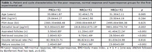
Table 1. Patient and cycle characteristics for the poor response, normal response and hyperresponse groups for the first experimental set
After the large-scale analysis of miRNA, we identified 22 miRNAs with amplification exclusively in the PR or HR groups when compared with the NR group. From the 22 miRNAs, 11 had poor dissociation curves, probably due to non-specific amplifications or primer dimer amplifications. Therefore, those products were excluded from further analysis (Table 2). Of the remaining 11 miRNAs, miR-99a-5p, miR-181d-5p, miR-221-3p, miR-92a-1-5p, and miR-1302 were only detected in the HR groups, while miR-150-5p, miR-223-3p, let-7d-3p, miR-891a-5p, miR-99a-3p and, miR-200c-5p were only detected in the PR group (Table 2).
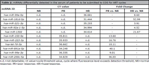
Table 2. miRNAs differentially detected in the serum of patients to be submitted to COS for ART cycles
The lists of predicted targets for each miRNA in both HR and PR groups were obtained and the gene set enrichment analysis (GSEA) was performed. The GSEA revealed that miRNAs from the HR group potentially modulate key members of GnRH, estrogen, prolactin, and oxytocin signaling pathways, as well as progesterone-mediated oocyte maturation and female pregnancy (Figures 1 A and 2). It is worth highlighting that miR-181d-5p targets comprised more than 40% of the targets in the HR group. On the other hand, miRNAs from the PR group potentially modulate members of the prolactin and estrogen signaling pathways in utero, embryo and post-embryonic development, and the genes involved in stem cell pluripotency and endometrial cancer (Figures 1 B and 2). miR-150-5p and miR-223-3p represent more than 49% and 32% of the targets, respectively.
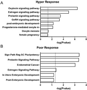
Figure 1. Gene set enrichment analysis of differentially detected miRNAs in HR and PR groups. The bars represent biological processes and signaling pathways enriched among the predicted target genes of the miRNAs from HR and PR groups. Values on the x-axis are represented as -log p values.
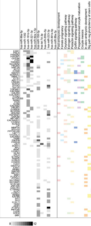
Figure 2. miRNAs from HR and PR groups and their target genes. Representative list of miRNA targets and enriched processes. The grey boxes indicate the number of prediction algorithms that consider that interaction possible (from 8 to 12) using the miRWalk web tool. Colored boxes indicate that each target gene is annotated in the given ontological category according to the DAVID platform.
DISCUSSION
The main objective of treatment individualization in IVF is to offer every single woman the best treatment tailored to her unique characteristics, thus maximizing success, eliminating iatrogenic risks such as OHSS, and minimizing the risk of cycle cancellation. However, the success of individualized COS depends on finding a reliable method for predicting the ovarian response to stimulation.
For the present study, potential miRNA biomarkers of the response to ovarian stimulation were identified in the serum of patients undergoing ART, before COS. Extensive research has been carried out in a search for markers that can be used to facilitate COS individualization. Previous studies have focused on the assessment of miRNAs as molecular markers in assisted reproduction. It has been found that a poor response to COS is associated with altered miRNA expression, specifically with elevated miR-21-5p expression (Karakaya et al., 2015) or miRNA-15a-5p (Zhang et al., 2017). These studies, however, analysed the exression on miRNAs in granulosa cells, which can only be retrived during ovum pick-up, which limits the clinical usefulness of these biomarkers for the management of COS.
In this manner, the study of altered expression of miRNAs in serum may be more useful. Zhao et al. (2015) evaluated the expression of serum miRNAs and their predictive value for OHSS in patients with polycystic ovarian syndrome (PCOS). Two miRNAs were differently expressed in PCOS patients likely to suffer from severe OHSS. The value of the aforementioned article is indisputable, since it is well-known that PCOS patients who undergo COS are at a high risk of OHSS. However, for the application of an individualised stimulus treatment, other patient categories should also be investigated.
In our study, we aimed at identifying miRNAs in the serum of patients with different responses to COS. Several miRNAs, expressed only in poor and hyperresponders, were identified in a large-scale analysis by the qPCR array platform. miRNAs analyses of target genes indicated that these molecules are key player candidates. Moreover, the levels of this miRNA positively correlate with important clinical aspects, such as the number of retrieved oocytes.
Altered expression of miR-181d-5p has been described in various types of cancer, including ovarian carcinoma (Lee et al., 2012; Neel & Lebrun, 2013; Pal et al., 2015; Mahdian-Shakib et al., 2016; Widodo et al., 2016). It is also known that the regulation of this miRNA expression in endometrial stromal cells plays a role in the decidualisation process (Estella et al., 2012). In addition, miR-181d-5p serum levels are predictive of non-alcoholic fatty liver disease, and can be used in the differential diagnosis between autoimmune pancreatitis and pancreatic carcinoma (Celikbilek et al., 2014; Akamatsu et al., 2016). However, the biological function of miR-181d-5p in ovarian physiology is still to be unveiled.
A study by Khan et al. (2015) suggests that miR-150 expression, detected only in the PR group, is decreased during ovarian development in mice. In addition, it has been shown that miR-150 is expressed in higher levels in atretic rather than healthy follicles in cattle, and may play a role in bovine oocyte maturation (Donadeu et al., 2017). Importantly, miR-150 expression is decreased in patients with endometriosis treated with simvastatin (Cosar et al., 2018).
Thus, miRNA biomarkers continue to emerge as potential tools for the management of infertility. Future analyses for the detection of these miRNAS in a large number of samples could contribute to their validation as diagnostic tools for the prediction of ovarian response to COS.
REFERENCES
Akamatsu M, Makino N, Ikeda Y, Matsuda A, Ito M, Kakizaki Y, Saito Y, Ishizawa T, Kobayashi T, Furukawa T, Ueno Y. Specific MAPK-Associated MicroRNAs in Serum Differentiate Pancreatic Cancer from Autoimmune Pancreatitis. PLoS One. 2016;11:e0158669. PMID: 27380024 DOI: 10.1371/journal.pone.0158669
Medline Crossref
Asirvatham AJ, Magner WJ, Tomasi TB. miRNA regulation of cytokine genes. Cytokine. 2009;45:58-69. PMID: 19121586 DOI: 10.1016/j.cyto.2008.11.010
Medline
Boothroyd C, Karia S, Andreadis N, Rombauts L, Johnson N, Chapman M; Australasian CREI Consensus Expert Panel on Trial evidence (ACCEPT) group. Consensus statement on prevention and detection of ovarian hyperstimulation syndrome. Aust N Z J Obstet Gynaecol. 2015;55:523-34. PMID: 26597569 DOI: 10.1111/ajo.12406
Medline Crossref
Celikbilek M, Baskol M, Taheri S, Deniz K, Dogan S, Zararsiz G, Gursoy S, Guven K, Ozbakır O, Dundar M ,Yucesoy M. Circulating microRNAs in patients with non-alcoholic fatty liver disease. World J Hepatol. 2014;6:613-20. PMID: 25232454 DOI: 10.4254/wjh.v6.i8.613
Medline Crossref
Corbett S, Shmorgun D, Claman P; REPRODUCTIVE ENDOCRINOLOGY INFERTILITY COMMITTEE; SPECIAL CONTRIBUTOR. The prevention of ovarian hyperstimulation syndrome. J Obstet Gynaecol Can. 2014;36:1024-33. PMID: 25574681 DOI: 10.1016/S1701-2163(15)30417-5
Medline Crossref
Cosar E, Mamillapalli R, Moridi I, Duleba A, Taylor HS. Serum MicroRNA Biomarkers Regulated by Simvastatin in a Primate Model of Endometriosis. Reprod Sci. 2018:1933719118765971. In Press.
Medline Crossref
Donadeu FX, Mohammed BT, Ioannidis J. A miRNA target network putatively involved in follicular atresia. Domest Anim Endocrinol. 2017;58:76-83.
Medline Crossref
Estella C, Herrer I, Moreno-Moya JM, Quiñonero A, Martínez S, Pellicer A, Simón C. miRNA signature and Dicer requirement during human endometrial stromal decidualization in vitro. PLoS One. 2012;7:e41080.
Medline Crossref
Ferraretti AP, La Marca A, Fauser BC, Tarlatzis B, Nargund G, Gianaroli L; ESHRE working group on Poor Ovarian Response Definition. ESHRE consensus on the definition of 'poor response' to ovarian stimulation for in vitro fertilization: the Bologna criteria. Hum Reprod. 2011;26:1616-24.
Medline Crossref
Karakaya C, Guzeloglu-Kayisli O, Uyar A, Kallen AN, Babayev E, Bozkurt N, Unsal E, Karabacak O, Seli E. Poor ovarian response in women undergoing in vitro fertilization is associated with altered microRNA expression in cumulus cells. Fertil Steril. 2015;103:1469-76e1-3.
Medline Crossref
Khan HA, Zhao Y, Wang L, Li Q, Du YA, Dan Y, Huo LJ. Identification of miRNAs during mouse postnatal ovarian development and superovulation. J Ovarian Res. 2015;8:44.
Medline Crossref
Lee RC, Feinbaum RL, Ambros V. The C. elegans heterochronic gene lin-4 encodes small RNAs with antisense complementarity to lin-14. Cell. 1993;75:843-54.
Medline Crossref
Lee H, Park CS, Deftereos G, Morihara J, Stern JE, Hawes SE, Swisher E, Kiviat NB, Feng Q. MicroRNA expression in ovarian carcinoma and its correlation with clinicopathological features. World J Surg Oncol. 2012;10:174.
Medline Crossref
Lensen SF, Wilkinson J, Leijdekkers JA, La Marca A, Mol BWJ, Marjoribanks J, Torrance H, Broekmans FJ. Individualised gonadotropin dose selection using markers of ovarian reserve for women undergoing in vitro fertilisation plus intracytoplasmic sperm injection (IVF/ICSI). Cochrane Database Syst Rev. 2018;2:CD012693.
Medline Crossref
Mahdian-Shakib A, Dorostkar R, Tat M, Hashemzadeh MS, Saidi N. Differential role of microRNAs in prognosis, diagnosis, and therapy of ovarian cancer. Biomed Pharmacother. 2016;84:592-600.
Medline Crossref
McCallie B, Schoolcraft WB, Katz-Jaffe MG. Aberration of blastocyst microRNA expression is associated with human infertility. Fertil Steril. 2010;93:2374-82.
Medline Crossref
Mouillet JF, Ouyang Y, Coyne CB, Sadovsky Y. MicroRNAs in placental health and disease. Am J Obstet Gynecol. 2015;213:S163-72.
Medline Crossref
Neel JC, Lebrun JJ. Activin and TGFβ regulate expression of the microRNA-181 family to promote cell migration and invasion in breast cancer cells. Cell Signal. 2013;25:1556-66.
Medline Crossref
Nyboe Andersen A, Nelson SM, Fauser BC, García-Velasco JA, Klein BM, Arce JC; ESTHER-1 study group. Individualized versus conventional ovarian stimulation for in vitro fertilization: a multicenter, randomized, controlled, assessor-blinded, phase 3 noninferiority trial. Fertil Steril. 2017;107:387-96.e4.
Medline Crossref
Oudshoorn SC, van Tilborg TC, Eijkemans MJC, Oosterhuis GJE, Friederich J, van Hooff MHA, van Santbrink EJP, Brinkhuis EA, Smeenk JMJ, Kwee J, de Koning CH, Groen H, Lambalk CB, Mol BWJ, Broekmans FJM ,Torrance HL; OPTIMIST study group. Individualized versus standard FSH dosing in women starting IVF/ICSI: an RCT. Part 2: The predicted hyper responder. Hum Reprod. 2017;32:2506-14.
Medline Crossref
Pal MK, Jaiswar SP, Dwivedi VN, Tripathi AK, Dwivedi A, Sankhwar P. MicroRNA: a new and promising potential biomarker for diagnosis and prognosis of ovarian cancer. Cancer Biol Med. 2015;12:328-41. PMID: 26779370 DOI: 10.7497/j.issn.2095-3941.2015.0024
Medline Crossref
Palermo GD, Colombero LT, Rosenwaks Z. The human sperm centrosome is responsible for normal syngamy and early embryonic development. Rev Reprod. 1997;2:19-27. PMID: 9414462 DOI: 10.1530/ror.0.0020019
Medline Crossref
Polyzos NP, Drakopoulos P, Parra J, Pellicer A, Santos-Ribeiro S, Tournaye H, Bosch E, Garcia-Velasco J. Cumulative live birth rates according to the number of oocytes retrieved after the first ovarian stimulation for in vitro fertilization/intracytoplasmic sperm injection: a multicenter multinational analysis including ∼ 15,000 women. Fertil Steril. 2018;110:661-70.e1.
Medline Crossref
Steptoe PC, Edwards RG. Birth after the reimplantation of a human embryo. Lancet. 1978;2:366. PMID: 79723 DOI: 10.1016/s0140-6736(78)92957-4
Medline Crossref
Steward RG, Lan L, Shah AA, Yeh JS, Price TM, Goldfarb JM, Muasher SJ. Oocyte number as a predictor for ovarian hyperstimulation syndrome and live birth: an analysis of 256,381 in vitro fertilization cycles. Fertil Steril. 2014;101:967-73.
Medline Crossref
Su RW, Fazleabas AT. Implantation and Establishment of Pregnancy in Human and Nonhuman Primates. Adv Anat Embryol Cell Biol. 2015;216:189-213. PMID: 26450500 DOI: 10.1007/978-3-319-15856-3_10
Medline Crossref
Sullivan EA, Zegers-Hochschild F, Mansour R, Ishihara O, de Mouzon J, Nygren KG, Adamson GD. International Committee for Monitoring Assisted Reproductive Technologies (ICMART) world report: assisted reproductive technology 2004. Hum Reprod. 2013;28:1375-90. PMID: 23442757 DOI: 10.1093/humrep/det036
Medline Crossref
Sunkara SK, Rittenberg V, Raine-Fenning N, Bhattacharya S, Zamora J, Coomarasamy A. Association between the number of eggs and live birth in IVF treatment: an analysis of 400 135 treatment cycles. Hum Reprod. 2011;26:1768-74.
Medline Crossref
Sunkara SK, LaMarca A, Polyzos NP, Seed PT, Khalaf Y. Live birth and perinatal outcomes following stimulated and unstimulated IVF: analysis of over two decades of a nationwide data. Hum Reprod. 2016;31:2261-7.
Medline Crossref
Thomou T, Mori MA, Dreyfuss JM, Konishi M, Sakaguchi M, Wolfrum C, Rao TN, Winnay JN, Garcia-Martin R, Grinspoon SK, Gorden P, Kahn CR. Adipose-derived circulating miRNAs regulate gene expression in other tissues. Nature. 2017a;542:450-5.
Medline Crossref
Thomou T, Mori MA, Dreyfuss JM, Konishi M, Sakaguchi M, Wolfrum C, Rao TN, Winnay JN, Garcia-Martin R, Grinspoon SK, Gorden P, Kahn CR. Corrigendum: Adipose-derived circulating miRNAs regulate gene expression in other tissues. Nature. 2017b;545:252.
Medline Crossref
Thouas GA, Dominguez F, Green MP, Vilella F, Simon C, Gardner DK. Soluble ligands and their receptors in human embryo development and implantation. Endocr Rev. 2015;36:92-130.
Medline Crossref
van Tilborg TC, Oudshoorn SC, Eijkemans MJC, Mochtar MH, van Golde RJT, Hoek A, Kuchenbecker WKH, Fleischer K, de Bruin JP, Groen H, van Wely M, Lambalk CB, Laven JSE, Mol BWJ, Broekmans FJM, Torrance HL; OPTIMIST study group. Individualized FSH dosing based on ovarian reserve testing in women starting IVF/ICSI: a multicentre trial and cost-effectiveness analysis. Hum Reprod. 2017a;32:2485-95.
Medline Crossref
van Tilborg TC, Torrance HL, Oudshoorn SC, Eijkemans MJC, Koks CAM, Verhoeve HR, Nap AW, Scheffer GJ, Manger AP, Schoot BC, Sluijmer AV, Verhoeff A, Groen H, Laven JSE, Mol BWJ, Broekmans FJM; OPTIMIST study group. Individualized versus standard FSH dosing in women starting IVF/ICSI: an RCT. Part 1: The predicted poor responder. Hum Reprod. 2017b;32:2496-505.
Medline Crossref
Wang K, Zhang S, Weber J, Baxter D, Galas DJ. Export of microRNAs and microRNA-protective protein by mammalian cells. Nucleic Acids Res. 2010a;38:7248-59.
Medline Crossref
Wang GK, Zhu JQ, Zhang JT, Li Q, Li Y, He J, Qin YW, Jing Q. Circulating microRNA: a novel potential biomarker for early diagnosis of acute myocardial infarction in humans. Eur Heart J. 2010b;31:659-66.
Medline Crossref
Weber JA, Baxter DH, Zhang S, Huang DY, Huang KH, Lee MJ, Galas DJ, Wang K. The microRNA spectrum in 12 body fluids. Clin Chem. 2010;56:1733-41.
Medline Crossref
Widodo, Djati MS, Rifa'i M. Role of MicroRNAs in carcinogenesis that potential for biomarker of endometrial cancer. Ann Med Surg (Lond). 2016;7:9-13.
Medline Crossref
Wightman B, Ha I, Ruvkun G. Posttranscriptional regulation of the heterochronic gene lin-14 by lin-4 mediates temporal pattern formation in C. elegans. Cell. 1993;75:855-62.
Medline Crossref
Zhang K, Zhong W, Li WP, Chen ZJ, Zhang C. miR-15a-5p levels correlate with poor ovarian response in human follicular fluid. Reproduction. 2017;154:483-96.
Medline Crossref
Zhao C, Liu X, Shi Z, Zhang J, Zhang J, Jia X, Ling X. Role of serum miRNAs in the prediction of ovarian hyperstimulation syndrome in polycystic ovarian syndrome patients. Cell Physiol Biochem. 2015;35:1086-94.
Medline Crossref