JBRA Assist. Reprod. 2021;25(1):115-121
ORIGINAL ARTICLE
doi: 10.5935/1518-0557.20200062
The usefulness of metaphase I oocytes in women who undergo controlled ovarian hyperstimulation for intracytoplasmic sperm injection
1Department of Obstetrics and Gynecology, Universidade Federal do Pará, Belém, Pará, Brazil
2Clínica de Reprodução Assistida Pronatus, Belém, PA, Brazil
3Grupo de Pesquisa Bilibio, Universidade Federal do Pará, Belém, PA, Brazil
4Programa de Pós Graduação de Ciências Médicas da Universidade Federal do Rio Grande do Sul, Porto Alegre, Rio Grande do Sul, Brazil.
Received March 10, 2020
Accepted September 14, 2020
CONFLICT OF INTEREST
The authors have no conflicts of interest to declare.
Convention This study was accepted for an e-poster presentation at the 36th ESHRE Annual Meeting, which will be held virtually from 5 to 8 July 2020.
Funding Sources
We thank the support of the Federal University of Pará (UFPA).
ABSTRACT
Objective: The aim of this study was to evaluate the fertilization and blastocyst
formation rates of oocytes in metaphase I (MI) obtained from women who
underwent controlled ovarian hyperstimulation (COH) for intracytoplasmic
injection.
Methods: A prospective cohort study that included women from whom at least 1 MI and 1
MII oocyte were obtained after COH was performed. We collected 1,907 oocytes
from 164 women (1291 MII, 352 MI and 258 prophase I or atretic). After
oocyte classification, the MII and MI oocytes were incubated for 4
hours.
Results: After 4 hours, the rescue maturation rate was 57.2%; 205 MI oocytes matured
to MII oocytes in vitro (rescued MI-MII group), and 153
remained in MI (arrested MI group). The normal fertilization rates were
directly associated with oocyte maturation, with rates of 79.1%, 60.2%, and
31.9% in MII, MI-MII and MI oocytes, respectively
(p<0.001). Group arrested MI had an odds ratio (OR) of
7.6 (CI 5.2 - 11.2, p<0.001) for abnormal fertilization
compared with Group MII. The blastocyst formation rate was directly
associated with oocyte maturation, at 36.4% for MII, 11.4% for MI-MII and
0.6% for MI.
Conclusion: Oocytes collected at the MI stage after OCH that did not mature to MII after
rescue maturation had a blastocyst formation rate of only 0.6%, while those
in MII and MI-MII had rates of 36.4% and 11.4%, respectively. However, we
found a pregnancy with the birth of a healthy baby from a blastocyst formed
after intracytoplasmic sperm injection (ICSI) of an MI oocyte.
Keywords: blastocyst, metaphase I, ICSI, IVF, oocyte
INTRODUCTION
Due to the increasing number of couples seeking to conceive via assisted reproduction
technology, particularly in cases of in vitro fertilization (IVF),
and due to the increasing number of women over 40 years of age who desire a new or
first pregnancy, specialists have sought to identify the strategy that yields the
best outcome (Huang & Rosenwaks, 2014).
Controlled ovarian hyperstimulation (COH) was adopted to increase pregnancy rates.
With COH, multiple follicles can develop. This yields multiple oocytes that can be
aspirated and fertilized and enables the selection of the best embryos for embryonic
transfer (Ingerslev et al.,
2001). However, the ovarian response to stimulation is variable and
depends mainly on each woman's ovarian reserve (Huddleston et al., 2010). With increasing maternal age,
the quantity and quality of obtainable oocytes decreases; thus, age is considered
the main influencer of ovarian response and oocyte quality and maturation (Su et al., 2017). IVF can
overcome many causes of infertility, but it cannot reverse fertility decline in
older women (Baker et al.,
2010; Malizia et al.,
2009).
The unresponsiveness of some oocytes to ovarian stimulation has not yet been
explained, but it is possible that the different stages of follicle development
yield oocytes with varying degrees of maturation after stimulation. During
aspiration, oocytes are collected from heterogeneous groups of follicles, and mature
oocytes in metaphase II (MII) or meiotically immature oocytes in metaphase I (MI) or
prophase I (PI) may be captured (Stouffer &
Zelinski-Wooten, 2004). The proportion of immature oocytes among
collected oocytes generally ranges from 10 to 20%, and a high number of immature
oocytes can negatively affect the ICSI outcomes of mature oocytes from the same
woman (Braga et al.,
2020).
Follicular maturation is extremely important for the generation of viable embryos for
implantation and, consequently, for the success of IVF. To be fertilized, oocytes
should ideally reach maturity, as oocytes in PI and MI are too morphologically
immature to be fertilized for the formation of good-quality embryos. MI-stage
oocytes can spontaneously undergo in vitro meiotic maturation to MII-stage oocytes
within a few hours after collection (Strassburger et al., 2004), but they generally remain unused
because their rates of fertilization, blastocyst formation and implantation are
lower than those of MII oocytes that mature in vivo (Strassburger et al., 2004; Alcoba et al., 2015).
The pregnancy rate is directly associated to the number of embryos in the blastocyst
stage that are formed during the IVF cycle, and this number depends on the quantity
and quality of MII oocytes collected (Ben-Nagi et al., 2019). Since the number of these oocytes is
inversely proportional to the age of the patient, the use of MI oocytes for IVF,
especially for couples of advanced age, can be an alternative. Thus, when a small
number of aspirated MII oocytes are available due to the patient's low response to
ovarian stimulation, the use of in vitro-matured MI oocytes can be
considered (Braga et al.,
2010).
The aim of this study was to evaluate the fertilization and blastocyst formation
rates of oocytes in MI from women who underwent COH for intracytoplasmic injection
(ICSI).
MATERIALS AND METHODS
A prospective cohort study was performed among women who underwent COH for
intracytoplasmic sperm injection (ICSI) during the study period, June 2018 to June
2019, at the Pronatus Reproductive Medicine Center, Belém, Pará State, Brazil. The
National Ethics and Research Committee on Human Beings and by the Ethics Committee
of Institute of Health Sciences, Federal University of Pará under protocol number
CAEE 55625115.0.0000.0018 (institutional review board equivalent), approved the
study.
The study included women from whom at least one MI oocyte and one MII oocyte was
harvested after COH. As the objective of the study was to evaluate oocyte quality
through fertilization rates and blastocyst formation rates, the exclusion criteria
were having had chemotherapy, radiotherapy, or severe male factor (sperm
concentration < 5 million /ml after seminal processing with the swim-up
method).
The probable causes of infertility were assessed for all couples and were as follows:
tubal factor (defined as an abnormality by hysterosalpingography or
videolaparoscopy); male factor (defined as a sperm count less than 15 million/ml and
excluding those with less than 5 million/ml after seminal processing with the
swim-up method); low ovarian reserve (defined as <5 antral follicles evaluated by
transvaginal ultrasound performed on the 2nd or 3rd day of the
menstrual cycle); repeat abortion (two or more consecutive abortions); polycystic
ovary syndrome; endometriosis (presence of endometriosis on videolaparoscopy or
endometrioma on imaging studies); and undetermined cause.
The ovarian stimulation protocol began on the second day of the menstrual cycle after
transvaginal ultrasound was performed to determine the antral follicle count;
recombinant follicle-stimulating hormone (FSH, Elonva 150®, Organon) was
applied subcutaneously. After day 6 of application, transvaginal ultrasound was
performed every two days to monitor follicle development. When one of the largest
follicles reached 14mm, gonadotrophin-releasing hormone (GnRH) antagonist
(Orgalutran®, Organon) was administered daily, subcutaneously, until the use
of recombinant human chorionic gonadotropin (rhCG). After day 9 of induction,
recombinant FSH (Puregon®, Organon) at a dose of 150 IU per day was used
until rhCG administration. When three or more follicles reached 17mm in diameter,
oocyte maturation was stimulated with rhCG (Ovidrel 250mcg, Serono). Transvaginal
ultrasound-guided oocyte recovery was performed 35 hours after hCG application.
Oocytes located in the follicular fluid, together with the cumulus cells, were
collected and transferred to a plate containing HAM buffered culture medium
(Modified HAM's F10, Irvine Scientific) and mineral oil (Light Oil - Irvine
Scientific). After screening, all oocytes were subjected to a pure 100µL
hyaluronidase solution (Ingase-hyaluronidase enzyme solution, 80IU/ml, Ingamed) for
30 seconds to remove cumulus cells. Then, all oocytes were transferred to a plate
with buffered culture medium and supplemented with complete multipurpose handling
medium (MHM-c, Irvine Scientific), covered with mineral oil for mechanical removal
of corona cells. After total removal of corona cells, we classified the oocyte
maturation with a magnifying glass, according to the presence of the first polar
corpuscle (MII), the absence of the polar corpuscle (MI) and presence of germinal
vesicles (prophase I - PI), rupture or atresia. A total of 1,907 oocytes from 164
patients were included for evaluation, and after denudation, there were 1,291 MII,
352 MI and 175 PI and 83 atretic follicles. For this study, the evaluation of oocyte
maturity, the same senior embryologist from the team performed ICSI and embryo
culture to blastocyst, and the image capture of each evaluation was carried out for
further discussion if necessary.
After oocyte classification and counting, we transferred only those classified as MII
and MI to a plate with a balanced single-step embryonic culture medium (Global
Total), where we incubated them after denudation for up to 4 hours. We set a maximum
time of 4 hours as the incubation limit for stabilization of mature MII oocytes and
the rescued in vitro-maturation time for immature MI prior to
ICSI.
After 4 hours in the single-step balanced embryonic culture medium (Global Total),
1,649 oocytes were fertilized by ICSI; 1,291 were in the MII stage (MII group). Of
the 358 oocytes in the MI stage, 205 were rescued in vitro-matured MII oocytes
(rescued MI-MII group), and 153 remained in stage MI (arrested MI). To proceed with
the ICSI, we collected semen samples on the same day of the oocyte recovery process
with sexual abstinence of up to a maximum of 5 days. We prepared the sperm by means
of the swim-up procedure, and performed embryonic culture in a Thermo Scientific
CO2 Incubator.
After 17 hours of ICSI, we assessed the fertilization rates of the MII, MI-MII and MI
groups (we considered it as an adequate fertilization when the embryo had 2
pronuclei (PN), and we considered it a fertilization failure when the embryo had 1PN
or ≥ 3PN).
On days 5 and 6, we assessed the rates of blastocyst formation in the MII, MI-MII and
MI groups, and we considered only blastocysts that were suitable for fresh transfer
or cryopreservation. The parameters for the blastocyst assessment were as follows:
blastocyst classification according to the developmental stage (1: initial, 2:
blastocyst, 3: expanded, 4: hatched/hatching), internal cell mass (A: prominent, B:
easily discernible, C: difficult to distinguish), and trophectoderm (A: many cells
forming a cohesive epithelium, B: few cells forming a loose epithelium, C: few
cells).
Continuous variables with a normal distribution and equal variances were evaluated
using ANOVA for independent samples. We used the chi-squared test for categorical
variables, and a logistic regression model to determine the associations between the
characteristics studied using the Pearson and chi-square correlations and calculated
according to Sheskin. We calculated the odds ratio and 95% confidence interval
according to Altman. The threshold for statistical significance was 5%. We ran the
statistical tests using the Statistical Package for the Social Sciences 20 (SPSS
Inc., Chicago, IL, USA) and MedCalc. Our results showed sufficient power, with the
results of Fisher’s exact test and a mid-P test between 99.98% and 100%, to analyze
the fertilization rate and blastocyst formation rate. Therefore, our findings are
robust and valuable, when considering a power greater than 80% to be adequate.
RESULTS
A total of 1,649 oocytes were subjected to ICSI and divided into the following 3
groups: 1,291 MII-stage oocytes in the MII group, 205 rescue in vitro-matured
oocytes in the MI-MII group and 153 MI-stage arrested oocytes in the MI group. The in vitro maturation rate was 57.2%. The clinical and oocyte
characteristics of the study cohort are shown in Table 1.
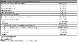
Table 1. Clinical and oocyte characteristics of the study group
The comparison of the normal fertilization rates among the MII, MI-MII and MI groups is shown in Table 2. The normal fertilization rate was directly related to oocyte maturation: the normal fertilization rates of the MII, MI-MII, and MI groups were 79.1%, 60.2% and 31.9%, respectively (p<0.001). MI oocytes had an OR of 7.6 (CI 5.2 - 11.2, p<0.001) for abnormal fertilization compared with MII oocytes and an OR of 4.0 (CI 2.5 - 6.3, p<0.001) compared with MI-MII oocytes. In addition, MI-MII oocytes had an OR of 1.9 (CI 1.4 - 2.6, p<0.001) for abnormal fertilization compared with MII oocytes.
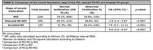
Table 2. Comparison of the normal fertilization rates of the MII, rescued MI-MII and arrested MI groups
The embryo polyploidy rate (≥ 3PN) for the stage of oocyte maturation is shown in Table 3. The risk of embryo polyploidy was higher in the MI group, with an OR of 2.6 (CI 1.28-5.41, p=0.008), than in the MII group. There was no increase in the risk of MI-MII versus MII oocyte polyploidy.
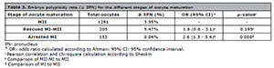
Table 3. Embryo polyploidy rate (≥ 3PN) for the different stages of oocyte maturation
The blastocyst formation rate according to oocyte maturation is shown in Table 4. The blastocyst formation rate decreased as oocyte maturation decreased, with blastocyst formation rates of 36.4%, 11.4% and 0.6% in the MII, MI-MII, and MI groups, respectively. MI-MII oocytes had an OR of 4.5 (CI 2.9 - 7.1, p<0.001) for not forming a blastocyst compared with MII oocytes. In addition, MI oocytes had an OR of 86.9 (CI 12.1 - 623.4, p<0.001) for not forming a blastocyst compared with MI oocytes and an OR of 19.2 (IC 2.5 - 143.8, p=0.004) for not forming a blastocyst compared with MI-MII oocytes. Although the rate of blastocyst formation from the MI oocytes was extremely low, one blastocyst formed and led to a pregnancy that resulted in the birth of a healthy baby with a normal karyotype.
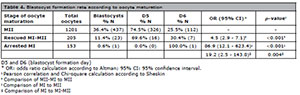
Table 4. Blastocyst formation rate according to oocyte maturation
The proportions of oocytes collected at each maturation stage according to female age group are shown in Table 5. We found no differences in the maturation rates of oocytes collected from the different age groups evaluated.
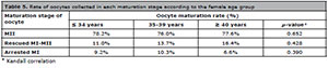
Table 5. Rate of oocytes collected in each maturation stage according to the female age group
The blastocyst formation rates according to female age group are shown in Table 6. There was a decrease in the rate of blastocyst formation from MII oocytes among women over 40 years of age (28.6% versus 37.5% and 38.4% for women over 40 years versus women 35 to 39 years and under 34 years, respectively, p=0.030). We found no differences in the rates of formation of blastocysts from MI-MII oocytes among the age groups evaluated.

Table 6. Blastocyst formation rate according to the female age group
DISCUSSION
In the present study, we evaluated whether rescue maturation after incubation of MI
oocytes collected in COH cycles could be used for ICSI by evaluating the
fertilization rate, polyploidy rate and blastocyst formation rate. We performed an
early denudation to assess oocyte maturation, and from this, we were able to
evaluate the maturation rate and consequent embryonic quality, since early
denudation does not appear to compromise ICSE cycle outcomes (Naji et al., 2018). We found a rescued
maturation rate of 57% of MI oocytes after 4 hours of incubation in culture medium,
and these rates were similar to those of other studies that reported rates between
43 and 54% in the first 4-6 hours (Shu et
al., 2007; Vanhoutte et al., 2005; Álvarez
et al., 2013). Regarding incubation time, Strassburger et al. (2004) found that 13% of MI oocytes
subjected to rescue maturation matured within 1 hour and that another 41% matured
within 2.5 hours, revealing that most MI oocytes (54%) matured in
vitro to MII oocytes after 2.5 hours of incubation; however, the
performance of these oocytes with respect to embryonic development appears to be
reduced compared with MII oocytes (Braga et
al., 2020).
When evaluating the normal fertilization rate, we found that the MI oocytes that had
not matured after 4 hours of incubation for rescue maturation had the lowest normal
fertilization rate (31.9%) compared with oocytes of other stages, particularly MII
oocytes (79.1%). We found that an MI oocyte was 7.6 times more likely to have
fertilization failure than an MII oocyte. A low normal fertilization rate of
immature oocytes has been reported by other studies, and among MI oocytes, the
fertilization rate was lower than that of in vitro-matured MI
oocytes (Alcoba et al., 2015; De Vincentiis et al.,
2013; Strassburger et
al., 2004; 2010; Shu et al., 2007). The results
of this study indicate that oocyte maturation has a direct influence on
fertilization rates. The increased fertilization rate of MI-MII oocytes compared to
immature MI oocytes seems to be related to the fact that they may not form meiotic
spindles during cell division, they may exhibit failed cytoplasmic maturation, or
they may fail to enter metaphase (Windt et
al., 2001; Santiquet et al., 2017; Sen
& Caiazza, 2013).
During their growth phase, oocytes accumulate proteins and RNAs to complete meiotic
division and maintain cellular homeostasis. Any change in this process can lead to
delayed and failed embryonic development (Jones et al., 2008). Studies evaluating MI-MII oocytes
revealed that nuclear maturation occurs before the cytoplasm reaches full maturity,
implying defects in their cytoplasmic and cytoskeletal organization, which alter the
morphogenesis of the meiotic spindle and result in embryos with nuclear
disorganization, chromosomal abnormalities, triploidy and polyploidy (Sanfins et al., 2004; Wang et al., 2001). Triploidy
appears to be the most common abnormality and is characterized by the presence of
three (3PN) instead of two (2PN) haploid chromosomal complements. During ICSI, which
excludes the possibility of polyspermy, polyploidy can occur due to one of the
following: fertilization of a haploid egg by a diploid sperm or fertilization of a
diploid egg by a haploid sperm (Rosenbusch,
2008). We found a high polyploidy rate after ICSI with MI oocytes, with a
2.6-fold higher risk compared with MII oocytes. Polyploid embryo formation may
indicate a dysfunctional oocyte, and high rates of polyploid embryo formation are
associated with a lower quality embryo cohort with a higher risk of implantation
failure and abortion (Figueira et
al., 2011).
In our study, we noticed that the blastocyst formation rate was highest among embryos
from MII oocytes (36.4%), that the blastocyst formation rate among embryos from
MI-MII oocytes was 11.4%, and embryos from MI oocytes had the lowest rate, at 0.6%.
Previous studies have demonstrated the influence of oocyte maturation on the rate of
blastocyst formation. Immature oocytes, in addition to having a lower fertilization
rate and lower blastocyst formation rate, have higher implantation failure and
abortion rates (Kedem et al.,
2018; Son et al.,
2013; Zhao et al.,
2009; Shu et al.,
2007). De Vos et al. (1999) and Alcoba et al.
(2015) did not evaluate the rate of blastocyst formation in their
studies; they evaluated the cleavage rate in D2 and D3, and found that the cleavage
rates were similar between embryos from MII oocytes and those from MI-MII oocytes,
demonstrating that in vitro-matured oocytes also have the ability
to generate good-quality embryos. However, we know that the evaluation of embryos in
the cleavage stage does not represent the true embryonic capacity; and therefore,
embryo cultivation to the blastocyst phase has become the most commonly used
approach by most assisted reproduction centers (Alcoba et al., 2015; Piqueras et al., 2017). Thus, although the cleavage
rate seems to be similar between embryos derived from in
vivo-matured (MII) and in vitro-matured (MI-MII) oocytes,
as described in the studies cited above, the results of our study demonstrate that
the blastocyst formation rate from MI-MII oocytes is significantly lower than that
from MII oocytes.
In addition, immature MI oocytes have a blastocyst formation rate of less than 1%,
and thus, immature in vitro MI oocytes have very low developmental
potential for progression to the blastocyst stage, and should not be used. Although
this is one of the conclusions of this study, it is important to emphasize that the
one blastocyst that did form from an immature MI oocyte generated a pregnancy that
culminated in the birth of a healthy baby. For this couple who had only that
particular embryo for transfer, the statistics on the rate of blastocyst formation
were not relevant, as they managed to have a healthy baby even after the
fertilization of an immature MI oocyte. In a review of the current literature, we
found one publication on ICSI outcomes with MI oocytes that were injected without
extrusion of their PB for a single embryo transfer. This embryo originated from one
oocyte that had arrested at the MI stage during in vitro maturation, and this oocyte that was injected and fertilized, underwent cleavage,
was transferred, had implanted and developed to a full-term pregnancy with a healthy
neonate (Strassburger et al.,
2004). Furthermore, the rates of normal fertilization (2PN) after ICSI of
MI oocytes (25-58%) in previous reports are similar to our observed rate of 31.9%
(Álvarez et al., 2013; Shu et al., 2007; Strassburger et al., 2004; 2010). This demonstrates that the MI
oocyte can complete the extrusion of its PB even after ICSI; however, the quality of
embryo development may be low because of DNA disorganization. One study that
examined the chromosomal content of embryos resulting from MI oocytes after COH
demonstrated that 70.2% of embryos from rescued MI-MII oocytes and 97.2% from
arrested MI are genetically abnormal, with 0% and 6.4% of them showing polyploidy,
respectively, thus demonstrating that it is possible to form euploid embryos after
ICSI of MI oocytes. However, in the aforementioned study, they analyzed probes for
only three chromosomes and indicated that a complete chromosomal analysis may
increase the rates of aneuploidy (Strassburger et al., 2010).
We also evaluated the ages of women in relation to the blastocyst formation rate. We
found a decrease in the rate of blastocyst formation from MII oocytes in the age
group of patients older than 40 years but no difference in the rate of blastocyst
formation from MI-MII oocytes. The rate of blastocyst formation from MII oocytes was
similar to that found in other studies (Macklon et al., 2002; Durand et al., 2016; Cimadomo et al., 2018), and the woman’s age was a major
factor in the success of IVF due to the decreased oocyte quality, decreased
blastocyst formation rate and increased risk of aneuploidy in this age group (Su et al., 2017; Macklon et al., 2002).
However, maternal age does not seem to influence the rate of blastocyst formation
from MI-MII oocytes, suggesting that they may be used for ICSI. The use of MI-MII
oocytes may be of particular importance in women with low ovarian reserve because,
despite having a lower fertilization rate and a lower blastocyst formation rate than
MII oocytes, MI-MII oocytes are still capable of generating a good blastocyst for
transfer. On the other hand, MI oocytes that have not rescue matured in vitro to MII
oocytes should not be used, as the fertilization rate is low, with a high polyploidy
rate, and especially because the rate of blastocyst formation is approximately zero,
with a high risk of embryonic aneuploidy.
CONCLUSION
In summary, in the present study, we found that oocytes collected at the MI stage
after COH that do not rescue mature in vitro to MII oocytes have a reduced
fertilization rate, a high incidence of embryo polyploidy and a blastocyst formation
rate of only 0.6%. Therefore, the use of immature MI oocytes is not justified and
should be discouraged. On the other hand, although the blastocyst formation rate
from rescue in vitro-matured MI-MII oocytes has been observed to be significantly
lower than that from MII oocytes, the use of MI-MII oocytes should be encouraged,
especially in women with a low MII oocyte yield, as these oocytes may increase the
number of blastocysts available for embryo transfer.
REFERENCES
Alcoba DD, Pimentel AM, Brum IS, Corleta HE. Developmental potential
of in vitro or in vivo matured oocytes. Zygote. 2015;23:93-8.
Medline Crossref
Álvarez C, García-Garrido C, Taronger R, González de Merlo G. In
vitro maturation, fertilization, embryo development & clinical outcome of
human metaphase-I oocytes retrieved from stimulated intracytoplasmic sperm
injection cycles. Indian J Med Res. 2013;137:331-8.
Medline
Baker VL, Luke B, Brown MB, Alvero R, Frattarelli JL, Usadi R,
Grainger DA, Armstrong AY. Multivariate analysis of factors affecting
probability of pregnancy and live birth with in vitro fertilization: an analysis
of the Society for Assisted Reproductive Technology Clinic Outcomes Reporting
System. Fertil Steril. 2010;94:1410-6.
Medline Crossref
Ben-Nagi J, Jones B, Naja R, Amer A, Sunkara S, SenGupta S, Serhal
P. Live birth rate is associated with oocyte yield and number of biopsied and
suitable blastocysts to transfer in preimplantation genetic testing (PGT) cycles
for monogenic disorders and chromosomal structural rearrangements. Eur J Obstet
Gynecol Reprod Biol X. 2019;4:100055.
Medline Crossref
Braga DP, Figueira Rde C, Ferreira RC, Pasqualotto FF, Iaconelli A
Jr, Borges E Jr. Contribution of in-vitro maturation in ovarian stimulation
cycles of poor-responder patients. Reprod Biomed Online. 2010;20:335-40.
Medline Crossref
Braga DPAF, Zanetti BF, Setti AS, Iaconelli A Jr, Borges E Jr.
Immature oocyte incidence: Contributing factors and effects on mature sibling
oocytes in intracytoplasmic sperm injection cycles. JBRA Assist Reprod.
2020;24:70-6.
Medline Crossref
Cimadomo D, Scarica C, Maggiulli R, Orlando G, Soscia D, Albricci L,
Romano S, Sanges F, Ubaldi FM, Rienzi L. Continuous embryo culture elicits
higher blastulation but similar cumulative delivery rates than sequential: a
large prospective study. J Assist Reprod Genet. 2018;35:1329-38.
Medline Crossref
De Vincentiis S, De Martino E, Buffone MG, Brugo-Olmedo S. Use of
metaphase I oocytes matured in vitro is associated with embryo multinucleation.
Fertil Steril. 2013;99:414-21.
Medline Crossref
De Vos A, Van de Velde H, Joris H, Van Steirteghem A. In-vitro
matured metaphase-I oocytes have a lower fertilization rate but similar embryo
quality as mature metaphase-II oocytes after intracytoplasmic sperm injection.
Hum Reprod. 1999;14:1859-63.
Medline Crossref
Durand M, Sermondade N, Herbemont C, Benard J, Gronier H, Boujenah
J, Cédrin-Durnerin I, Poncelet C, Grynberg M, Sifer C. Embryo development in two
single-step media: Analysis of 2059 sibling oocytes. Gynecol Obstet Fertil.
2016;44:163-7.
Medline Crossref
Figueira RC, Setti AS, Braga DP, Iaconelli A Jr, Borges E Jr.
Prognostic value of triploid zygotes on intracytoplasmic sperm injection
outcomes. J Assist Reprod Genet. 2011;28:879-83.
Medline Crossref
Huang JY, Rosenwaks Z. Assisted reproductive techniques. Methods Mol
Biol. 2014;1154:171-231.
Medline Crossref
Huddleston HG, Cedars MI, Sohn SH, Giudice LC, Fujimoto VY. Racial
and ethnic disparities in reproductive endocrinology and infertility. Am J
Obstet Gynecol. 2010;202:413-9.
Medline Crossref
Ingerslev HJ, Højgaard A, Hindkjaer J, Kesmodel U. A
randomized study comparing IVF in the unstimulated cycle with IVF following
clomiphene citrate. Hum Reprod. 2001;16:696-702.
Medline Crossref
Jones GM, Cram DS, Song B, Magli MC, Gianaroli L, Lacham-Kaplan O,
Findlay JK, Jenkin G, Trounson AO. Gene expression profiling of human oocytes
following in vivo or in vitro maturation. Hum Reprod. 2008;23:1138-44.
Medline Crossref
Kedem A, Yerushalmi GM, Brengauz M, Raanani H, Orvieto R, Hourvitz
A, Meirow D. Outcome of immature oocytes collection of 119 cancer patients
during ovarian tissue harvesting for fertility preservation. J Assist Reprod
Genet. 2018;35:851-6.
Medline Crossref
Macklon NS, Pieters MH, Hassan MA, Jeucken PH, Eijkemans MJ, Fauser
BC. A prospective randomized comparison of sequential versus monoculture systems
for in-vitro human blastocyst development. Hum Reprod. 2002;17:2700-5.
Medline Crossref
Malizia BA, Hacker MR, Penzias AS. Cumulative live-birth rates after
in vitro fertilization. N Engl J Med. 2009;360:236-43.
Medline Crossref
Naji O, Moska N, Dajani Y, El-Shirif A, El-Ashkar H, Hosni MM,
Khalil M, Khalaf Y, Bolton V, El-Toukhy T. Early oocyte denudation does not
compromise ICSI cycle outcome: a large retrospective cohort study. Reprod Biomed
Online. 2018;37:18-24.
Medline Crossref
Piqueras P, Gallardo M, Hebles M, Jiménez JM, Migueles B, Montero L,
Sánchez-Martín F, Sánchez-Martín P. Live birth after replacement of an embryo
obtained from a spontaneously in vitro matured metaphase-I oocyte. Syst Biol
Reprod Med. 2017;63:209-11.
Medline Crossref
Rosenbusch BE. Mechanisms giving rise to triploid zygotes during
assisted reproduction. Fertil Steril. 2008;90:49-55.
Medline Crossref
Sanfins A, Plancha CE, Overstrom EW, Albertini DF. Meiotic spindle
morphogenesis in in vivo and in vitro matured mouse oocytes: insights into the
relationship between nuclear and cytoplasmic quality. Hum Reprod.
2004;19:2889-99.
Medline Crossref
Santiquet NW, Greene AF, Becker J, Barfield JP, Schoolcraft WB,
Krisher RL. A pre-in vitro maturation medium containing cumulus oocyte complex
ligand-receptor signaling molecules maintains meiotic arrest, supports the
cumulus oocyte complex and improves oocyte developmental competence. Mol Hum
Reprod. 2017;23:594-606.
Medline Crossref
Sen A, Caiazza F. Oocyte maturation: a story of arrest and release.
Front Biosci (Schol Ed). 2013;5:451-77.
Medline Crossref
Shu Y, Gebhardt J, Watt J, Lyon J, Dasig D, Behr B. Fertilization,
embryo development, and clinical outcome of immature oocytes from stimulated
intracytoplasmic sperm injection cycles. Fertil Steril. 2007;87:1022-7.
Medline Crossref
Son WY, Chung JT, Das M, Buckett W, Demirtas E, Holzer H.
Fertilization, embryo development, and clinical outcome of immature oocytes
obtained from natural cycle in vitro fertilization. J Assist Reprod Genet.
2013;30:43-7.
Medline Crossref
Stouffer RL, Zelinski-Wooten MB. Overriding follicle selection in
controlled ovarian stimulation protocols: quality vs quantity. Reprod Biol
Endocrinol. 2004;2:32.
Medline Crossref
Strassburger D, Friedler S, Raziel A, Kasterstein E, Schachter M,
Ron-El R. The outcome of ICSI of immature MI oocytes and rescued in vitro
matured MII oocytes. Hum Reprod. 2004;19:1587-90.
Medline Crossref
Strassburger D, Goldstein A, Friedler S, Raziel A, Kasterstein E,
Mashevich M, Schachter M, Ron-El R, Reish O. The cytogenetic constitution of
embryos derived from immature (metaphase I) oocytes obtained after ovarian
hyperstimulation. Fertil Steril. 2010;94:971-8.
Medline Crossref
Su YT, Lin PY, Huang FJ, Kung FT, Lin YJ, Tsai YR, Lan KC. Age is a
major prognosticator in extremely low oocyte retrieval cycles. Taiwan J Obstet
Gynecol. 2017;56:175-80.
Medline Crossref
Vanhoutte L, De Sutter P, Van der Elst J, Dhont M. Clinical benefit
of metaphase I oocytes. Reprod Biol Endocrinol. 2005;3:71.
Medline Crossref
Wang WH, Meng L, Hackett RJ, Keefe DL. Developmental ability of
human oocytes with or without birefringent spindles imaged by Polscope before
insemination. Hum Reprod. 2001;16:1464-8.
Medline Crossref
Windt ML, Coetzee K, Kruger TF, Marino H, Kitshoff MS, Sousa M.
Ultrastructural evaluation of recurrent and in-vitro maturation resistant
metaphase I arrested oocytes. Hum Reprod. 2001;16:2394-8.
Medline Crossref
Zhao JZ, Chen X, Wang PY, Zhou W, Lin JJ, Zhang W, Huang XF, Lin WQ,
Yang HY, Chen Y. Outcome of pregnancy in women with polycystic ovary syndrome
treated by in vitro maturation of immature oocytes. Zhonghua Fu Chan Ke Za Zhi.
2009;44:409-12.
Medline