JBRA Assisted Reproduction 2021;25(2):202-208
ORIGINAL ARTICLE
doi: 10.5935/1518-0557.20200067
Unbalance of Se and nutritional status in male infertility
1Nutrition department, State University of Ceará, Fortaleza, CE, Brazil
2Reproduction Human Center Evangelista Torquato, Fortaleza, CE, Brazil
3Nutrition department, Federal University of Sergipe, Aracaju, SE, Brazil
The authors have no conflicts of interest to declare.
ABSTRACT
Objective: To evaluate the selenium status and oxidative stress in male infertility
cases selected from a private human reproduction center in the state of
Ceará, Brazil.
Methods: The present study had a cross-sectional quantitative approach, carried out
between January and October 2013 at a Human Reproduction Center. The studied
population was composed of 49 male individuals seen at the clinic, aged
between 18 and 60 years. Blood samples were collected to measure serum
selenium concentrations, erythrocyte activity and glutathione peroxidase.
After medical diagnosis, the participants were divided into fertile and
infertile groups. Blood samples were collected for establishing Se
concentrations in plasma and erythrocytes, and measurements of the enzymatic
activity of glutathione peroxidase in the erythrocytes.
Result: it resulted in 53.1% of fertile men and 46.9% of infertile men. The average
age of the fertile group was 34.1 years and the infertile group was 37.3
years. Regarding the assessment of nutritional status, the scatter diagram
of the infertility group showed a higher body mass index and waist
circumference, showing that this group has a higher risk of global and
abdominal obesity compared to the fertile group
(p<0.0001, respectively). There were similarities
between the groups regarding caloric intake, macronutrient and selenium
intake.
Conclusion: We can conclude that the serum values of selenium, in excess and in
deficiency, can be harmful to male fertility.
Keywords: male infertility, selenium, oxidative stress, overweight
INTRODUCTION
Infertility is defined as the inability of a couple to achieve conception or to bring
a pregnancy to term after one year or more of regular, unprotected intercourse, and
it has been recognized as a public health problem worldwide (World Health
Organization - WHO, 2000; Agarwal et al., 2015). It
affects 10-15% of couples in reproductive age (Winters & Walsh, 2014). Some studies show that environmental,
physiological and genetic factors are related to 25% of male factor infertility due
to semen quality, including oxidative stress, which has frequently been associated
with the problem (Kolesnikova et
al., 2015; Aitken,
2016).
Oxidative stress adversely affects sperm function by altering membrane fluidity,
permeability and sperm functional competence (Kumar
& Singh, 2018; Riaz et
al., 2016; Aitken et
al., 2016). Sperm membranes are characterized by relatively
greater concentrations of polyunsaturated fatty acids that are extremely sensitive
to the attack of reactive oxygen species (ROS) (Treulen et al., 2015). Oxidative stress attacks not
only the fluidity of the sperm plasma membrane, but also the deoxyribonucleic acid
(DNA) integrity in the sperm nucleus, making the sperm unable to fertilize an egg or
start a viable pregnancy (Bisht & Dada,
2017; Hashemi et al.,
2018).
Essential trace minerals such as selenium (Se) not only act as antioxidants but also
play vital roles in multiple metabolic processes (Türk et al., 2014; Ahsan et al., 2014). Se is an essential trace mineral
recognized as fundamental for human reproduction, spermatogenesis, promotion of
normal testicular development and preserving spermatozoa motility (Adeoye et al., 2018).
One of Se’s biochemical functions is linked to the enzyme glutathione peroxidase
(GPx), a selenoprotein with an antioxidant function that protects the sperm membrane
against oxidative stress. GPx acts as an Se reservoir, which is used in emerging
needs. Due to its powerful antioxidant effect, Se in the GPx needs to be better
investigated as a nutritional factor for the protection of men's health in relation
to male infertility, considering that oxidative stress compromises sperm motility,
vitality and function (Qazi et al.,
2019).
The primary source of Se for men is through food. Thus, the Se present in foodstuff
can be influenced by the environment and soils, especially those with a high
concentration of Se, which can theoretically produce foods richer in this nutrient,
enabling its adequate intake. Based on this, the consumption of this mineral in
Brazilian diets varies according to region (Tureck et al., 2017).
Since Se is a mineral essential for improving reproductive health and Ceará (state of
Brazilian Northeast) has a Se-rich soil, the present study’s goal was to evaluate
Se, nutritional status and their association with human reproduction in Fortaleza,
Ceará- Brazil.
MATERIAL AND METHODS
Study design
The present study was a quantitative cross-sectional approach, carried out
between January and October of 2013 at a Human Reproduction Center in Fortaleza,
capital of the state of Ceará, Brazil. The studied population consisted of male
individuals seen at the clinic, aged between 18 and 60 years without chronic
diseases, non-smokers, without the use of vitamin and mineral supplements and
hormonal medications. The study was conducted according to the ethical
principles and guidelines for the protection of human participants in research,
which was approved by the Ethics Committee for research of the State University
of Ceará (protocol number 5882212.9.00005534 issued on 27/12/2012).
Data collection
We collected sociodemographic data to characterize the sample. Weight (kg) and
height (m) were measured according to the Frisancho methodology (Frisancho, 1990). We calculated the Body Mass
Index (BMI) by dividing the body weight by height (kg/m²). Nutritional diagnosis
was assessed according to the classification for adults (WHO, 2000). Waist circumference (WC) was measured using a
flexible, tension-sensitive, non-stretchable measuring tape (Gulick II) placed
directly onto the skin. Measurements were taken from the midpoint between the
iliac crest and the lowest rib (WHO,
2011).
We assessed dietary intake using the 24-h recall method. Three non-consecutive
24-h dietary recalls were collected, which included two weekdays and one weekend
day (Fisberg et al.,
2005). We used the computer software NutWin (version 2.5, 2002) (fed
with the USDA database); the UNIFESP-EPM was used for food intake calculation,
with adequacy established by the Dietary Reference Intakes (DRIs) (Institute of Medicine - IOM, 2005). The
amounts indicated by the subjects were transformed into homemade measures and
analyzed (Pinheiro et al.,
2008). The Se values were adjusted for energy (IOM, 2000).
Semen analysis was completed by an accredited laboratory according to the
standards set by the WHO (2010), which
consisted of the macroscopic and microscopic evaluations of the semen.
After medical diagnosis, the participants were divided into fertile and infertile
groups. Blood samples were collected for the determination of Se concentrations
in plasma and erythrocytes, and we measured their glutathione peroxidase (GPx)
enzymatic activity in erythrocytes.
The plasma and erythrocyte Se levels were determined by atomic absorption
spectrometry using the hydride generation coupled to the quartz cell HITACHI
(Z-5000 model), adapted from Martens et
al. (2015). GPx activity in erythrocyte was assessed
using the kinetic method described by Paglia
& Valentine (1967).
We used the commercially available kit (RANSEL 505 - RANDOX Laboratories Ltd, UK
- reference range: 27.5 to 73.6 U/g Hb) according to the manufacturer's
instructions.
Statistical analysis
We processed the data using the free software R (version 2.15.0). To compare the
mean parameters of the fertile and the infertile groups, we used the Student
t-test and ANOVA, and in both cases, we obtained the p-value by the exact test
via Bootstrap, ensuring the robustness against the lack of normality. To
correlate dietary intake of Se and dietary energy, we used the Kolmogorov -
Smirnov test and applied the Pearson’s correlation to assess whether there was a
statistically significant correlation between energy and Se. After that, we ran
a linear regression analysis between the energy (independent variable) and Se
(dependent variable) to make the adjustment for energy. All analyses were
considered statistically significant if p<0.05.
RESULTS
We assessed the fertility of 50 male subjects. One participant left the study,
yielding 53.1% fertile men and 46.9% infertile men. The mean age of the fertile
group was 34.1 (7.6) years and the infertile group was 37.3 (8.1) years.
Regarding nutritional status assessment, the scatter diagram of the infertility group
showed higher BMI and WC, showing that this group presents a higher risk of global
and abdominal obesity as compared to the fertile group
(p<0.0001, respectively) (Table
1) (Figure 1). There were
similarities between the groups regarding caloric intake and that of macronutrients
and Se (Table 1).
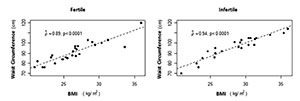
Figure 1. Scatter plot between waist circumference (cm) and body mass intake
(Kg/m2) by group
There was no significant difference for serum Se (Table 1). According to Van Dael &
Deelstra (1993), the plasma values should be in the range of 60 to 120
μg/L. In this sense, 42.31% of the fertile and 56.52% of the infertile were within
this range. In the infertile group, the mean selenium concentration in erythrocytes
was higher than in the fertile group (Table
1). According to Van Dael & Deelstra
(1993), the reference value of the erythrocyte Se should be in the range
of 90-190 μg/L. We had 42.31% of the subjects in the fertile group and 56.52% of the
infertile group within this range.
There was a positive correlation between Se intake and motility index (%)
(p=0.0407) at the 5% level. The higher the intake of Se, the
higher the motility index (%) in the fertile group. (Figure 2). However, there was a negative correlation between WC and Se
intake (g/day) in the infertile group (r=-0, 42 p=0.0456) (Figure 3).
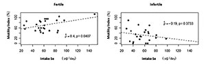
Figure 2. Scatter plot between motility index (%) and intake Se (µg/day) by
group
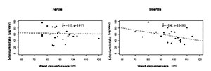
Figure 3. Scatter plot between selenium intake (µg/day) and waist
circumference (cm) by group
On the other hand, there was a significant negative linear correlation
(p<0.0001) between the Kruger morphology (%) and serum Se in
the fertile group. Thus, the higher the plasma Se levels in this group, the lower
the Kruger morphology (%) (Figure 4).
Another result of the Kruger's strict sperm morphology (%) showed a significant
positive linear correlation (p=0.0081), at the 5% level, with the
motility index (%) in infertile men (Figure 5).
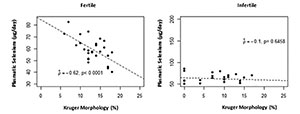
Figure 4. Scatter plot between plasmatic selenium (µg/day) and Kruger morphology
(%) by group
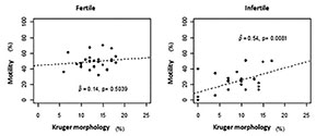
Figure 5. Scatter plot between motility (%) and Kruger morphology (%) by group
Between groups there was no difference in GPx activity (Table 1). In addition, there was a positive correlation (p=0.0305) between ejaculate volume and GPx in the infertile group (Figure 6).
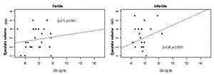
Figure 6. Scatter plot between ejaculate volume (ml) and GPx (Ug/Hb) by group
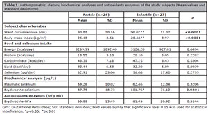
Table 1. Anthropometric, dietary, biochemical analyses and antioxidants enzymes of the
study subjects (Mean values and standard deviations)
DISCUSSION
Selenium biomarkers were associated with male fertility because among fertile men,
serum Se levels was inversely associated with the Kruger morphology rate (%), and
among the infertile, erythrocyte Se was at higher levels, revealing that serum
selenium imbalance, both excess and reduction influence male fertility. GPx
activity, also called GPx1, was preserved in both groups without statistical
difference between them. Fertile and infertile men were studied for Se
concentrations (plasm and erythrocyte).
Trace elements such as selenium are involved in many vital reproductive health and
performance processes. However, studies are scarce on the concentration of Se in
various body fluids/tissues for optimal reproductive performance. Adequate
concentrations of this mineral in the reproductive tissues are fundamental for
several processes associated with fertility such as spermatogenesis, maturation and
motility of spermatozoids, besides maintenance and improvement of GPx antioxidant
activity. Some selenoenzymes such as selenoprotein P (SelP) and the GPx family
(especially the GPx 4 isoform) are involved in fertility and reproductive processes
(Qazi et al., 2019). Thomson (2004) stated that the
consumption of 45-50 μg/day is able to optimize the activity of GPx and SelP. Thus,
considering the consumption from both groups in this study we can justify the
appropriate activity of the GPx enzyme in these groups (Thomson, 2004).
The selenium from food is rich in organic forms of this mineral which has been
related to better assimilation being more bioavailable and less toxic. Vitamin
B6 also participates in the optimization of the GPx system from
organic forms of selenium. Therefore, proper nutrition can contribute to fertility.
Consumption and adequate concentrations of selenium are linked to testosterone
biosynthesis whose mechanism is related to the expression and antioxidant action of
SelP in the cells of Leydig and cytosolic GPx acting on the neutralization of
H2O2 produced in this biosynthesis (Qazi et al., 2019).
Se deficient and Se excess diets have been reported to result in reduced spermatozoa
motility, concentration, and fertility. Diets excessive or deficient in Se affect
the gross as well as histological morphology of the testis. Sperm morphology is
considered one of the most important seminal parameters, being the best indicator of
male fertility. At appropriate levels of serum Se, sperm morphology presents good
quality, but as there is inadequacy in consumption, that is, it exceeds the
recommended value, this morphology becomes impaired. Therefore, although sperm
morphology is a controversial parameter, it is an important result regarding the
fertility of men who underwent antioxidant treatment (Qazi et al., 2019).
Studies observed by Riaz et al.
(2016) and Lovercamp et
al. (2013) show that high levels of plasma Se resulted in a
sperm morphology reduction and consequently its motility in fertile men. Our results
are consistent with the findings and the same effect was observed in the fertile
group. According to WHO (2010), the strict
Kruger morphology criteria has a directly association to motility. In the infertile
group, the mean erythrocyte Se value was higher than the fertile.
These data suggest that elevated levels of erythrocyte Se decrease motility because
the effect of motile onset was probably mediated by secretions of accessory sex
glands or during sperm maturation or ejaculation, since there was little evidence of
toxic effect upon ingestion (Dorostghoal et
al., 2017).
Although no difference in food intake was found between groups analysed, the
fertility group showed a positive correlation between Se intake and sperm motility
(%). These results are in agreement with Foresta et al. (2002), Klein
(2004), Moslemi & Tavanbakhsh
(2011) that showed the relationship between Se ingestion and increased
sperm motility.
Besides that, there was a positive correlation between ejaculate volume and GPx in a
fertile group. This is in accordance with the study of Türk et al. (2014) confirming the presence of
GPx in the ejaculate mainly coming from the testes and epididymis, thus regulated by
the ingestion of Se.
Thus, decreased GPx activity during Se deficiency and increased lipid peroxidation,
indicate increased levels of free radicals which disrupts the normal spermatogenic
process and might be responsible for reduced sperm motility and viability leading to
reduced fertility and fewer litters. The GPx is responsible for over 75% of
antioxidant activity in spermatozoa. In Se excess, increased lipid peroxidation
could be attributed to the ability of selenite to form a highly reactive species, a
selenopersulfide which generates free radical, superoxide as well as other Reactive
Oxygen Species (Shalini & Bansal, 2007; Shalini & Bansal, 2008).
Besides these aspects, it can be verified that the nutritional status indicated
higher levels of BMI and WC in the infertile group. Obesity seems to have a
deleterious effect on semen quality by changes in epididymal function, reducing the
biomarkers of normal accessory gland function. In addition, the ejaculated volume
(mL), sperm concentration (million / mL), and total sperm count (millions /
ejaculate) are inversely correlated with BMI. Obese men were 19 times more likely to
have a lower total sperm count (millions / ejaculate) than eutrophic (Bieniek et al., 2016; Oliveira et al., 2017; Wang et al., 2017; Liu & Ding, 2017). The results are similar
to studies designed by Borges Jr. et
al. (2015) and Schlichthorst et al. (2016) that showed a relationship of obesity
and infertility with men with mean age of 37 years. The increased WC is associated
with obesity-related health risk that can reduce male fertility (Hayden et al., 2018; Kasum et al., 2016). Se may be
a protective factor in male fertility, since the higher the Se intake, the smaller
the waist circumference is in the infertile group, and higher the sperm motility
index in the fertile group.
Our study demonstrates the possible interferences of obesity, deficiency and excess
selenium in male fertility. Thus the action of dietary selenium appears to be
U-shaped with respect to reproductive capacity and its markers. Further studies are
needed to clarify the mechanisms involved in this process. It also makes an
important contribution by calling attention to the use of supplements with Se
megadoses present in antioxidant treatments.
CONCLUSION
From the above findings, we can conclude that fertile and infertile men were suitable
for Se concentrations. Serum values of Se, both excess and deficiency can be
detrimental to male fertility. In the fertile group, Se consumption was positively
correlated with sperm motility and, in the infertile group, negatively correlated
with WC.
ACKNOWLEDGMENT
The authors would like to thank the Torquato Evangelista Human Reproduction Center,
the Laboratory of Biochemistry and Food Analysis of the State University of Ceara -
UECE; the Laboratory of Mineral Nutrition of the University of São Paulo - USP, the
Center for Experimental Biology of the University of Fortaleza - UNIFOR, the
Laboratory of Pharmacology of Inflammation and Cancer - LAFICA and the Pasteur
Diagnostic Laboratory for support in the development of the research.
REFERENCES
Adeoye O, Olawumi J, Opeyemi A, Christiania O. Review on the role of
glutathione on oxidative stress and infertility. JBRA Assist Reprod.
2018;22:61-6. PMID: 29266896 DOI: 10.5935/1518-0557.20180003
Medline Crossref
Agarwal A, Mulgund A, Hamada A, Chyatte MR. A unique view on male
infertility around the globe. Reprod Biol Endocrinol. 2015;13:37. PMID: 25928197
DOI: 10.1186/s12958-015-0032-1
Medline Crossref
Ahsan U, Kamran Z, Raza I, Ahmada S, Babar W, Riaz MH, Iqbal Z. Role
of selenium in male reproduction - a review. Anim Reprod Sci. 2014;146:55-62.
PMID: 24613013 DOI: 10.1016/j.anireprosci.2014.01.009
Medline Crossref
Aitken RJ. Oxidative stress and the etiology of male infertility. J
Assist Reprod Genet. 2016;33:1691-2. PMID: 27544275 DOI:
10.1007/s10815-016-0791-4
Medline Crossref
Aitken RJ, Gibb Z, Baker MA, Drevet J, Gharagozloo P. Causes and
consequences of oxidative stress in spermatozoa. Reprod Fertil Dev.
2016;28:1-10. PMID: 27062870 DOI: 10.1071/RD15325
Medline Crossref
Bieniek JM, Kashanian JA, Deibert CM, Grober ED, Lo KC, Brannigan
RE, Sandlow JI, Jarvi KA. Influence of increasing body mass index on semen and
reproductive hormonal parameters in a multi-institutional cohort of subfertile
men. Fertil Steril. 2016;106:1070-5. PMID: 27460460 DOI:
10.1016/j.fertnstert.2016.06.041
Medline Crossref
Bisht S, Dada R. Oxidative stress: Major executioner in disease
pathology, role in sperm DNA damage and preventive strategies. Front Biosci
(Schol Ed). 2017;9:420-47. PMID: 28410127 DOI: 10.2741/s495
Medline Crossref
Borges E Jr, Setti AS, Braga DP, Figueira Rde C, Iaconelli A Jr.
Decline in semen quality among infertile men in Brazil during the past 10 years.
Int Braz J Urol. 2015;41:757-63. DOI:
10.1590/S1677-5538.IBJU.2014.0186
Crossref
Dorostghoal M, Kazeminejad SR, Shahbazian N, Pourmehdi M, Jabbari A.
Oxidative stress status and sperm DNA fragmentation in fertile and infertile
men. Andrologia. 2017;10:e12762. PMID: 28124476 DOI:
10.1111/and.12762
Medline Crossref
Foresta C, Bettella A, Merico M, Garolla A, Ferlin A, Rossato M. Use
of recombinant human follicle-stimulating hormone in the treatment of male
factor infertility. Fertil Steril. 2002;77:238-44. PMID: 11821078 DOI:
10.1016/S0015-0282(01)02966-1
Medline Crossref
Hashemi MM, Behnampour N, Nejabat M, Tabandeh A, Ghazi-Moghaddam B,
Joshaghani HR. Impact of Seminal Plasma Trace Elements on Human Sperm Motility
Parameters. Rom J Intern Med. 2018;56:15-20. PMID: 28865234 DOI:
10.1515/rjim-2017-0034
Crossref
Hayden RP, Flannigan R, Schlegel PN. The Role of Lifestyle in Male
Infertility: Diet, Physical Activity, and Body Habitus. Curr Urol Rep.
2018;19:56. PMID: 29774489 DOI: 10.1007/s11934-018-0805-0
Medline Crossref
IOM - Institute of Medicine. Dietary Reference Intakes for Energy,
Carbohydrate, Fiber, Fat, Fatty Acids, Cholesterol, Protein, and Amino Acids.
Washington, DC: National Academies Press; 2005. Available at: DOI:
10.17226/10490
Crossref
Kasum M, Anić-Jurica S, Čehić E, Klepac-Pulanić T, Juras J, Žužul K.
Influence of male obesity on fertility. Acta Clin Croat. 2016;2:301-8. PMID:
28394547 DOI: 10.20471/acc.2016.55.02.18
Medline Crossref
Klein EA. Selenium: epidemiology and basic science. J Urol.
2004;17:S50-3. PMID: 14713754 DOI:
10.1097/01.ju.0000107837.66277.e9
Medline Crossref
Kolesnikova LI, Kolesnikov SI, Kurashova NA, Bairova TA. Causes and
factors of male infertility. Vestn Ross Akad Med Nauk. 2015;70:579-84. PMID:
26846084 DOI: 10.15690/vramn.v70.i5.1445
Medline Crossref
Kumar N, Singh AK. Reactive oxygen species in seminal plasma as a
cause of male infertility. J Gynecol Obstet Hum Reprod. 2018;47:565-72. PMID:
30016715 DOI: 10.1016/j.jogoh.2018.06.008
Medline Crossref
Liu Y, Ding Z. Obesity, a serious etiologic factor for male
subfertility in modern society. Reproduction. 2017;154:R123-31. PMID: 28747541
DOI: 10.1530/REP-17-0161
Medline Crossref
Lovercamp KW, Stewart KR, Lin X, Flowers WL. Effect of dietary
selenium on boar sperm quality. Anim Reprod Sci. 2013;138:268-75. PMID: 23523235
DOI: 10.1016/j.anireprosci.2013.02.016
Medline Crossref
Martens IB, Cardoso BR, Hare DJ, Niedzwiecki MM, Lajolo FM, Martens
A, Cozzolino SM. Selenium status in preschool children receiving a Brazil
nut-enriched diet. Nutrition. 2015;11:1339-43. PMID: 26429652 DOI:
10.1016/j.nut.2015.05.005
Medline Crossref
Moslemi MK, Tavanbakhsh S. Selenium-vitamin E supplementation in
infertile men: effects on semen parameters and pregnancy rate. Int J Gen Med.
2011;4:99-104. PMID: 21403799 DOI: 10.2147/IJGM.S16275
Medline Crossref
Oliveira PF, Sousa M, Silva BM, Monteiro M, Alves MG. Obesity,
energy balance and spermatogenesis. Reproduction. 2017;153:R173-85. PMID:
28283671 DOI: 10.1530/REP-17-0018
Medline Crossref
Paglia DE, Valentine WN. Studies on the quantitative and qualitative
characterization of erythrocyte glutathione peroxidase. J Lab Clin Med.
1967;70:158-69. PMID: 6066618
Medline
Qazi IH, Angel C, Yang H, Zoidis E, Pan B, Wu Z, Ming Z, Zeng CJ,
Meng Q, Han H, Zhou G. Role of Selenium and Selenoproteins in Male Reproductive
Function: A Review of Past and Present Evidences. Antioxidants (Basel).
2019;8:268. PMID: 31382427 DOI: 10.3390/antiox8080268
Medline Crossref
Riaz M, Mahmood Z, Shahid M, Saeed MU, Tahir IM, Shah SA, Munir N,
El-Ghorab A. Impact of reactive oxygen species on antioxidant capacity of male
reproductive system. Int J Immunopathol Pharmacol. 2016;29:421-5. PMID: 26684624
DOI: 10.1177/0394632015608994
Medline Crossref
Schlichthorst M, Sanci LA, Hocking JS. Health and lifestyle factors
associated with sexual difficulties in men - results from a study of Australian
men aged 18 to 55 years. BMC Public Health. 2016;16:1043. PMID: 28185600 DOI:
10.1186/s12889-016-3705-6
Medline Crossref
Shalini S, Bansal MP. Alterations in selenium status influences
reproductive potential of male mice by modulation of transcription factor
NFkappaB. Biometals. 2007;20:49-59. PMID: 16758115 DOI:
10.1007/s10534-006-9014-2
Medline Crossref
Shalini S, Bansal MP. Dietary selenium deficiency as well as excess
supplementation induces multiple defects in mouse epididymal spermatozoa:
understanding the role of selenium in male fertility. Int J Androl.
2008:31:438-49. PMID: 17651402 DOI:
10.1111/j.1365-2605.2007.00789.x
Medline Crossref
Thomson CD. Assessment of requirements for selenium and adequacy of
selenium status: a review. Eur J Clin Nutr. 2004;58:391-402. PMID: 14985676 DOI:
10.1038/sj.ejcn.1601800
Medline Crossref
Treulen F, Uribe P, Boguen R, Villegas JV. Mitochondrial
permeability transition increases reactive oxygen species production and induces
DNA fragmentation in human spermatozoa. Hum Reprod. 2015;30:767-76. PMID:
25662811 DOI: 10.1093/humrep/dev015
Medline Crossref
Tureck C, Locateli G, Corrêa VG, Koehnlein EA. Evaluation of the
Brazilian population’s intake of antioxidant nutrients and their relation with
the nutritional status. Rev Bras Epidemiol. 2017;20:30-42. PMID: 28513792 DOI:
10.1590/1980-5497201700010003
Medline Crossref
Türk S, Mändar R, Mahlapuu R, Viitak A, Punab M, Kullisaar T. Male
infertility: decreased levels of selenium, zinc and antioxidants. J Trace Elem
Med Biol. 2014;28:179-85. PMID: 24462254 DOI:
10.1016/j.jtemb.2013.12.005
Medline Crossref
Van Dael P, Deelstra H. Selenium. Int J Vitam Nutr Res.
1993;63:312-6. PMID: 8157441
Medline
Wang EY, Huang Y, Du QY, Yao GD, Sun YP. Body mass index effects
sperm quality: a retrospective study in Northern China. Asian J Androl.
2017;19:234-7. PMID: 26732109 DOI: 10.4103/1008-682X.169996
Medline Crossref
Winters BR, Walsh TJ. The epidemiology of male infertility. Urol
Clin North Am. 2014;41:195-204. PMID: 24286777 DOI:
10.1016/j.ucl.2013.08.006
Medline Crossref