JBRA Assist. Reprod. 2022;26(2):199-207
ORIGINAL ARTICLE
doi: 10.5935/1518-0557.20210008
Associations among morphological parameters, clinical factors and euploid blastocyst formation
1Department of Obstetrics and Gynecology, Universidade Federal do Pará, Belém, PA, Brazil
2Clínica de Reprodução Assistida Pronatus, Belém, PA, Brazil
3Grupo de Pesquisa Bilibio, Universidade Federal do Pará, Belém, PA, Brazil
4Programa de Pós-Graduação de Ciências Médicas da Universidade Federal do Rio Grande do Sul, Porto Alegre, RS, Brazil
Authors reported no conflict of interest associated with this study.
ABSTRACT
Objective: To evaluate the association among embryonic morphological parameters, clinical factors and euploid blastocyst formation.
Methods: This prospective cohort study included 422 blastocysts from 135 patients who had undergone preimplantation genetic analysis after intracytoplasmic sperm injection (ICSI).
Results: Of 422 blastocysts, 200 (47.4%) were euploid and 222 (52.6%) aneuploid. Women aged older than 38 years were more likely to develop aneuploid embryos (OR: 3.4, CI: 2.2-5.4, p<0.001). Poor ovarian reserve (OR: 3.3, p<0.001), increased male age (39.0 versus 40.7, p=0.019), and decrease in sperm percentage with normal morphology (2.5% vs. 1.9%, p=0.047) were associated with aneuploidy. Type C trophectoderm (TE) and type C inner cell mass were associated with a high risk of embryo aneuploidy, with OR of 4.1 (CI: 2.2-7.7, p<0.001) and 1.7 (CI: 1.01-3.0, p=0.048), respectively. Logistic regression analysis revealed maternal age and type C TE as the main risk factors for aneuploidy. Among combinations of factors, the best marker for the risk of aneuploidy was maternal age older than 38 years, combined with a type-C embryo with trophectoderm, which showed a positive predictive value of 88.6% and a specificity of 97.5%.
Conclusions: Trophectoderm and type-C inner cell mass are the main embryo risk factors for aneuploidy, explaining approximately 71% and 60% of the risk, respectively. Among clinical factors, advanced maternal and paternal age (older than 38 and 36 years, respectively), antral follicles (<5), and a low percentage of sperm with normal morphology increased the risk of embryonic aneuploidy.
Keywords: aneuploidy, PGT-A, trophectoderm, blastocyst, inner cell mass
INTRODUCTION
Aneuploidy is the most common type of chromosomal abnormality and the leading cause of implantation failure, miscarriage and congenital abnormalities in humans (Lee et al., 2015). Approximately 30-35% of miscarriages in women over 35 are due to chromosomal abnormalities (Hodes-Wertz et al., 2012). Preimplantation genetic testing for aneuploidies (PGT-A) can be used to select an euploid embryo for transfer, to prevent chromosomal abnormality-induced abortions due to embryonic aneuploidies after in vitro fertilization (IVF) (Brezina et al., 2012).
PGT-A improves embryo transfer implantation rates and reduces spontaneous abortion rates, particularly in patients at an increased risk of producing aneuploid embryos (Lee et al., 2015). PGT-A increases embryo implantation rates to 70-80%; as a result, many couples, especially those at an increased risk of forming aneuploid embryos, due to advanced maternal age for instance, or who fear generating aneuploid embryos, are seeking this technology (Lee et al., 2015). However, despite these advantages, approximately 45%-50% of biopsied embryos are aneuploid or mosaic-i.e. many embryos that are subjected to PGT-A are not viable, causing an increase in costs for couples due to the need to analyze several embryos (Platteau et al., 2006; Friedenthal et al., 2018; Lawrenz et al. 2019; Friedenthal et al., 2020).
Although preimplantation genetic analysis can help select embryos for transfer, it does not change the final pregnancy rate and may even decrease this rate due to the aggressive manipulation of embryos during biopsy (Zhang et al., 2016; Guzman et al., 2019; Munné et al., 2019). As such, couples undergoing IVF treatment, in addition to having difficulty achieving pregnancy, now also need to decide whether to undergo PGT-A to avoid the risk of pregnancy with an aneuploid embryo. To do so, these couples need to bear the high costs of this technology, which on the one hand, prevents the transfer of aneuploid embryos, but on the other hand, yields a similar or even lower pregnancy rate than in couples who do not choose an embryonic biopsy (Zhang et al., 2016; Guzman et al., 2019; Munné et al., 2019). It is noteworthy that the costs of PGT-A depend on the number of biopsied embryos, and approximately 50% of them are aneuploid (Gazzo et al., 2020).
Researchers have attempted to correlate embryonic morphology with euploidy, to try to predict which embryos are most suitable for biopsy. Some found a higher risk of aneuploidy in embryos with a higher number of blastomeres at the cleavage stage (Kroener et al., 2015), and others found that blastocysts with higher inner cell mass (ICM) and trophectoderm (TE) scores are more likely to be euploid (Barash et al., 2017; Wang et al., 2018). Having embryonic culture in time-lapse incubators, in addition to the morphological aspect, and embryonic morphokinetics has also begun to be evaluated as a possible predictor of the formation of euploid blastocysts (Zaninovic et al., 2017). However, due to its high cost, few centers have access to this time-lapse technology; thus, the morphological criteria for embryonic classification and selection are still the most commonly used.
In addition to embryonic morphology, clinical factors have been associated with the risk of aneuploidy, especially advanced maternal and paternal ages, and low ovarian reserves (García-Ferreyra et al., 2015; Shahine et al., 2016). Other clinical factors associated with infertility, such as endometriosis (Juneau et al., 2017), recurrent miscarriage (Hodes-Wertz et al., 2012), polycystic ovary syndrome (PCOS) (Wang et al., 2016) and male factors (Mazzilli et al., 2017), have also been investigated to assess the risk of aneuploid embryo formation. Many of these clinical factors have shown conflicting results and need to be further evaluated regarding their association with embryonic aneuploidy.
Therefore, due to the increased use of PGT-A, the need to find markers associated with embryonic euploidy to better select embryos, and especially the need to better help couples regarding embryonic euploidy prognoses in different situations, the aim of this study was to evaluate the association among morphological parameters, clinical factors and euploid blastocyst formation determined by PGT-A.
MATERIALS AND METHODS
Patients
We ran a prospective cohort study with 430 blastocyst embryos that underwent PGT-A, by next-generation sequencing (NGS) from 135 patients who underwent intracytoplasmic sperm injection (ICSI) during the study period, from June 2018 to June 2019, at the Pronatus Reproductive Medicine Center, Belém, Pará State - Brazil. The National Ethics in Research Committee approved the study and by the Ethics Committee of the Instituto de Ciências da Saúde da Universidade Federal do Pará under CAAE number 12779919.0000.0018.
All couples were evaluated for the probable cause of infertility according to the following criteria: tubal factor (defined by change on hysterosalpingography or videolaparoscopy); male factor (defined as sperm count below 15 million/ml - according to the criteria of the World Health Organization) (Cooper et al., 2010); a low ovarian reserve (defined as <5 antral follicles evaluated by transvaginal ultrasound performed on the 2nd or 3rd day of the menstrual cycle); repeat abortion (two or more consecutive abortions); polycystic ovary syndrome (PCOS); endometriosis (presence of endometriosis foci on videolaparoscopy or endometrioma on imaging studies); and undetermined cause.
Ovarian stimulation
The ovarian stimulation protocol began on the second day of the menstrual cycle, after transvaginal ultrasound was performed to determine the antral follicle count; recombinant follicle-stimulating hormone (FSH, Elonva 150®, Organon) was injected subcutaneously. After day 6 of the application, transvaginal ultrasound was performed every two days to monitor follicle development. When one of the largest follicles reached 14 mm, the gonadotrophin-releasing hormone (GnRH) antagonist (Orgalutran®, Organon) was administered daily and subcutaneously until the use of recombinant human chorionic gonadotropin (rhCG). After day 9 of the induction, we used recombinant FSH (Puregon®, Organon) at a dose of 150 IU per day, until rhCG was used. When three or more follicles reached 17 mm in diameter, we stimulated oocyte maturation with rhCG (Ovidrel 250 mcg; Serono). Transvaginal ultrasound-guided oocyte recovery was performed 35h after hCG application. After denudation, we classified the collected oocytes into metaphase II (MII), metaphase I (MI), germinal vesicle (GV), ruptured or atretic.
Assessment of fertilization, embryo quality and embryo biopsy
We subjected all MII oocytes to ICSI, and we performed embryonic culture in a Thermo Scientific CO2 Incubator. We used the CSCM-C IRVINE® culture medium for the Single Step system-type with the pH adjusted to 7.3 to obtain a CO2 pressure of 7.4%, and the embryonic development was carried out according to the Istanbul consensus (Alpha Scientists in Reproductive and Embryology, 2011).
The same embryologist performed all embryonic evaluations and biopsies. Additionally, in all evaluations, we photographed the embryos and stored the images in a database so that, if necessary, we could run a retrospective evaluation of the embryonic classification.
On day 1 (D1, evaluation 17 hours post insemination (HPI)), the ideal fertilized oocyte was spherical and there were two polar corpuscles and two centrally located, juxtaposed pronuclei of uniform size, with distinct membranes. The nuclear precursor corpuscles classification was divided into three categories (1: symmetrical, 2: asymmetrical, 3: abnormal).
On D2 and D3 (D2: assessment 44 HPI, D3: assessment 68 HPI), embryonic cleavage along with the blastomere count, degree of fragmentation, cell size and presence of multinucleation were observed and classified as grade 1 (<10% fragmentation, stage-specific cell size, no multinucleation), grade 2 (10-25% fragmentation, stage-specific cell size for most cells, no evidence of multinucleation) and grade 3 (severe fragmentation > 25%, cell size not stage-specific, evidence of multinucleation).
On the morning of day 5 (D5), approximately 116+2 HPI, blastocyst classification was performed by assessing the developmental stage (1: initial, 2: blastocyst, 3: expanded, 4: hatched/hatching), ICM (A: prominent, B: easily discernible, C: difficult to distinguish), and TE (A: many cells forming a cohesive epithelium, B: few cells forming a loose epithelium, C: few cells). Expanded blastocysts were deemed usable and considered for biopsy if they displayed a single adequately cellular and compact ICM. Expanded blastocysts were biopsied as soon as they met these criteria. Notably, if a blastocyst was collapsed at the time of assessment, it was re-evaluated 1-2h later. All embryos not meeting these criteria on the morning of day 5 remained in culture until the morning of day 6, at which point they were again assessed.
Blastocyst biopsy
Only blastocysts that presented at least 3 degrees of expansion (complete cavity), a detectable internal cell mass and detectable trophectoderm cells were biopsied. Approximately 5-8 trophectoderm cells were excised using a laser (OCTAX Laser Shot™ System - Infrared Diode Laser - 1.48 µm Wave Length) and 1- to 2,800-mm pulses to break apart cell junctions in the trophectoderm layer for tissue removal. All embryos were vitrified on average 30 minutes after the biopsy. The removed cells were then sent for aneuploidy analysis (PGT-A).
For the PGT-A analysis, 5-8 cells of the trophectoderm from each blastocyst were analyzed. The test detects numerical chromosomal abnormalities and aneuploidies in the 24 chromosomal types by next-generation sequencing (NGS). This analysis checks for gains and losses of small chromosomal fragments. We used Ion ReproSeq PGS and Ion Chef System kits (Thermo Fisher Scientific, USA) for the examination, and we used the Ion Reporter software for the analysis, in which the readings were aligned using the latest compilation of human genome data (hg19) (Thermos Fisher Scientific, USA).
After the results, we ran statistical analyses to assess the association of the risk of aneuploidy with the characteristics of embryonic development and the couple’s clinical infertility factors.
Statistical analysis
We assessed continuous variables with a normal distribution and equal variances using the Student’s t-test for independent samples. We used the Chi-squared test for categorical variables. We ran a logistic regression model to determine the association between embryonic developmental characteristics, couple clinical factors and aneuploidy. The threshold for statistical significance was 5%. We performed the statistical tests using the Statistical Package for the Social Sciences 20 (SPSS, Inc., Chicago, IL, USA). We ran a receiver operating characteristic (ROC) curve analysis according to the area under the ROC curve (AUC) and compared the according to the 95% confidence interval by the Hanley and McNeil method using MedCalc. The best cutoff to maximize sensitivity and specificity was selected according to the ROC curve. The sensitivity, specificity, positive predictive value, negative predictive value, positive likelihood ratio and negative likelihood ratio were determined after choosing the best cutoff point for aneuploidy risk. Our results showed sufficient power, Fisher’s test and mid-P test results between 99.98% and 100%, to analyze the fertilization rate and blastocyst formation rate. Therefore, our findings are robust and valuable, considering an adequate power greater than 80%.
RESULTS
Of the 430 embryos subjected to PGT-A from 135 couples who underwent ICSI, 8 embryos were excluded from the analysis due to failed DNA amplification; in all, 200 (47.4%) were euploid (euploid group), and 222 (52.6%) were aneuploid (aneuploid group). The most frequent aneuploidies found were trisomy of 16 (1.9%), 9 (1.9%), 21 (1.4%) and 6 (0.9%); and monosomies of 15 (1.6%), 16 (1.6%), 21 (1.4%) and 7 (1.4%). Most aneuploid embryos showed two or more chromosomal changes. Four blastocysts had mosaicism (included in the aneuploid group), and two embryos with amplification failure were re-biopsied, both of which were aneuploid.
Table 1 depicts the effects of maternal age, ovarian reserve, paternal age and semen quality on the risk of embryonic aneuploidy. Advanced maternal age was associated with the risk of aneuploidy (33.9 years in the euploid group versus 36.4 years in the aneuploid group, p<0.001). We found that women over 38 years of age had an odds ratio (OR) of 3.4 (CI: 2.2-5.4, p<0.001) for forming aneuploid embryos. We also found that infertility time was associated with risk of aneuploidy, but this risk is probably due to the increase in the age of couples secondary to this time. Consequently, the infertility time was not analyzed in the logistic regression. In addition, a low ovarian reserve with an antral follicle count (AFC) of less than 5 had an OR of 3.3 (CI: 1.5-7.0, p<0.001) for forming aneuploid embryos. An advanced paternal age was also associated with a higher risk of aneuploid embryo formation (39.0 years in the euploid group versus 40.7 years in the aneuploid group, p=0.019).
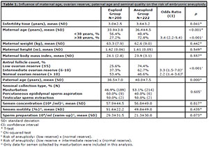
Table 1. Influence of maternal age, ovarian reserve, paternal age and seminal quality on the risk of embryonic aneuploidy.
To assess whether or not the increased paternal age could cause a false association, we performed multivariate analysis to assess the influence of each of these factors (paternal age and maternal age) with the risk of embryo aneuploidy. We found that these factors were independently associated with the risk of aneuploidy (dependent variable: aneuploidy; independent variables: maternal age (B=0.106; Sig<0.001; Exp(B)=1.112), paternal age (B=0.026; Sig=0.025; Exp(B)=1.026), and constant (B=-4.767; Sig<0.001; Exp(B)=0.009).
Of the parameters analyzed regarding the seminal quality of samples collected by masturbation, only a decrease in the number of sperm with normal morphology was associated with the risk of aneuploidy (2.5% of normal forms in the euploid group versus 1.9% in the aneuploid group, p=0.047). Other factors (type of collection, concentration, motility, and sperm preparation) were not associated with embryonic aneuploidy.
Table 2 depicts the influence of infertility factors on the risk of embryonic aneuploidy. None of the factors analyzed (tubal factor, male factor, repeat abortion, endometriosis, PCOS, cryopreserved oocyte) were associated with the risk of aneuploidy.
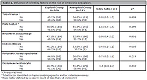
Table 2. Influence of infertility factors on the risk of embryonic aneuploidy.
Table 3 shows the influence of embryonic morphological quality on D1 (assessment 17 h after ICSI), D2 and D3 on the risk of embryonic aneuploidy. None of the morphological parameters on D1 (pro-nucleus, type of nuclear precursor corpuscles, polar corpuscles), D2 and D3 (number of blastomeres, embryonic classification) showed an association with the risk of embryonic aneuploidy.
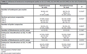
Table 3. Influence of embryonic morphological quality on D1 (17 HPI), D2 (44 HPI) and D3 (HPI) on the risk of embryonic aneuploidy
The influence of the morphological quality of blastocysts on the risk of embryonic aneuploidy is shown in Table 4. Type-C TE was associated with a high risk of embryonic aneuploidy; the risk of aneuploidy was 36.5%, 51.5% and 69.7% for types A, B, and C TE, respectively (p<0.001). In comparing types (A + B) TE versus type C TE, we found an OR of 2.7 (CI: 1.6-4.3, p<0.001) for the risk of aneuploidy, and in comparing type A TE versus type C TE, we found an OR of 4.1 (CI: 2.2-7.7, p<0.001) for the risk of aneuploidy. In the evaluation of the ICM, only a type-C ICM compared with a type-A ICM was associated with a higher risk of aneuploidy, with an OR of 1.7 (CI: 1.01-3.0, p=0.048). The degree of embryonic expansion and the day of embryonic biopsy were not associated with the risk of embryonic aneuploidy.
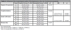
Table 4. Influence of the morphological quality of the blastocyst on the risk of embryonic aneuploidy
The logistic regression analysis results regarding factors of aneuploidy risk (dependent variable: aneuploidy; independent variables: maternal age, ovarian reserve, paternal age, sperm with normal morphology, TE quality, and ICM quality) are shown in Table 5. Logistic regression analysis showed that the main factors for the risk of aneuploidy are maternal age (OR 1.1 per year of age), type B TE (risk: 3.5), and type C TE (risk: 4.8).
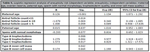
Table 5. Logistic regression analysis of aneuploidy risk (dependent variable: aneuploidy; independent variables: maternal age, ovarian reserve, paternal age, sperm with normal morphology, trophectoderm quality, and inner cell mass quality)
To better assess the role of maternal and paternal age in aneuploidy risk, ROC curve analysis was performed to determine the best maternal and paternal age cutoffs for determining the frequency of aneuploidy. The ROC curve showed that the best cutoff point for maternal age was 38 years (AUC:0.709, p<0.001, sensitivity: 46.5, specificity: 82.4, +LR: 3.68, -LR: 0.57), as shown in Figure 1; the best cutoff age point for male age was 36 years, as shown in Figure 2 (AUC: 0.591, p<0.001, sensitivity: 64.7, specificity: 49.5, +LR: 1.28, -LR: 0.71).
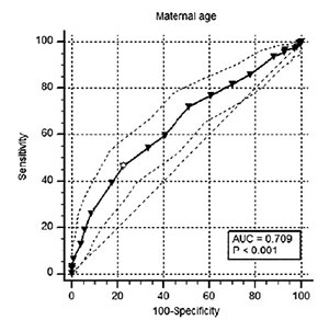
Figure 1. Evaluation of the best maternal age cut off (>38 years) to predict the risk of aneuploidy.
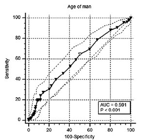
Figure 2. Evaluation of the best paternal age cut off (>36 years) to predict the risk of aneuploidy.
To assess the associations of the identified markers with embryonic aneuploidy, we analyzed the predictive value, likelihood ratio, sensitivity, and specificity for diagnosis of embryonic aneuploidy of the risk factors separately and in association (Table 6). Of the independent factors, maternal age and antral follicle count less than five had the best positive predictive value (72.7% and 72.5%, respectively), with high specificities (81.5% and 94.0%, respectively). Among the combinations of factors, the best marker for the risk of aneuploidy was maternal age older than 38 years, associated with an embryo with trophectoderm type C, with a positive predictive value of 88.6% and a specificity of 97.5%.
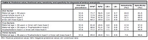
Table 6. Predictive value, likelihood ratio, sensitivity, and specificity for diagnosis of embryo aneuploidy
DISCUSSION
We found that approximately 53% of the embryos analyzed were aneuploid, similar to the findings of other studies (Platteau et al., 2006; Friedenthal et al. 2018; Lawrenz et al., 2019). We sought to evaluate whether alterations in embryonic and clinical morphology factors of the couple were predictive of forming aneuploid blastocysts. Of the embryonic morphology factors, poor blastocyst TE and ICM quality were associated with a higher risk of aneuploidy, whereas morphology factors during cleavage, blastocyst expansion and blastocyst formation were not associated with the risk of aneuploidy. Of the clinical factors, maternal age and antral follicle count less than five had the best positive predictive value, with high specificities.
The transfer of embryos in the blastocyst phase, especially those with better morphology, increases the rate of pregnancy per transfer cycle due to a better embryo selection enabled by prolonged cultivation (Alfarawati et al., 2011). PGT-A, which enables better embryo selection, has raised costs without effectively increasing final pregnancy rates (Munné et al., 2019), and even causing this rate to decrease (Zhang et al., 2016; Guzman et al., 2019; Munné et al., 2019). Next-generation sequencing for PGT-A has led to an increase in reports of chromosomal mosaicism in trophectoderm biopsies (Sachdev et al., 2017; Vera-Rodriguez & Rubio, 2017). Although these embryos may lead to healthy live births, they are linked to poorer clinical outcomes when compared with euploid blastocysts (Munné et al., 2017); thus, we included mosaic embryos in the aneuploid group.
Assessing predictive factors of the risk of aneuploid embryo formation can help us select the embryos to be biopsied and, most importantly, provide patients with more precise information about the real possibility of having an euploid embryo according to their case for each embryo. We found that a type C TE blastocyst had a high risk of being aneuploid (69.7%), with an OR of 4.0, whereas a type A TE blastocyst had a risk of only 36.5%. In addition, compared to ICM type A blastocysts, ICM type C blastocysts had an OR of 1.7 for aneuploidy. Other studies have also shown that blastocyst-stage embryo morphology is associated with euploidy, especially concerning ICM and TE parameters (Wang et al., 2016; Barash et al., 2017). The reduced number of TE cells can be attributed to cell division failure due to organelle and chromosomal abnormalities, especially aneuploidy (Iwasawa et al., 2019).
When we assessed the risk of aneuploidy in terms of embryonic quality in the cleavage phase, we found no association with aneuploidy. Although some studies have found an association between aneuploidy and the cleavage stage (Kroener et al., 2015), more recent studies have not found this association (Barash et al., 2017). The embryonic morphology in the cleavage phase is associated with blastocyst formation (Lawrenz et al., 2019), but not with blastocyst euploidy. The explanation is that the embryos that became blastocysts are those with the best morphology at the cleavage stage, resulting in a very similar quality at the time of the blastocyst biopsy.
Maternal age is known to increase the risk of embryonic aneuploidy and miscarriage, and decrease embryonic implantation rate due to an increased risk of chromosome disruption failure during oogenesis (Ziebe et al., 2001). Our results confirm this association, as we found that women over 38 years of age had a 3.4-fold greater risk of having aneuploid embryos, which agrees with the results of previous studies (Shahine et al., 2016). To better assess the role of maternal age in aneuploidy risk, we performed a ROC curve analysis and found that age 38 was the best cutoff point for predicting aneuploidy risk, suggesting that this could serve as an indication for further tests, such as PGT-A. In addition to maternal age, low ovarian reserve has been associated with the risk of aneuploidy, albeit with conflicting results (Morin et al., 2018). We found that patients with a low ovarian reserve (AFC less than five) had an approximately 73% risk of aneuploid embryos; according to logistic regression, patients with AFC greater than eleven had a lower risk of aneuploidy.
The relationship between paternal age and risk of aneuploidy is very controversial in the literature; some studies have found an association (García-Ferreyra et al., 2015; Capelouto et al., 2018), while others have not (Carrasquillo et al., 2019). Our study found an association between increased paternal age and the risk of aneuploidy, and when we evaluated the ROC curve, 36 years was the best cutoff point to predict the risk of aneuploidy. The role of the male factor of infertility often ends up being overlooked as a potential aneuploidy factor. Of all the sperm data evaluated, the only factor we found that was associated with the risk of aneuploidy was a reduction in the percentage of sperm with normal morphology, which corroborates the findings of previous studies (Coban et al., 2018). Our result reinforces the suggestion that men with a reduction in the percentage of sperm with normal morphology should take steps to decrease oxidative stress, which may influence sperm quality and morphology (Smits et al., 2019).
In addition to maternal age, paternal age, ovarian reserve, and seminal quality, other factors in the couple’s clinical history could increase the risk of aneuploidy. Some studies have attempted to evaluate PCOS as a possible risk factor for aneuploidy but found no association (Wang et al., 2016), which is in accordance with our findings. Another disease widely studied as a potential risk factor for embryonic aneuploidy is endometriosis; however, we found no association of endometriosis with aneuploidy, similar to other studies (Juneau et al., 2017). In addition, in patients with recurrent miscarriage, the cause could be a high rate of aneuploid embryo formation (Hodes-Wertz et al., 2012; Shahine et al., 2016); however, we did not find this association. Our results indicate that implantation failures and miscarriages that may occur in patients with PCOS, endometriosis and recurrent miscarriage are possibly due to factors other than embryonic aneuploidy, such as implantation, immunologic and thrombophilic factors.
In the logistic regression analysis to identify the factor with the greatest influence on the risk of aneuploidy, we found that the main factors associated were maternal age, with a risk of 1.1 per year, and TE quality, as patients with type C TE had a 4.8-fold greater risk of having aneuploid blastocysts.
Although ours is a cohort study, the number of embryos evaluated as well as the clinical data are representative. Our findings have shown that evaluating embryonic morphology and couple’s clinical factors is important and could enable us to better understand embryonic dynamics and the risk of aneuploidy. On the other hand, the influence of paternal age, seminal alterations and some diseases, such as endometriosis and PCOS, needs to be better evaluated in studies with more suitable designs for this purpose. When we evaluated the best markers to predict aneuploidy, we found that of the independent factors, maternal age and antral follicle count less than five had the best positive predictive value (72.7% and 72.5%, respectively), with high specificities (81.5% and 94.0%, respectively). When we examined the associations of markers to assess the risk of aneuploidy, the best marker for the risk of aneuploidy was maternal age above 38 years combined with an embryo with trophectoderm type C, with a positive predictive value of 88.6% and specificity of 97.5%. These results can be useful in informing patients and even the embryology laboratory team.
CONCLUSION
Our study shows that trophectoderm and inner cell mass type C are the major embryo risk factors for aneuploidy, with aneuploidy risks of approximately 71% and 60%, respectively. In addition, the analysis of clinical factors showed that advanced maternal and paternal ages, antral follicle counts less than five, and reduction in the percentage of sperm with normal morphology increase the risk of embryo aneuploidy. Maternal age greater than 38 years is the best cut off for predicting the risk of aneuploidy (72.7%), and if this marker is associated with an embryo with trophectoderm type C, the positive predictive value is 88.6% with a specificity of 97.5%. These findings are of great importance for the clinical practices of assisted reproduction centers, as they may assist in the selection of embryos to be submitted to PGT-A and in educating patients regarding the real possibility of having an euploid embryo according to the embryonic and clinical characteristics of the couple.
ACKNOWLEDGEMENTS
We thank the Federal University of Pará (UFPA) for their support.
REFERENCES
Alfarawati S, Fragouli E, Colls P, Stevens J, Gutiérrez-Mateo C, Schoolcraft WB, Katz-Jaffe MG, Wells D. The relationship between blastocyst morphology, chromosomal abnormality, and embryo gender. Fertil Steril. 2011;95:520-4. PMID: 20537630 DOI: 10.1016/j.fertnstert.2010.04.003
Medline Crossref
Alpha Scientists in Reproductive Medicine and ESHRE Special Interest Group of Embryology. The Istanbul consensus workshop on embryo assessment: proceedings of an expert meeting. Hum Reprod. 2011;26:1270-83. PMID: 21502182 DOI: 10.1093/humrep/der037
Medline Crossref
Barash OO, Ivani KA, Willman SP, Rosenbluth EM, Wachs DS, Hinckley MD, Pittenger Reid S, Weckstein LN. Association between growth dynamics, morphological parameters, the chromosomal status of the blastocysts, and clinical outcomes in IVF PGS cycles with single embryo transfer. J Assist Reprod Genet. 2017;34:1007-16. PMID: 28560610 DOI: 10.1007/s10815-017-0944-0
Medline Crossref
Brezina PR, Brezina DS, Kearns WG. Preimplantation genetic testing. BMJ. 2012;345:e5908. PMID: 22990995 DOI: 10.1136/bmj.e5908
Medline Crossref
Capelouto SM, Nagy ZP, Shapiro DB, Archer SR, Ellis DP, Smith AK, Spencer JB, Hipp HS. Impact of male partner characteristics and semen parameters on in vitro fertilization and obstetric outcomes in a frozen oocyte donor model. Fertil Steril. 2018;110:859-69. PMID: 30316432 DOI: 10.1016/j.fertnstert.2018.06.003
Medline Crossref
Carrasquillo RJ, Kohn TP, Cinnioglu C, Rubio C, Simon C, Ramasamy R, Al-Asmar N. Advanced paternal age does not affect embryo aneuploidy following blastocyst biopsy in egg donor cycles. J Assist Reprod Genet. 2019;36:2039-45. PMID: 31385121 DOI: 10.1007/s10815-019-01549-z
Medline Crossref
Coban O, Serdarogullari M, Onar Sekerci Z, Bilgin EM, Serakinci N. Evaluation of the impact of sperm morphology on embryo aneuploidy rates in a donor oocyte program. Syst Biol Reprod Med. 2018;64:169-73. PMID: 29353506 DOI: 10.1080/19396368.2018.1428384
Medline Crossref
Cooper TG, Noonan E, von Eckardstein S, Auger J, Baker HW, Behre HM, Haugen TB, Kruger T, Wang C, Mbizvo MT, Vogelsong KM. World Health Organization reference values for human semen characteristics. Hum Reprod Update. 2010;16:231-45. PMID: 19934213 DOI: 10.1093/humupd/dmp048
Medline Crossref
Friedenthal J, Maxwell SM, Munné S, Kramer Y, McCulloh DH, McCaffrey C, Grifo JA. Next generation sequencing for preimplantation genetic screening improves pregnancy outcomes compared with array comparative genomic hybridization in single thawed euploid embryo transfer cycles. Fertil Steril. 2018;109:627-32. PMID: 29605407 DOI: 10.1016/j.fertnstert.2017.12.017
Medline Crossref
Friedenthal J, Maxwell SM, Tiegs AW, Besser AG, McCaffrey C, Munné S, Noyes N, Grifo JA. Clinical error rates of next generation sequencing and array comparative genomic hybridization with single thawed euploid embryo transfer. Eur J Med Genet. 2020;63:103852. PMID: 31972371 DOI: 10.1016/j.ejmg.2020.103852
Medline Crossref
García-Ferreyra J, Luna D, Villegas L, Romero R, Zavala P, Hilario R, Dueñas-Chacón J. High Aneuploidy Rates Observed in Embryos Derived from Donated Oocytes are Related to Male Aging and High Percentages of Sperm DNA Fragmentation. Clin Med Insights Reprod Health. 2015;9:21-7. PMID: 26604851 DOI: 10.4137/CMRH.S32769
Medline Crossref
Gazzo E, Peña F, Valdéz F, Chung A, Velit M, Ascenzo M, Escudero E. Blastocyst contractions are strongly related with aneuploidy, lower implantation rates, and slow-cleaving embryos: a time lapse study. JBRA Assist Reprod. 2020;24:77-81. PMID: 31524340 DOI: 10.5935/1518-0557.20190053
Medline Crossref
Guzman L, Nuñez D, López R, Inoue N, Portella J, Vizcarra F, Noriega-Portella L, Noriega-Hoces L, Munné S. The number of biopsied trophectoderm cells may affect pregnancy outcomes. J Assist Reprod Genet. 2019;36:145-51. PMID: 30328573 DOI: 10.1007/s10815-018-1331-1
Medline Crossref
Hodes-Wertz B, Grifo J, Ghadir S, Kaplan B, Laskin CA, Glassner M, Munné S. Idiopathic recurrent miscarriage is caused mostly by aneuploid embryos. Fertil Steril. 2012;98:675-80. PMID: 22683012 DOI: 10.1016/j.fertnstert.2012.05.025
Medline Crossref
Iwasawa T, Takahashi K, Goto M, Anzai M, Shirasawa H, Sato W, Kumazawa Y, Terada Y. Human frozen-thawed blastocyst morphokinetics observed using time-lapse cinematography reflects the number of trophectoderm cells. PLoS One. 2019;14:e0210992. PMID: 30650134 DOI: 10.1371/journal.pone.0210992
Medline Crossref
Juneau C, Kraus E, Werner M, Franasiak J, Morin S, Patounakis G, Molinaro T, de Ziegler D, Scott RT. Patients with endometriosis have aneuploidy rates equivalent to their age-matched peers in the in vitro fertilization population. Fertil Steril. 2017;108:284-8. PMID: 28651959 DOI: 10.1016/j.fertnstert.2017.05.038
Medline Crossref
Kroener LL, Ambartsumyan G, Pisarska MD, Briton-Jones C, Surrey M, Hill D. Increased blastomere number in cleavage-stage embryos is associated with higher aneuploidy. Fertil Steril . 2015;103:694-8. PMID: 25557243 DOI: 10.1016/j.fertnstert.2014.12.090
Medline Crossref
Lawrenz B, El Khatib I, Liñán A, Bayram A, Arnanz A, Chopra R, De Munck N, Fatemi HM. The clinicians' dilemma with mosaicism-an insight from inner cell mass biopsies. Hum Reprod. 2019;34:998-1010. PMID: 31114858 DOI: 10.1093/humrep/dez055
Medline Crossref
Lee E, Illingworth P, Wilton L, Chambers GM. The clinical effectiveness of preimplantation genetic diagnosis for aneuploidy in all 24 chromosomes (PGD-A): systematic review. Hum Reprod. 2015;30:473-83. PMID: 25432917 DOI: 10.1093/humrep/deu303
Medline Crossref
Mazzilli R, Cimadomo D, Vaiarelli A, Capalbo A, Dovere L, Alviggi E, Dusi L, Foresta C, Lombardo F, Lenzi A, Tournaye H, Alviggi C, Rienzi L, Ubaldi FM. Effect of the male factor on the clinical outcome of intracytoplasmic sperm injection combined with preimplantation aneuploidy testing: observational longitudinal cohort study of 1,219 consecutive cycles. Fertil Steril. 2017;108:961-72.e3. PMID: 28985908 DOI: 10.1016/j.fertnstert.2017.08.033
Medline Crossref
Morin SJ, Kaser DJ, Franasiak JM. The dilemma of aneuploidy screening on low responders. Curr Opin Obstet Gynecol. 2018;30:179-84. PMID: 29697414 DOI: 10.1097/GCO.0000000000000449
Medline Crossref
Munné S, Blazek J, Large M, Martinez-Ortiz PA, Nisson H, Liu E, Tarozzi N, Borini A, Becker A, Zhang J, Maxwell S, Grifo J, Babariya D, Wells D, Fragouli E. Detailed investigation into the cytogenetic constitution and pregnancy outcome of replacing mosaic blastocysts detected with the use of high-resolution next-generation sequencing. Fertil Steril. 2017;108:62-71.e8. PMID: 28579407 DOI: 10.1016/j.fertnstert.2017.05.002
Medline Crossref
Munné S, Kaplan B, Frattarelli JL, Child T, Nakhuda G, Shamma FN, Silverberg K, Kalista T, Handyside AH, Katz-Jaffe M, Wells D, Gordon T, Stock-Myer S, Willman S; STAR Study Group. Preimplantation genetic testing for aneuploidy versus morphology as selection criteria for single frozen-thawed embryo transfer in good-prognosis patients: a multicenter randomized clinical trial. Fertil Steril. 2019;112:1071-9.e7. PMID: 31551155 DOI: 10.1016/j.fertnstert.2019.07.1346
Medline Crossref
Platteau P, Staessen C, Michiels A, Van Steirteghem A, Liebaers I, Devroey P. Which patients with recurrent implantation failure after IVF benefit from PGD for aneuploidy screening? Reprod Biomed Online. 2006;12:334-9. PMID: 16569323 DOI: 10.1016/S1472-6483(10)61006-1
Medline Crossref
Sachdev NM, Maxwell SM, Besser AG, Grifo JA. Diagnosis and clinical management of embryonic mosaicism. Fertil Steril. 2017;107:6-11. PMID: 27842993 DOI: 10.1016/j.fertnstert.2016.10.006
Medline Crossref
Shahine LK, Marshall L, Lamb JD, Hickok LR. Higher rates of aneuploidy in blastocysts and higher risk of no embryo transfer in recurrent pregnancy loss patients with diminished ovarian reserve undergoing in vitro fertilization. Fertil Steril. 2016;106:1124-8. PMID: 27371114 DOI: 10.1016/j.fertnstert.2016.06.016
Medline Crossref
Smits RM, Mackenzie-Proctor R, Yazdani A, Stankiewicz MT, Jordan V, Showell MG. Antioxidants for male subfertility. Cochrane Database Syst Rev. 2019;3:CD007411. PMID: 30866036 DOI: 10.1002/14651858.CD007411.pub4
Medline Crossref
Vera-Rodriguez M, Rubio C. Assessing the true incidence of mosaicism in preimplantation embryos. Fertil Steril. 2017;107:1107-12. PMID: 28433370 DOI: 10.1016/j.fertnstert.2017.03.019
Medline Crossref
Wang A, Kort J, Behr B, Westphal LM. Euploidy in relation to blastocyst sex and morphology. J Assist Reprod Genet. 2018;35:1565-72. PMID: 30030712 DOI: 10.1007/s10815-018-1262-x
Medline Crossref
Wang Q, Luo L, Lei Q, Lin MM, Huang X, Chen MH, Zeng YH, Zhou CQ. Low aneuploidy rate in early pregnancy loss abortuses from patients with polycystic ovary syndrome. Reprod Biomed Online. 2016;33:85-92. PMID: 27157933 DOI: 10.1016/j.rbmo.2016.04.006
Medline Crossref
Zaninovic N, Irani M, Meseguer M. Assessment of embryo morphology and developmental dynamics by time-lapse microscopy: is there a relation to implantation and ploidy? Fertil Steril. 2017;108:722-9. PMID: 29101997 DOI: 10.1016/j.fertnstert.2017.10.002
Medline Crossref
Zhang S, Luo K, Cheng D, Tan Y, Lu C, He H, Gu Y, Lu G, Gong F, Lin G. Number of biopsied trophectoderm cells is likely to affect the implantation potential of blastocysts with poor trophectoderm quality. Fertil Steril . 2016;105:1222-7.e4. PMID: 26820770 DOI: 10.1016/j.fertnstert.2016.01.011
Medline Crossref
Ziebe S, Loft A, Petersen JH, Andersen AG, Lindenberg S, Petersen K, Andersen AN. Embryo quality and developmental potential is compromised by age. Acta Obstet Gynecol Scand. 2001;80:169-74. PMID: 11167214 DOI: 10.1034/j.1600-0412.2001.080002169.x
Medline Crossref