JBRA Assist. Reprod. 2026;30(1):203-212
ORIGINAL ARTICLE
doi: 10.5935/1518-0557.20250181
Zingerone Mitigates Testicular Dysfunction Induced by Cisplatin
1Student Research committee, Ahvaz Jundishapur University of Medical Sciences, Ahvaz, Iran
2Cellular and Molecular Research Center, Medical Basic Sciences Research Institute, Ahvaz Jundishapur University of Medical Sciences, Ahvaz, Iran
3Department of Anatomical Sciences, Faculty of Medicine, Ahvaz Jundishapur University of Medical Sciences, Ahvaz, Iran
4Department of Anatomical Sciences, School of Medicine, Dezful University of Medical Sciences, Dezful, Iran
5Toxicology Research Center, Medical Basic Sciences Research Institute, Ahvaz Jundishapur University of Medical Sciences, Ahvaz, Iran
6Department of Anatomy, School of Medicine, Arak University of Medical Sciences, Arak, Iran
CONFLICT OF INTEREST
No potential conflict of interest was reported by the author(s).
ABSTRACT
Objective: Cisplatin is one of the most widely used antitumor drugs globally, particularly in treating various solid tumors. The reproductive system is impacted by cisplatin toxic effects. This study aims to understand how Zingerone affects spermatogenesis defects in mice.
Methods: In the present experimental laboratory study, the 48 male NMRI mice (6 to 8 weeks of age, 25 to 30g weight) were treated with Cisplatin (7 mg/kg) for 5 days and zingerone for 30 days at concentrations of 10, 20, and 40 mg/kg before cisplatin administration. After the treatment period, the testicles were dissected immediately following sacrifice. Morphometric parameters, serum testosterone concentration, histology, Bax/Bcl-2 ratio, and testis weight have been assessed. To determine levels of oxidative stress, malondialdehyde contents and antioxidant levels were evaluated.
Results: Cisplatin-induced structural damages enhanced the Bax/Bcl-2 ratio, and reduced testosterone levels and testis weight. Cisplatin caused oxidative stress by enhancing malondialdehyde contents in the mouse testicles. Zingerone dose-dependently reduced the Bax/Bcl-2 ratio and reversed the histological changes, testosterone levels, and antioxidant capacity.
Conclusions: According to the results of the present study, Pretreatment with zingerone can improve testosterone production by preventing apoptosis and oxidative stress in the testicles of mice that have undergone cisplatin intoxication.
Keywords: zingerone, cisplatin, oxidative stress, apoptosis, reproductive system
INTRODUCTION
Infertility is one of the main critical issues associated with cancer treatment (Tian En et al., 2020). In young male patients, chemotherapy agents affect fertility by impairing testicular function (Okada & Fujisawa, 2019; Yumura et al., 2023). Cisplatin (CP) is used to manage various malignancies, such as testicular and ovarian cancers (Helm & States, 2009; Zoń & Bednarek, 2023). CP induces apoptosis by generating oxidative stress and interacting with DNA (Dermitzakis et al., 2016). It causes testicular damage characterized by germ cell apoptosis, impaired steroidogenesis, and histological alterations (A A Aly & Eid, 2020; Nna et al., 2020; Saher et al., 2023). Infertility can result from CP treatment due to its damaging effects on Leydig cells, which are crucial for testosterone production. This impairment in testosterone synthesis disrupts spermatogenesis (Datrianto et al., 2021; Demir & Altındağ, 2022). Oxidative stress is a key factor that regulates apoptosis, and various mechanisms of apoptosis modulation by oxidative stress have been established (Sharma et al., 2023).
According to studies, apoptosis plays a crucial role in regulating testicular function (Rotimi & Singh, 2022). Excessive apoptosis in spermatogenic cells can lead to infertility (Vardiyan et al., 2020). The primary regulators of cell survival and apoptosis are Bcl-2 and Bax (Hatok & Racay, 2016). Extracting bioactive molecules from plants can improve sperm quality, testosterone secretion, and fertility indices (Shahedi et al., 2021; Yaghutian Nezhad et al., 2021; Behairy et al., 2022; Rotimi et al., 2023). Among many natural substances, flavonoids have garnered significant attention for their potential to treat male reproductive system dysfunction (Farombi et al., 2012; Ye et al., 2020).
Flavonoids, as secondary metabolites of polyphenolic plants and fungi, possess a phenyl-benzopyran structure. They are natural antioxidants with potential benefits such as anti-inflammatory, immune-stimulating, antiviral, anticancer, anti-apoptotic, and anti-allergic properties (Al-Khayri et al., 2022). Ginger, one of the most commonly used herbs, is a traditional medicine employed globally (El-Seedi et al., 2019). Ginger contains a significant amount of Zingerone (ZG), which is believed to be responsible for its pharmacological properties. ZG exhibits potent antioxidant, anti-inflammatory, anticancer, antidiabetic, antihypertensive, antimicrobial, antithrombotic, anxiolytic, anti-ulcer, and appetite-stimulant properties (Ahmad et al., 2015).
In addition to its antioxidant properties, ZG is used to treat various diseases due to its ability to scavenge free radicals. As a result, ZG can reduce reactive oxygen species (ROS) while maintaining its antioxidant qualities. ZG mitigates cisplatin-induced kidney damage by attenuating oxidative stress, suppressing apoptotic gene expression, and reducing inflammatory factors (Kandemir et al., 2019). In a rat model of CIS-induced nephrotoxicity, ZG demonstrated nephroprotective effects, primarily by suppressing oxidative stress and inflammation (Alibakhshi et al., 2018). It is essential to identify a compound that can mitigate the adverse effects of CP on the human reproductive system. To date, the effects of Zingerone on cisplatin-induced cytotoxicity have not been thoroughly investigated. The purpose of this research is to analyze how ZG protects against CP-induced testicular toxicity in mice by evaluating oxidative stress and apoptosis.
MATERIAL AND METHODS
Animals and Research Design (Figure 1)
For this experiment, a total of 48 adult male NMRI mice, aged 6 to 8 weeks and weighing between 25 to 30 grams each, were purchased from the Animal Care Center in Ahvaz, Iran. The animals were housed under standard laboratory conditions with a humidity level of 50±5%, a 12-hour light/dark cycle, and a temperature of 22±5 °C throughout the study. The research was conducted in 2023 at the Cellular and Molecular Research Center of Ahvaz Jundishapur University of Medical Sciences, Ahvaz, Iran. This project adhered to the guidelines established by the Animal Ethics Committee of our institution (approval code: IR.AJUMS.ABHC.REC.1402.006).
The mice were randomly assigned to six groups (eight animals per group), as described below:
- Control: Received 0.2 ml of normal saline intraperitoneally (IP) for 35 days.
- CP: Administered 7 mg/kg of cisplatin (CP) intraperitoneally (IP) on days 30-35.
- ZG: received Zingerone (ZG) orally at a dose of 40 mg/kg for 35 days.
- CP+ZG (10, 20 and 40): Received ZG orally at doses of 10, 20, or 40 mg/kg for 35 days, along with 7 mg/kg CP (IP) on days 30-35.
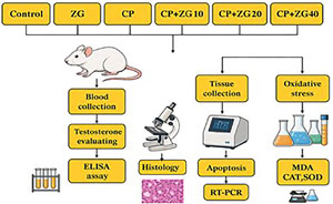
Figure 1. Schematic illustration detailing the experimental design of the study.
ZG and CP were purchased from Sigma Company. Both compounds were dissolved in normal saline. The dosing protocols for ZG (Sigma) and CP (Sigma) were based on previous studies (Aldemir et al., 2014; Hassan et al., 2019). One day after the final treatment, the mice were euthanized using high doses of ketamine and xylazine. Following euthanasia, the testes were promptly dissected. The left testes were fixed in Bouin’s solution for histological examination and right testes were stored at -80°C for other assessments, while the left testes were fixed in Bouin’s solution for histological examination.
Histology and Morphometry
Six microscopy slides per animal were stained with hematoxylin and eosin (H&E) to analyze histological criteria, such as detachment of germ cells and vacuolization of the germinal epithelium. The average percentage of each characteristic was calculated for every treatment group. Two researchers, blinded to the group allocation, independently analyzed the slides. The diameter of the seminiferous tubules and the height of the seminiferous epithelium were calculated using Motic software. For each animal in the experiment, the researchers assessed 100 tubules (Bahrami et al., 2018).
Evaluation of lipid peroxidation
After centrifuging the testicular homogenate, 500 μL of the supernatant was mixed with 10% trichloroacetic acid and centrifuged again at 7000×g for 10 minutes. The resulting supernatant was then transferred to two milliliters of trichloroacetic acid. Each sample was centrifuged at 7500×g for 10 minutes after adding N-butanol. The optical density (OD) of the supernatant was measured at 535 nm using a spectrophotometer.
SOD and CAT activity
The Ransod kit (SD125, United Kingdom) was used to determine superoxide dismutase (SOD) activity. This method involves the reaction with the superoxide anion, producing a water-soluble formazan dye. SOD inhibits xanthine oxidase, creating a linear relationship between its activity and the rate of reduction. The inhibition activity of SOD was measured at 505 nm using a spectrophotometer. Catalase (CAT) activity was assessed by measuring the rate of complex formation between hydrogen peroxide and ammonium molybdate.
Testosterone evaluating
Blood samples were collected directly from the heart using heparinized tubes, then centrifuged at 400 g for 20 minutes to separate the serum. After centrifugation, the samples were stored at -80°C until analysis. A mouse testosterone ELISA assay kit was utilized to detect serum testosterone levels (Monobind, USA).
Real-time PCR
RNA was extracted from testicular tissue using the RNeasy kit (Qiagen, Germany). To synthesize complementary DNA (cDNA), the extracted RNA was reverse-transcribed using a reverse transcription kit (Qiagen, Germany). The PCR reaction mixture consisted of SYBR Green Master Mix (Qiagen), DEPC-treated water, cDNA, and forward and reverse primers. Real-time PCR (RT-PCR) was performed over 45 cycles, with the following steps: initial denaturation at 95°C for 50 seconds, followed by 45 cycles of denaturation at 95°C for 30 seconds, and annealing/extension at 60°C for 35 seconds. The relative gene expression levels were normalized to GAPDH as the reference gene. Data analysis was conducted using REST software (2009).
Statistical analyses
Statistical analyses were conducted using the Statistical Package for the Social Sciences (SPSS) software (version 21.0, Chicago, IL, USA). Data were evaluated through one-way analysis of variance (ANOVA), followed by post hoc Tukey’s or LSD tests. Statistical significance was determined at a p-value<0.05, and results were expressed as mean±standard deviation (SD).
RESULTS
Body and relative testis weight
Body weights did not significantly differ among the groups. However, mice exposed to cisplatin (CP) showed a significant decrease in the ratio of testis weight to body weight (relative testis weight) compared to the control group (p<0.01). Treatment with Zingerone (ZG) dose-dependently increased the relative testis weight in CP-treated animals (p<0.05, Figure 2).
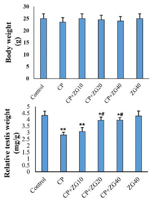
Figure 2. Effects of ZG on (A) body weight and (B) relative testes weight in CP-treated mice. Data are illustrated in mean±SD (n=6). *p<0.05, **p<0.01, #p<0.05. Symbols indicate comparison to the control (*) and CP (#) groups (one-way ANOVA followed by Tukey’s post-hoc test).
Testosterone assay
In CP-treated mice, testosterone levels were significantly reduced (p<0.001). However, testosterone levels were significantly increased in the CP+ZG20 and CP+ZG 40 groups compared to the CP-only group (p<0.01). The CP+ZG10 group also showed a slight but significant increase in testosterone levels compared to the CP-treated mice (p<0.05, Figure 3).
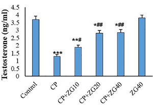
Figure 3. Effects of ZG on serum testosterone concentration in CP-treated mice. CP administration significantly decreased testosterone levels (***p<0.001 vs control). Co-treatment with ZG at 20 and 40 mg/kg (CP +ZG20 and CP +ZG40 groups) significantly restored testosterone levels compared to CP group (##p<0.01), while CP +ZG10 showed a modest increase. Data are presented as mean±SD (n=6). *p<0.05, **p<0.01, ***p<0.001 vs control group; #p<0.05, ##p<0.01 vs CP group (one-way ANOVA followed by Tukey’s post-hoc test).
Histology and Morphometry
The control and ZG40 groups displayed normal histological features, characterized by well-preserved seminiferous epithelium. In contrast, CP-treated mice exhibited sloughing and vacuolization of the seminiferous tubules, leading to atrophy of the germinal epithelium. Treatment with ZG restored normal tissue architecture and seminiferous parameters, as illustrated in Figures 4 and 5.
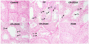
Figure 4. Photomicrograph from testicular tissues. Control group exhibits typical seminiferous epithelium; CP group shows vacuolization (V) and sluing (S) of the germinal epithelium, and extensive degenerative changes (D); The CP+ZG10 and CP+ZG20 groups shows moderate degenerative changes and vacuolization in g; and CP+ZG40 group displays minimal degenerative changes and vacuolization in the germinal epithelium. ZG40 group shows typical germinal epithelium. H&E staining; Magnifications: ×250.
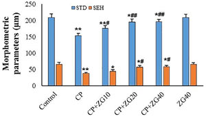
Figure 5. Histopathological evaluation of testicular tissue in: Control, CP-treated, CP +ZG10, CP +ZG 20, CP +ZG40, and ZG-alone groups. Two blinded investigators independently analyzed 100 seminiferous tubules per animal using Motic software to measure tubule diameter and epithelial height. Data are presented as mean±SD (n=6 groups). *p<0.05 and **p<0.01 vs control group; #p<0.05 and ##p<0.01 vs CP group (one-way ANOVA with Tukey’s post-hoc test).
Expression of apoptosis-related genes
The Bax/Bcl-2 ratio was significantly increased in CP-treated mice (p<0.001). However, a significant reduction in the Bax/Bcl-2 ratio was observed in the CP+ZG20 and CP+ZG40 groups compared to the CP-treated group (p<0.01). Furthermore, the Bax/Bcl-2 ratio in the CP+ZG20 and CP+ZG40 groups was significantly lower than that in the CP+ZG10 group (Figure 6).
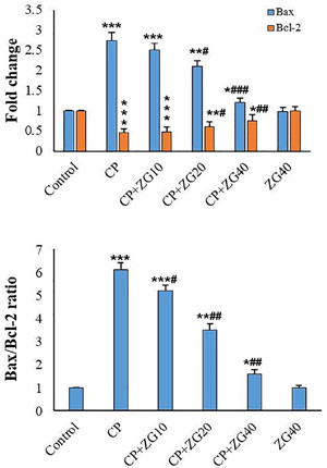
Figure 6. Effects of ZG on the Bax/Bcl-2 ratio in testicular tissue of CP-treated mice. The Bax/Bcl-2 ratio was significantly elevated in CP-treated mice compared to controls (***p<0.001). ZG co-treatment at 20 and 40 mg/kg (CP +ZG20 and CP +ZG40 groups) significantly reduced the ratio compared to CP-treated mice (##p<0.01), with CP +ZG40 showing the most pronounced effect. Notably, the ratio in CP +ZG20 and CP +ZG40 groups was significantly lower than in CP +ZG10 group (p<0.05). Data represent mean±SD (n=6 groups). *p<0.05, **p<0.01, ***p<0.001 vs. control; #p<0.05, ##p<0.01, ###p<0.001 vs. CP group (one-way ANOVA with Tukey’s post-hoc test).
MDA and antioxidant levels
Administration of CP significantly reduced testicular SOD and CAT enzyme activity compared to the control group (p<0.001). Additionally, MDA levels were significantly increased in the CP group compared to the control animals (p<0.001). Pre-treatment with ZG20 and ZG40 significantly increased testicular SOD and CAT levels compared to the CP group (p<0.05 and p<0.01, respectively). However, administration of ZG to normal animals did not result in significant changes in antioxidant indices compared to the control group. In CP-treated mice, ZG administration dose-dependently reduced testicular MDA levels compared to the CP group (Figure 7).
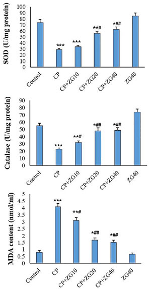
Figure 7. Effects of ZG on oxidative stress markers in testicular tissue of CP-treated mice. SOD activity, CAT activity, and MDA levels. CP administration significantly reduced SOD and CAT activities (p<0.001 vs. control) while increasing MDA content (p<0.001). ZG pretreatment at 20 and 40 mg/kg (CP +ZG20 and CP +ZG40 groups) significantly restored SOD (#p<0.05) and CAT activity (##p<0.01), and dose-dependently reduced MDA levels (#p<0.05, ##p<0.01) compared to CP group. ZG alone showed no significant effects on antioxidant indices versus controls. Data represent mean±SD (n=6 groups). *p<0.05, **p<0.01, ***p<0.001 vs. control group; #p<0.05, ##p<0.01 vs. CP group (one-way ANOVA with Tukey’s post-hoc test).
DISCUSSION
This study aimed to investigate the protective effects of ZG against CP-induced testicular damage. Our findings demonstrated that CP caused structural changes in testicular tissue, increased cell death and oxidative stress, and reduced testosterone levels and testicular weights. However, pre-treatment with ZG significantly mitigated these adverse effects in a dose-dependent manner.
The reduction in testicular weights highlights the toxic effects of CP on mouse testes. The reversal of testis weight following ZG administration suggests that ZG may prevent CP-induced germ cell loss. This hypothesis is supported by morphometric findings. CP treatment reduced the diameter of seminiferous tubules and the height of the germinal epithelium, whereas ZG treatment improved these morphometric parameters in CP-injected mice, indicating enhanced spermatogenesis. These results align with previous studies (Abdullah & Bondagji, 2011; Yahyazadeh et al., 2020).
In a previous study, ZG was shown to improve sperm quality, reduce malondialdehyde (MDA) levels, and increase superoxide dismutase (SOD) and glutathione (GSH) levels in testicular tissue (Rafiee et al., 2019). In the current study, ZG pretreatment effectively reduced MDA levels in the testicles of CP-treated mice. Similarly, Mohammadi et al. (2014) demonstrated that ZG alleviated oxidative damage in testicular tissue induced by cyclophosphamide. Additionally, Çağlayan et al. (2019) reported that ZG administration improved the oxidative/antioxidative balance, enhanced sperm quality, and increased testosterone levels in vancomycin-intoxicated subjects.
Recent studies have elucidated that cisplatin (CP)-induced testicular toxicity primarily involves three interlinked molecular pathways: CP administration triggers NADPH oxidase (NOX4)-dependent ROS overproduction, depleting antioxidant defenses (SOD2, CAT) and impairing steroidogenic enzymes (StAR, CYP11A1), ultimately reducing testosterone synthesis, CP upregulates pro-apoptotic Bax while suppressing anti-apoptotic Bcl-2 in germ cells, leading to cytochrome c release and caspase-3 activation and Disruption of Steroidogenesis (Cregan et al., 2013; A A Aly & Eid, 2020; Sharma et al., 2023).
Lowered levels of testosterone in the CP group could indicate damage to Leydig cells and dysfunction in steroidogenesis. Leydig cells, which are responsible for testosterone production, are particularly vulnerable to oxidative stress (Awny et al., 2021). Previous studies have demonstrated that oxidative stress induces apoptosis in Leydig cells (Sun et al., 2017). Therefore, the observed decrease in testosterone levels following CP exposure may be attributed to Leydig cell damage.
The seminiferous tubules exhibit histological changes as a result of reduced testosterone levels (Asadi et al., 2017). Additionally, it has been established that declining testosterone levels can lead to the induction of apoptosis in germ cells (Yadav et al., 2022). The presence of vacuoles in the germinal epithelium may indicate germ cell apoptosis caused by CP (Hasanin et al., 2018). This hypothesis is further supported by the increased Bax/Bcl-2 ratio in the testicular tissue. Mesbahzadeh et al. (2021) found that CP exposure reduced tubular diameter, testosterone levels, and increased seminiferous tubule apoptosis in rats. However, ZG was able to reverse these morphometric parameters, suggesting a reduction in germ cell apoptosis in CP-treated mice.
Following testicular damage induced by zinc oxide nanoparticles, Rafiee et al. (2019) observed that ZG improved histological changes and reduced apoptosis in testicular tissue. In this study, ZG demonstrated the potential to elevate testosterone levels, reduce vacuolarization in the seminiferous tubules, and decrease the Bax/Bcl-2 ratio. These findings suggest that ZG may attenuate CP-induced testicular damage by inhibiting germ cell apoptosis. This study was intentionally designed to evaluate ZG testicular protective effects; thus, systematic evaluation of off-target actions in non-reproductive tissues was not included. Future studies should address ZG organ-specific pharmacokinetics and potential dose-related adverse effects to fully establish its clinical translational potential.
CONCLUSION
This investigation has demonstrated that ZG attenuates testicular injury induced by CP in mice. ZG pretreatment increases testosterone levels, enhances antioxidant capacity, and suppresses apoptosis. Further research is necessary to elucidate the specific mechanisms by which ZG mitigates CP-induced toxicity.
Ethical approval
This project was conducted according to the guidelines established by the Animal Ethics Committee of our organization (approval code: IR.AJUMS.ABHC.REC.1402.006).
Acknowledgment
We thank our colleagues from the Cellular and Molecular Research Center. Student Research Committee of Ahvaz Jundishapur University granted this study (Grant No: 02s1).
REFERENCES
A A Aly H, G Eid B. Cisplatin induced testicular damage through mitochondria mediated apoptosis, inflammation and oxidative stress in rats: impact of resveratrol. Endocr J. 2020;67:969-80. PMID: 32507773 DOI: 10.1507/endocrj.EJ20-0149 Medline
Abdullah L, Bondagji N. Histopathological patterns of testicular biopsy in male infertility: A retrospective study from a tertiary care center in the western part of Saudi Arabia. Urol Ann. 2011;3:19-23. PMID: 21346828 DOI: 10.4103/0974-7796.75867 Medline
Ahmad B, Rehman MU, Amin I, Arif A, Rasool S, Bhat SA, Afzal I, Hussain I, Bilal S, Mir Mu. A A Review on Pharmacological Properties of Zingerone (4-(4-Hydroxy-3-methoxyphenyl)-2-butanone). ScientificWorldJournal. 2015;2015:816364. PMID: 26106644 DOI: 10.1155/2015/816364 Medline
Aldemir M, Okulu E, Kösemehmetoğlu K, Ener K, Topal F, Evirgen O, Gürleyik E, Avcı A. Evaluation of the protective effect of quercetin against cisplatin-induced renal and testis tissue damage and sperm parameters in rats. Andrologia. 2014;46:1089-97. PMID: 24266675 DOI: 10.1111/and.12197 Medline
Alibakhshi T, Khodayar MJ, Khorsandi L, Rashno M, Zeidooni L. Protective effects of zingerone on oxidative stress and inflammation in cisplatin-induced rat nephrotoxicity. Biomed Pharmacother. 2018;105:225-32. PMID: 29857302 DOI: 10.1016/j.biopha.2018.05.085 Medline
Al-Khayri JM, Sahana GR, Nagella P, Joseph BV, Alessa FM, Al-Mssallem MQ. Flavonoids as Potential Anti-Inflammatory Molecules: A Review. Molecules. 2022;27:2901. PMID: 35566252 DOI: 10.3390/molecules27092901 Medline
Asadi N, Bahmani M, Kheradmand A, Rafieian-Kopaei M. The Impact of Oxidative Stress on Testicular Function and the Role of Antioxidants in Improving it: A Review. J Clin Diagn Res. 2017;11:IE01-IE05. PMID: 28658802 DOI: 10.7860/JCDR/2017/23927.9886 Medline
Awny MM, Al-Mokaddem AK, Ali BM. Mangiferin mitigates di-(2-ethylhexyl) phthalate-induced testicular injury in rats by modulating oxidative stress-mediated signals, inflammatory cascades, apoptotic pathways, and steroidogenesis. Arch Biochem Biophys. 2021;711:108982. PMID: 34400143 DOI: 10.1016/j.abb.2021.108982 Medline
Bahrami N, Mehrzadi S, Goudarzi M, Mansouri E, Fatemi I. Lycopene abrogates di-(2-ethylhexyl) phthalate induced testicular injury by modulating oxidative, endocrine and inflammatory changes in mice. Life Sci. 2018;207:265-71. PMID: 29886059 DOI: 10.1016/j.lfs.2018.06.009 Medline
Behairy A, Hashem MM, Abo-El-Sooud K, El-Metwally AE, Hassan BA, Abd-Elhakim YM. Quercetin Abates Aluminum Trioxide Nanoparticles and Lead Acetate Induced Altered Sperm Quality, Testicular Oxidative Damage, and Sexual Hormones Disruption in Male Rats. Antioxidants (Basel). 2022;11:2133. PMID: 36358505 DOI: 10.3390/antiox11112133 Medline
Çağlayan C, Taslimi P, Demir Y, Küçükler S, Kandemir FM, Gulçin İ. The effects of zingerone against vancomycin-induced lung, liver, kidney and testis toxicity in rats: The behavior of some metabolic enzymes. J Biochem Mol Toxicol. 2019;33:e22381. PMID: 31454121 DOI: 10.1002/jbt.22381 Medline
Cregan IL, Dharmarajan AM, Fox SA. Mechanisms of cisplatin-induced cell death in malignant mesothelioma cells: role of inhibitor of apoptosis proteins (IAPs) and caspases. Int J Oncol. 2013;42:444-52. PMID: 23229133 DOI: 10.3892/ijo.2012.1715 Medline
Datrianto DS, Budipitojo T, Prihatno SA. Secretome improves testosterone and androgen-binding protein production in testicular dysfunction rats induced by cisplatin. J Adv Vet Anim Res. 2021;8:687-94. PMID: 35106310 DOI: 10.5455/javar.2021.h561 Medline
Demir M, Altındağ F. Sinapic acid ameliorates cisplatin-induced disruptions in testicular steroidogenesis and spermatogenesis by modulating androgen receptor, proliferating cell nuclear antigen and apoptosis in male rats. Andrologia. 2022;54:e14369. PMID: 34997775 DOI: 10.1111/and.14369 Medline
Dermitzakis EV, Kimiskidis VK, Lazaridis G, Alexopoulou Z, Timotheadou E, Papanikolaou A, Romanidou O, Georgiadis G, Kalogeras KT, Tsiptsios I, Tarlatzis B, Fountzilas G. The impact of paclitaxel and carboplatin chemotherapy on the autonomous nervous system of patients with ovarian cancer. BMC Neurol. 2016;16:190. PMID: 27716097 DOI: 10.1186/s12883-016-0710-4 Medline
El-Seedi HR, Khalifa SAM, Yosri N, Khatib A, Chen L, Saeed A, Efferth T, Verpoorte R. Plants mentioned in the Islamic Scriptures (Holy Qur’ân and Ahadith): Traditional uses and medicinal importance in contemporary times. J Ethnopharmacol. 2019;243:112007. PMID: 31170516 DOI: 10.1016/j.jep.2019.112007 Medline
Farombi EO, Adedara IA, Akinrinde SA, Ojo OO, Eboh AS. Protective effects of kolaviron and quercetin on cadmium-induced testicular damage and endocrine pathology in rats. Andrologia. 2012;44:273-84. PMID: 22356231 DOI: 10.1111/j.1439-0272.2012.01279.x Medline
Hasanin NA, Sayed NM, Ghoneim FM, Al-Sherief SA. Histological and Ultrastructure Study of the Testes of Acrylamide Exposed Adult Male Albino Rat and Evaluation of the Possible Protective Effect of Vitamin E Intake. J Microsc Ultrastruct. 2018;6:23-34. PMID: 30023264 DOI: 10.4103/JMAU.JMAU_7_18 Medline
Hassan E, El-Neweshy M, Hassan M, Noreldin A. Thymoquinone attenuates testicular and spermotoxicity following subchronic lead exposure in male rats: Possible mechanisms are involved. Life Sci. 2019;230:132-40. PMID: 31136753 DOI: 10.1016/j.lfs.2019.05.067 Medline
Hatok J, Racay P. Bcl-2 family proteins: master regulators of cell survival. Biomol Concepts. 2016;7:259-70. PMID: 27505095 DOI: 10.1515/bmc-2016-0015 Medline
Helm CW, States JC. Enhancing the efficacy of cisplatin in ovarian cancer treatment - could arsenic have a role. J Ovarian Res. 2009;2:2. PMID: 19144189 DOI: 10.1186/1757-2215-2-2 Medline
Kandemir FM, Yildirim S, Caglayan C, Kucukler S, Eser G. Protective effects of zingerone on cisplatin-induced nephrotoxicity in female rats. Environ Sci Pollut Res Int. 2019;26:22562-74. PMID: 31165450 DOI: 10.1007/s11356-019-05505-3 Medline
Mesbahzadeh B, Hassanzadeh-Taheri M, Aliparast MS, Baniasadi P, Hosseini M. The protective effect of crocin on cisplatin-induced testicular impairment in rats. BMC Urol. 2021;21:117. PMID: 34470647 DOI: 10.1186/s12894-021-00889-2 Medline
Mohammadi F, Nikzad H, Taghizadeh M, Taherian A, Azami-Tameh A, Hosseini SM, Moravveji A. Protective effect of Zingiber officinale extract on rat testis after cyclophosphamide treatment. Andrologia. 2014;46:680-6. PMID: 23889539 DOI: 10.1111/and.12135 Medline
Nna VU, Ujah GA, Suleiman JB, Mohamed M, Nwokocha C, Akpan TJ, Ekuma HC, Fubara VV, Kekung-Asu CB, Osim EE. Tert-butylhydroquinone preserve testicular steroidogenesis and spermatogenesis in cisplatin-intoxicated rats by targeting oxidative stress, inflammation and apoptosis. Toxicology. 2020;441:152528. PMID: 32565124 DOI: 10.1016/j.tox.2020.152528 Medline
Okada K, Fujisawa M. Recovery of Spermatogenesis Following Cancer Treatment with Cytotoxic Chemotherapy and Radiotherapy. World J Mens Health. 2019;37:166-74. PMID: 30588779 DOI: 10.5534/wjmh.180043 Medline
Rafiee Z, Khorsandi L, Nejad-Dehbashi F. Protective effect of Zingerone against mouse testicular damage induced by zinc oxide nanoparticles. Environ Sci Pollut Res Int. 2019;26:25814-24. PMID: 31270769 DOI: 10.1007/s11356-019-05818-3 Medline
Rotimi DE, Singh SK. Interaction between apoptosis and autophagy in testicular function. Andrologia. 2022;54:e14602. PMID: 36161318 DOI: 10.1111/and.14602 Medline
Rotimi DE, Elebiyo TC, Ojo OA. Therapeutic potential of rutin in male infertility: A mini review. J Integr Med. 2023;21:130-5. PMID: 36717303 DOI: 10.1016/j.joim.2023.01.004 Medline
Saher F, Ijaz MU, Hamza A, Ain QU, Hayat MF, Afsar T, Almajwal A, Shafique H, Razak S. Mitigative potential of rhoifolin against cisplatin prompted testicular toxicity: biochemical, spermatogenic and histological based analysis. Toxicol Res. 2023;12:814-23. PMID: 37915485 DOI: 10.1093/toxres/tfad073 Medline
Shahedi A, Talebi AR, Mirjalili A, Pourentezari M. Protective effects of curcumin on chromatin quality, sperm parameters, and apoptosis following testicular torsion-detorsion in mice. Clin Exp Reprod Med. 2021;48:27-33. PMID: 33648042 DOI: 10.5653/cerm.2020.03853 Medline
Sharma P, Kaushal N, Saleth LR, Ghavami S, Dhingra S, Kaur P. Oxidative stress-induced apoptosis and autophagy: Balancing the contrary forces in spermatogenesis. Biochim Biophys Acta Mol Basis Dis. 2023;1869:166742. PMID: 37146914 DOI: 10.1016/j.bbadis.2023.166742 Medline
Sun J, Wang H, Liu B, Shi W, Shi J, Zhang Z, Xing J. Rutin attenuates H2O2-induced oxidation damage and apoptosis in Leydig cells by activating PI3K/Akt signal pathways. Biomed Pharmacother. 2017;88:500-6. PMID: 28126675 DOI: 10.1016/j.biopha.2017.01.066 Medline
Tian En L, Brougham MFH, Wallace WHB, Mitchell RT. Impacts of platinum-based chemotherapy on subsequent testicular function and fertility in boys with cancer. Hum Reprod Update. 2020;26:874-85. PMID: 32935838 DOI: 10.1093/humupd/dmaa041 Medline
Vardiyan R, Ezati D, Anvari M, Ghasemi N, Talebi A. Effect of L-carnitine on the expression of the apoptotic genes Bcl-2 and Bax. Clin Exp Reprod Med. 2020;47:155-60. PMID: 32911587 DOI: 10.5653/cerm.2019.03440 Medline
Yadav A, Yadav K, Rajpoot A, Lal B, Mishra RK. Sub-chronic restraint stress exposure in adult rats: An insight into possible inhibitory mechanism on testicular function in relation to germ cell dynamics. Andrologia. 2022;54:e14575. PMID: 36056817 DOI: 10.1111/and.14575 Medline
Yaghutian Nezhad L, Mohseni Kouchesfahani H, Alaee S, Bakhtari A. Thymoquinone ameliorates bleomycin-induced reproductive toxicity in male Balb/c mice. Hum Exp Toxicol. 2021;40:S611-21. PMID: 34818114 DOI: 10.1177/09603271211048184 Medline
Yahyazadeh A, Altunkaynak BZ, Kaplan S. Biochemical, immunohistochemical and morphometrical investigation of the effect of thymoquinone on the rat testis following exposure to a 900-MHz electromagnetic field. Acta Histochem. 2020;122:151467. PMID: 31784235 DOI: 10.1016/j.acthis.2019.151467 Medline
Ye RJ, Yang JM, Hai DM, Liu N, Ma L, Lan XB, Niu JG, Zheng P, Yu JQ. Interplay between male reproductive system dysfunction and the therapeutic effect of flavonoids. Fitoterapia. 2020;147:104756. PMID: 33069836 DOI: 10.1016/j.fitote.2020.104756 Medline
Yumura Y, Takeshima T, Komeya M, Karibe J, Kuroda S, Saito T. Long-Term Fertility Function Sequelae in Young Male Cancer Survivors. World J Mens Health. 2023;41:255-71. PMID: 36593712 DOI: 10.5534/wjmh.220102 Medline
Zoń A, Bednarek I. Cisplatin in Ovarian Cancer Treatment-Known Limitations in Therapy Force New Solutions. Int J Mol Sci. 2023;24:7585. PMID: 37108749 DOI: 10.3390/ijms24087585 Medline