JBRA Assist. Reprod. 2026 2026;00(0):00-00
ORIGINAL ARTICLE
doi: 10.5935/1518-0557.20260059
Protective Role of All-Trans Retinoic Acid Against Ipsilateral Testicu-lar Ischemia-Reperfusion Injury in Male Wistar Rats
1Ladoke Akintola University of Technology (LAUTECH), Department of Physiology, Faculty of Basic Medical Sci-ences, College of Health Sciences, Ogbomoso, Nigeria
2Department of Animal Nutrition and Biotechnology, Faculty of Agricultural Sciences, LAUTECH, Ogbomoso
3Walden University, Department of Public Health, Faculty of Social Policy, Texas, USA
CONFLICT OF INTERESTS
ABSTRACT
Objective: Testicular torsion (TT) may cause testicular necrosis. Although restoration of blood flow is essential to prevent testicular loss, reperfusion may induce testicular ischemia-reperfusion injury (TIRI), leading to testicular damage and potentially long-term infertility. All-trans reti-noic acid (ATRA) has been shown to enhance the transcrip-tion of antioxidant enzymes and anti-apoptotic proteins, inhibit NF-κB-mediated transcription of inflammatory cyto-kines, and support germ-cell differentiation and spermato-genesis. This study investigated the protective effect of ATRA on the ipsilateral testis following unilateral testicular torsion/detorsion.
Methods: Twenty-four male Wistar rats were divided into four groups (n=6): Sham, Torsion/Detorsion (TD), AT-RA+TD (7.5mg/kg administered 3 days before TIRI induc-tion), and TD+ATRA (7.5mg/kg administered 3 days after TIRI induction). Torsion was induced for 1 hour and was followed immediately by detorsion.
Results: TD reduced the levels of reproductive hormones (testosterone and inhibin), Bcl-2, sperm indices, SOD, CAT, GPx, GST, GSH, and total protein, with concomitant increas-es in testicular MDA, NO, TNF-α, MPO, IL-6, NF-κB, Bax, caspase-3, DFI, and 8-OHdG levels. Histological evaluation of TD rats revealed reduced spermatogenesis indices, disrup-tion of the seminiferous tubules, germ-cell loss, and vascular congestion. ATRA treatment significantly improved antioxi-dant enzyme activity, downregulated NF-κB and Bax signal-ing, restored reproductive hormones and sperm parameters, and protected testicular and epididymal structures.
Conclusion: ATRA protects against ipsilateral testicu-lar damage following torsion/detorsion, as shown by res-toration of depleted antioxidant enzymes, downregulation of inflammatory and pro-apoptotic markers, and enhance-ment of spermatogenesis and hormonal balance. These findings support the potential role of ATRA in preserving fertility after testicular torsion/detorsion.
Keywords: testicular torsion/detorsion, all-trans retino-ic acid, ipsilateral testis, oxidative stress, inflammation, apoptosis
INTRODUCTION
Testicular torsion, characterized by sudden twisting of the spermatic cord that supplies the testis, may occur in association with increased testicular volume at the onset of puberty, testicular trauma (as seen in accidents affecting the scrotum), intense sports activities (such as cycling, football, or baseball), hyperactivity of the cremasteric muscle, or during sleep without previous trauma (Irekpita et al., 2011; Ibrahim et al., 2012; Njeze & Nzegwu, 2012; Korkes et al., 2012). It is usually accompanied by scrotal pain, testicular swelling, and erythema of the scrotal skin (Korkes et al., 2012). Prolonged testicular torsion results in necrosis of the testicular tissue; therefore, orchiectomy is recommended in nonviable testes. If a necrotic testis is not removed, damage to the contralateral testis may occur, potentially resulting in complete infertility in the long term (Kaur et al., 2021).
When testicular torsion is detected early, immediate surgical intervention, known as surgical detorsion, is performed to restore perfusion to the testicular tissue and relieve pain (Laher et al., 2020; Alawamlh et al., 2021). However, untwisting of the torsed testis results in a sudden influx of oxygen into the tissue, triggering reperfusion injury, a pathophysiological event that exacerbates tissue damage beyond the initial ischemic insult (Li, 2022; Maanvi et al., 2024). Reperfusion injury has been documented to induce tissue damage through activation of oxidative stress, inflammation, and apoptotic cascades (Ajike et al., 2025; Kalogeris et al., 2014; Granger & Kvietys, 2015). Reoxygenation of testicular tissue after ischemic injury results in the production of superoxide anions, which interact with other molecules to form peroxynitrite and other radicals. These species inflict oxidative damage on Leydig and Sertoli cells within the seminiferous tubules, disrupting spermatogenesis and steroidogenesis, both of which are essential for male fertility (Chovanec et al., 2021; Sharma et al., 2021; Adamczewska et al., 2022; Törzsök et al., 2022).
Reactive oxygen species (ROS) are known initiators of tissue damage after reperfusion. The sources of ROS include xanthine oxidase activity, leukocyte infiltration at the injury site, and mitochondrial electron transport chain dysfunction (Sies & Jones, 2020). ROS include hydrogen peroxide, hydroxyl radicals, and superoxide anions, which directly damage cellular membranes through lipid peroxidation, compromise protein and DNA integrity, and ultimately trigger germ-cell death and necrosis (Juan et al., 2021). Concurrently, ROS act as potent signaling molecules that activate redox-sensitive transcription factors, such as nuclear factor kappa B (NF-κB), initiating the expression of pro-inflammatory cytokines (Kostenko et al., 2023). This process stimulates neutrophil recruitment and infiltration, amplifying local oxidative stress and tissue damage through the release of proteolytic enzymes and generating a vicious cycle of destruction within the testicular tissue (Singhal & Kumar, 2022). Ajike et al. (2025) also reported that targeting ROS during both the ischemic and reperfusion phases may help reduce reperfusion injury, thereby supporting the rationale for targeting ROS production in the management of testicular ischemia-reperfusion injury.
Accordingly, studies have proposed that agents with antioxidant, anti-inflammatory, and anti-apoptotic properties may help halt the progression of ischemia-reperfusion injury in different organs and thereby confer protection (Kang et al., 2024). One such agent is all-trans retinoic acid (ATRA), a vitamin A metabolite that has been shown to possess antioxidant, anti-inflammatory, cytoprotective, and anti-apoptotic properties (Siddikuzzaman & Grace, 2013; Sodhi & Singh, 2013; Zhu et al., 2020). In addition to these properties, ATRA plays a specific role in spermatogenesis (Zhao et al., 2024). Because of this intrinsic role, ATRA may protect the testes against TIRI not only by mitigating oxidative stress, inflammation, and apoptosis, but also by protecting the germ-cell lineage within the seminiferous tubules. Previous studies have used ATRA to protect against ischemia-reperfusion injury in the brain (Kang et al., 2024), heart (Zhu et al., 2015), and liver (Rao et al., 2010). Therefore, this study investigated the role of ATRA in mitigating testicular ischemia-reperfusion injury.
MATERIALS AND METHODS
Animals
Twenty-four male Wistar rats (120-150 g) were obtained from the Faculty of Basic Medical Sciences, Ladoke Akintola University of Technology (LAUTECH), and housed in well-ventilated plastic cages in the animal facility of the Department of Physiology, LAUTECH, Ogbomoso, Nigeria, under standard environmental conditions. The rats had ad libitum access to food and water throughout the experiment. All experiments were conducted in accordance with guidelines approved by the Ethics Committee for Animal Care and Use in Research, Faculty of Basic Medical Sciences, LAUTECH (ERCFBMSLAUTECH: 033/05/2024).
All-trans retinoic acid (ATRA)
ATRA was purchased from Roche (Istanbul, Turkey) and dissolved in distilled water. ATRA was administered intraperitoneally for 3 days before or after torsion/ detorsion (TD). All-trans retinoic acid (ATRA) was prepared as a working solution by dissolving 322.4mg of the compound in 100 mL of distilled water. The solution was stored at -4 °C and aliquoted into small volumes to prevent degradation due to repeated freeze-thaw cycles. To achieve the required intraperitoneal dose of 7.5mg/kg body weight, injection volumes were calculated based on the prepared stock concentration (3.224mg/mL). Thus, for an animal with body weight W (kg), the required dose was calculated as follows: (7.5 x Wmg) / 3.224 mL.
Surgical Procedure for Induction of Testicular Ischemia-Reperfusion Injury
The rats were fasted for 12 hours, and the scrotal, perineal, and inguinal areas were shaved and cleaned with methylated spirit before the experiment. They were anesthetized intraperitoneally with ketamine (50mg/kg) and xylazine (10mg/kg). The procedure was performed as described by Afolabi et al. (2022a) and Ajike et al. (2025). After the rats were restrained on a dissecting board, the left testis was firmly grasped, and a high left scrotal incision was made to open the tunica vaginalis slightly and locate the testis. The edges of the tunica vaginalis were clamped with artery forceps to create a tissue plane. Gentle pressure was applied to exteriorize the left (ipsilateral) testis. The gubernaculum testis was identified and cut to free the left testis. The freed testis was twisted 720° clockwise to induce ischemia for 1 hour. Using an anchoring suture, the torsed testis was returned to the scrotum, and the incision site was closed with a 2-0 chromic suture. After 1 hour of torsion, the scrotum was reopened, the testis was untwisted, and reperfusion was established.
Experimental Design
The animals were randomized into four groups (n=6). Group 1 (Sham): rats underwent surgery without TIRI induction. Group 2 (Torsion+Detorsion [TD]): rats underwent torsion of the left testis for 1 hour, followed by detorsion. Group 3 (ATRA+Torsion+Detorsion [ATD]): rats were pretreated with 7.5mg/kg ATRA (i.p.) for 3 days before induction of testicular torsion for 1 hour, followed by detorsion. Group 4 (Torsion+Detorsion+ATRA [TDA]): rats underwent torsion of the left testis for 1 hour, followed by detorsion and treatment with 7.5mg/kg ATRA (i.p.) for 3 days. In the pretreated groups, animals were sacrificed 24 hours after reperfusion. In the post-treated group, animals were sacrificed after the third day of treatment. Animals were euthanized under anesthesia with ketamine (50mg/kg). Blood was withdrawn by retro-orbital puncture, and the ipsilateral testis was collected for biochemical assays. The ATRA dose was selected based on the study by Yucel et al. (2019).Serum Luteinizing Hormone, Follicle-Stimulating Hormone, Testosterone, and Inhibin Levels by ELISA Serum LH, FSH, testosterone, and inhibin levels were measured using ELISA kits (Elabscience Technology Co., Ltd, USA) according to the manufacturer’s instructions. Absorbance was measured at 450 nm using an SM-MR96A microplate reader (Surgifield Medical, England). Hormonal measurements were performed at the time of sacrifice. For pretreated animals, blood samples were collected 24 hours after reperfusion, whereas samples from post-treated animals were collected after completion of the 3-day ATRA treatment period.
Testicular Caspase-3, Bcl-2-Associated X Protein (Bax), and B-Cell Lymphoma 2 Protein Family (Bcl-2) Levels by ELISA
Tissue caspase-3 activity and Bax and Bcl-2 levels were assessed using ELISA kits (Elabscience Biotechnology Inc., USA) according to the manufacturer’s instructions and the antibody-antigen principle.
Testicular DNA Fragmentation Index (DFI) and 8-Hydroxy-2’-Deoxyguanosine (8-OHdG) Levels
Testicular DFI was determined by dividing the absorbance of the homogenate supernatant by the sum of the absorbance of the pellet and supernatant. The amount of fragmented DNA was expressed as a percentage (%) as reported by Sultana et al. (2020). DFI assessment was performed in testicular tissue cells obtained from the ipsilateral testis at the time of sacrifice. The testicular tissues were homogenized, and DNA fragmentation was quantified spectrophotometrically. This analysis reflects DNA damage in testicular germ cells rather than in mature ejaculated spermatozoa.8-OHdG, an index of oxidative DNA damage, was determined using an ELISA kit (Elabscience Biotechnology Co., Ltd, USA) according to the manufacturer’s instructions.
Determination of Inflammatory Markers
Testicular myeloperoxidase (MPO) activity was determined as the concentration of oxidized guaiacol generated during the catalytic oxidation of guaiacol by myeloperoxidase (Desser et al., 1972). In this reaction, MPO catalyzes the oxidation of guaiacol in the presence of hydrogen peroxide. Oxidized guaiacol has a brown color that is measured photometrically at 470 nm. The concentration of oxidized guaiacol produced in the reaction is proportional to the color intensity. Testicular nitrite (NO) was determined using Griess reagent, as previously reported by Ridnour et al. (2000). The absorbance of the nitrite-containing sample was measured at 548 nm relative to the reference sample. Testicular tumor necrosis factor-alpha (TNF-α), interleukin-6 (IL-6), and nuclear factor kappa B (NF-κB) levels were assayed in testicular tissue using ELISA kits (Elabscience Biotechnology Inc., USA) according to the manufacturer’s instructions.
Assessment of Oxidative Stress and Antioxidant Markers in Testicular Tissue
Malondialdehyde (MDA), superoxide dismutase (SOD), catalase (CAT), glutathione peroxidase (GPx), glutathione S-transferase (GST), glutathione (GSH), and total protein (TP) were used as markers of oxidation-reduction status in the testicular tissues. MDA levels, which reflect testicular lipid peroxidation, were determined by the method of Adegunlola et al. (2012) and expressed as μmol per milligram of tissue homogenate. SOD activity was measured in testicular tissue using the method of Misra & Fridovich (1972). SOD activity was quantified as the percentage inhibition of epinephrine autoxidation to adrenochrome and expressed as U per milligram of tissue homogenate. CAT activity was determined using the direct colorimetric method described by Sinha (1972), which is based on the reduction of dichromate in acetic acid to chromic acetate when heated in the presence of hydrogen peroxide. The produced chromic acetate was measured at 570-610 nm, and CAT activity was expressed as U per milligram of protein. GPx activity was determined using the method of Paglia & Valentine (1967), which is based on the reduction of organic peroxide (ROOH), with oxidation of reduced GSH to glutathione disulfide (GSSG). Oxidized glutathione is then reduced by glutathione reductase and NADPH to form NADP+. Because GPx is the limiting factor, the reduction in absorbance at 340 nm directly reflects GPx concentration. Testicular GST was determined using the method of Hamed et al. (2004), which is based on the relatively high activity of GST with 1-chloro-2,4-dinitrobenzene as the second substrate; absorbance was read against a blank at 340 nm. GST activity was expressed as µmol per minute per milliliter of tissue homogenate. GSH levels were determined by the method of Rahman et al. (2006), which involves oxidation of GSH by the sulfhydryl reagent 5,5’-dithio-bis(2-nitrobenzoic acid) (DTNB) to form the yellow derivative 5’-thio-2-nitrobenzoic acid (TNB), measured colorimetrically at 412 nm. GSH levels were expressed as millimoles per tissue homogenate. Testicular TP concentration was measured using the biuret method, as described by Gornall et al. (1949), which is based on the presence of peptide bonds in all proteins. These peptide bonds react with Cu2+ ions in alkaline solution to form a colored product, whose absorbance was measured spectrophotometrically at 540 nm. TP levels were expressed as grams per deciliter of tissue homogenate.
Determination of Epididymal Sperm Indices
The left caudal epididymis was collected for analysis of sperm count, motility, viability, and morphology.Epididymal fluid was prepared for analysis as described by Raji & Hart (2012). For sperm count determination, a small incision was made in the epididymis to release spermatozoa into a known volume of normal saline. The sample was further diluted to a dilution factor of 1:200. The solution was transferred to the chambers of an Improved Neubauer hemocytometer (LABART, Germany) and covered with a coverslip. Sperm count was examined under a light microscope at x40 magnification. Sperm cells were counted, calculated, and expressed as million/ mL. Sperm motility was determined by adding two drops of warm 2.9% sodium citrate to the sperm suspension. The solution was placed on a clean slide, covered with a coverslip, and examined under a microscope using a x40 objective. Sperm motility was expressed as a percentage. Sperm viability and morphology were evaluated using eosin-nigrosin staining. The specimen used for sperm motility assessment was retrieved, 25 µL of eosin-nigrosin stain was added, and a smear was prepared and air-dried. Slides were examined under a light microscope using a x40 objective. Living sperm cells remained unstained, whereas dead cells were stained. One hundred cells per slide were counted to determine the live/dead ratio. Normal and abnormal sperm cells were observed, classified, and expressed as percentages (%).
Histological Examination of Testicular Tissues
Testicular tissue was sectioned into pre-labeled cassettes (4 mm thickness) using a microtome and then submerged in 10% formal saline for 1 day for fixation. The tissues were processed and stained with hematoxylin and eosin according to the method of Avwioro et al. (2010). Testicular tissues were viewed under a light microscope (Omax 40x-2, 500x LED binocular laboratory compound microscope, M82EZ-C50S, China), and images were captured at x100 magnification. All cells were counted manually in approximately 5-10 seminiferous tubules per photomicrograph with assistance from an expert. Large round cells in the interstitial space outside the seminiferous tubules were counted as Leydig cells, and the mean count was calculated and recorded. Large irregularly shaped cells with pale nuclei, located from the basement membrane to the lumen of the seminiferous tubules, were counted and designated as Sertoli cells. The testicular biopsy score, an index of complete spermatogenesis, was determined using Johnsen’s scoring system as described by Johnsen (1970). Each evaluated seminiferous tubule was scored from 1 to 10 according to the presence or absence of specific cells in the seminiferous tubules. Johnsen’s scoring system is shown in the Table 1.
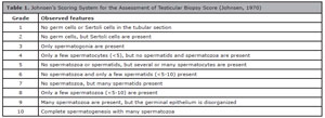
Table 1. Johnsen’s Scoring System for the Assessment of Testicular Biopsy Score (Johnsen, 1970)
Statistical Analysis
Data were expressed as mean±standard error of the mean (SEM) and analyzed using analysis of variance (ANOVA) in GraphPad Prism 5.0 (GraphPad Software, Inc., USA). Tukey’s post hoc test was used for multiple comparisons. A p-value<0.05 was considered statistically significant.
RESULTS
Antioxidant Enzyme Activities, MDA, and Nitrite Levels in Ipsilateral Testicular Tissue After ATRA Treatment
The activities of the testicular antioxidants CAT, total protein, SOD, GPx, GST, and GSH in each group are shown in Figs. 1-6. These activities were significantly decreased in the Torsion+Detorsion (TD) group 3 days after reperfusion compared with the sham group (p<0.05; p<0.01). Conversely, the ATRA-pretreated and ATRA-post-treated groups showed increased antioxidant activities (p<0.05; p<0.01) compared with the TD group. Testicular MDA and nitrite levels in each group are shown in Figs. 7-8. MDA and nitrite levels were increased in the TD group compared with the sham group (p<0.001). In contrast, MDA and nitrite levels were significantly decreased in the pretreated and post-treated groups compared with the TD group (p<0.001). This reduction approached the levels observed in sham rats.
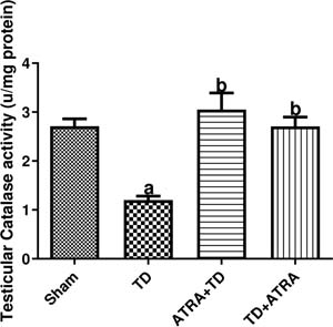
Figure 1. Effect of ATRA on testicular catalase activity in the ipsilateral Testis following TDData were presented as mean ± SEM (n=6).arepresents significant decrease at (p<0.05) compared with the sham group. brepresents significant increase at (p<0.01; p<0.05) compared with the TD group.
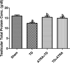
Figure 2. Effect of ATRA on testicular protein concentration in the ipsilateral Testis following TDData were presented as mean ± SEM (n=6). arepresents significant decrease at (p<0.01) compared with the sham group. brepresents significant increase at (p<0.05) com-pared with the TD group.
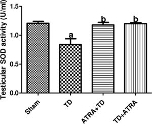
Figure 3. Effect of ATRA on testicular superoxide dismutase (SOD) activity in the ipsilateral Testis following TDData were presented as mean ± SEM (n=6). arepresents significant decrease at (p<0.01) compared with the sham group. brepresents significant increase at (p<0.05) com-pared with the TD group.
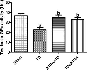
Figure 4. Effect of ATRA on testicular glutathione peroxidase (GPx) activity in the ipsilateral Testis following TDData were presented as mean ± SEM (n=6). a represents significant decrease at (p<0.01) compared with the sham group. b represents significant increase at (p<0.05) com-pared with the TD group.
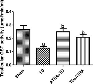
Figure 5. Effect of ATRA on testicular glutathione-S-transferase (GST) activity in the ipsilateral Testis following TDData were presented as mean ± SEM (n=6). arepresents significant decrease at (p<0.01) compared with the sham group. brepresents significant increase at (p<0.01; p<0.05) compared with the TD group.
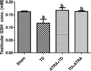
Figure 6. Effect of ATRA on testicular glutathione concentration in the ipsilateral Testis following TDData were presented as mean ± SEM (n=6). arepresents significant decrease at (p<0.05) compared with the sham group. brepresents significant increase at (p<0.05) com-pared with the TD group.
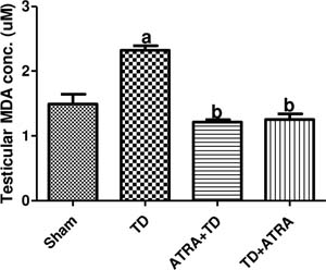
Figure 7. Effect of ATRA on testicular malondialdehyde (MDA) concentration in the ipsilateral Testis following TDData were presented as mean ± SEM (n=6). arepresents significant decrease at (p<0.001) compared with the sham group. brepresents significant increase at (p<0.001) com-pared with the TD group.
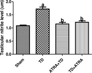
Figure 8. Effect of ATRA on testicular nitrite level in the ipsilateral Testis following TDData were presented as mean ± SEM (n=6). arepresents significant decrease at (p<0.001) compared with the sham group. brepresents significant increase at (p<0.001; p<0.01) compared with the TD group.
ATRA Attenuates Inflammation in Ipsilateral Tes-ticular Tissue
Testicular TNF-α, IL-6, NF-κB, and MPO levels in each group are shown in Figs. 9-12. These levels were significantly increased in the TD group 3 days after reperfusion compared with the sham group (p<0.05; p<0.01; p<0.001). The ATRA-pretreated (ATRA+TD) and ATRA-post-treated (TD+ATRA) groups showed significant decreases in testicular TNF-α, IL-6, NF-κB, and MPO levels compared with the TD group (p<0.05; p<0.01; p<0.001). MPO was significantly lower in the pretreatment group (ATRA+TD) than in the post-treatment group (TD+ATRA) (p<0.01), as shown in the graph.
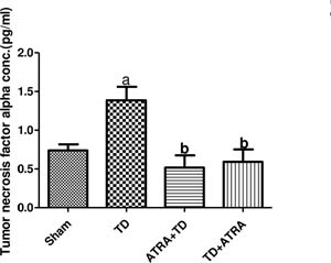
Figure 9. Effect of ATRA on tumor necrosis factor-alpha concentration in the ipsilateral Testis following TDData were presented as mean ± SEM (n=6). arepresents significant increase at (p<0.05) compared with the sham group. brepresents significant decrease at (p<0.01; p<0.05) compared with the TD group.
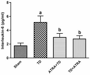
Figure 10. Effect of ATRA on interleukin-6 concentration in the ipsilateral Testis following TDData were presented as mean ± SEM (n=6). arepresents significant increase at (p<0.01) compared with the sham group. brepresents significant decrease at (p<0.05) com-pared with the TD group.
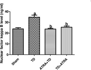
Figure 11. Effect of ATRA on nuclear factor keppa-B (NF kB) concentration in the ipsilateral Testis following TDData were presented as mean ± SEM (n=6). arepresents significant increase at (p<0.001) compared with the sham group. brepresents significant decrease at (p<0.001) com-pared with the TD group.
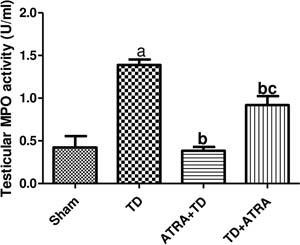
Figure 12. Effect of ATRA on testicular myeloperoxidase (MPO) activity in the ipsilateral Testis following TDData were presented as mean ± SEM (n=6). arepresents significant increase at (p<0.001) compared with the sham group. brepresents significant decrease at (p<0.001; p<0.05) compared with the TD group. crepresents significant increase at (p<0.01) compared with the ATRA+TD group.
Testicular Caspase-3 Activity and Bax and Bcl-2 Levels in Ipsilateral Testicular Tissue After ATRA Treatment
As shown in Figs. 13-15, three days after reperfusion, testicular caspase-3 activity and Bax levels were significantly elevated (p<0.05), whereas Bcl-2 levels were significantly reduced (p<0.05), in the TD group compared with the sham group. Caspase-3 activity and Bax levels were significantly decreased in the ATRA-pretreated (ATRA+TD) and ATRA-post-treated (TD+ATRA) groups compared with the TD group (p<0.001; p<0.05). Conversely, Bcl-2 levels were significantly increased in both the ATRA-pretreated (ATRA+TD) and ATRA-post-treated (TD+ATRA) groups compared with the TD group (p<0.05).
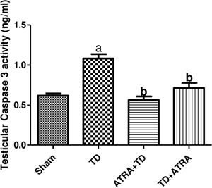
Figure 13. Effect of ATRA on testicular Caspase 3 activity in the ipsilateral Testis following TDData were presented as mean ± SEM (n=6). arepresents significant increase at (p<0.001) compared with the sham group. brepresents significant decrease at (p<0.001) com-pared with the TD group.
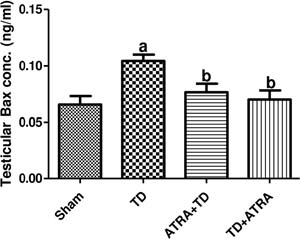
Figure 14. Effect of ATRA on testicular Bax concentration in the ipsilateral Testis following TDData were presented as mean ± SEM (n=6). arepresents significant increase at (p<0.05) compared with the sham group. brepresents significant decrease at (p<0.05) com-pared with the TD group.
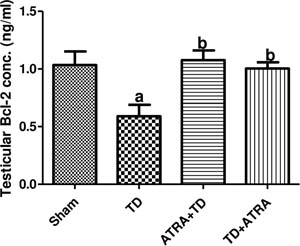
Figure 15. Effect of ATRA on testicular Bcl-2 concentration in the ipsilateral Testis following TDData were presented as mean ± SEM (n=6). arepresents significant decrease at (p<0.05) compared with the sham group. brepresents significant Increase at (p<0.05) com-pared with the TD group.
Reproductive Hormone Concentrations After Ip-silateral TIRI With ATRA Treatment
The levels of the reproductive hormones testosterone, LH, FSH, and inhibin in each group are shown in Figs. 16-19. Testosterone and inhibin levels were significantly decreased in the TD group (p<0.05; p<0.001), whereas LH and FSH levels were significantly increased compared with the sham group (p<0.01). ATRA significantly restored testosterone, LH, FSH, and inhibin levels in the ATRA+TD and TD+ATRA groups, except for testosterone, which was not restored in the TD+ATRA group.
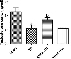
Figure 16. Effect of ATRA on testosterone concentration in the ipsilateral Testis following TDData were presented as mean ± SEM (n=6). arepresents significant decrease at (p<0.05) compared with the sham group. brepresents significant Increase at (p<0.05) com-pared with the TD group.
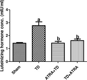
Figure 17. Effect of ATRA on Luteinizing concentration in the ipsilateral Testis following TDData were presented as mean ± SEM (n=6). arepresents significant increase at (p<0.01) compared with the sham group. brepresents significant decrease at (p<0.01) com-pared with the TD group
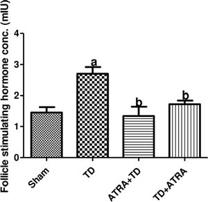
Figure 18. Effect of ATRA on Follicle stimulating hormone concentration in the ipsilateral Testis following TDData were presented as mean ± SEM (n=6). arepresents significant increase at (p<0.01) compared with the sham group. brepresents significant decrease at (p<0.05) com-pared with the TD group
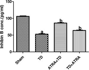
Figure 19. Effect of ATRA on inhibin concentration in the ipsilateral Testis following TDData were presented as mean ± SEM (n=6). arepresents significant decrease at (p<0.001) compared with the sham group. brepresents significant increase at (p<0.001) com-pared with the TD group
Testicular 8-OHdG and DNA Fragmentation Index in Ipsilateral Testicular Tissue After ATRA Treatment Oxidative damage markers were assessed using8-hydroxy-2-deoxyguanosine (8-OHdG) and testicular DNA fragmentation index (DFI) (Figs. 20 and 21). Both markers were significantly increased in the TD group compared with the sham-operated group (p<0.001). In the ATRA-pretreated (ATRA+TD) and ATRA-post-treated (TD+ATRA) groups, the elevated levels of 8-OHdG and DFI were significantly decreased toward normal compared with the TD group (p<0.01; p<0.001). Testicular DFI was significantly higher in the TD+ATRA group than in the ATRA+TD group (p<0.01).
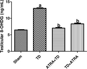
Figure 20. Effect of ATRA on 8-hydroxy-2’-deoxyguanosine (8-OHDG) concentration in the ipsilateral Testis following TDData were presented as mean ± SEM (n=6). arepresents significant increase at (p<0.001) compared with the sham group. brepresents significant decrease at (p<0.001) com-pared with the TD group
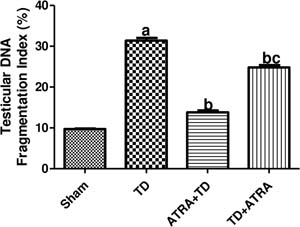
Figure 21. Effect of ATRA on testicular DNA fragmentation index concentration in the ipsilateral Testis following TDData were presented as mean ± SEM (n=6). arepresents significant increase at (p<0.001) compared with the sham group. brepresents significant decrease at (p<0.001; p<0.01) compared with the TD group. crepresents significant increase at (p<0.01) compared with the ATRA +TD group
Sperm Indices After Ipsilateral TIRI With ATRA Treatment
Sperm count, motility, and viability were significantly reduced in the TD group compared with the sham group (p<0.001). This damage was significantly ameliorated in the ATRA-pretreated (ATRA+TD) and ATRA-post-treated (TD+ATRA) groups (p<0.001), as shown in Table 2. Abnormal sperm morphology was significantly increased in the TD group compared with the sham group (p<0.001), whereas morphology was significantly improved in the ATRA-pretreated and ATRA-post-treated groups (p<0.001).
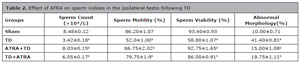
Table 2. Effect of ATRA on sperm indices in the ipsilateral testis following TD
Data are presented as mean±SEM (n=6).Sperm count: a significant decrease (p<0.001) compared with the sham group; b significant increase (p<0.001) compared with the TD group.Sperm motility: a significant decrease (p<0.001) compared with the sham group; b significant increase (p<0.001) com-pared with the TD group.Sperm viability: a significant decrease (p<0.001) compared with the sham group; b significant increase (p<0.001) com-pared with the TD group.Abnormal morphology: a significant increase (p<0.001) compared with the sham group; b significant decrease (p<0.001) compared with the TD group.
Spermatogenesis Indices After Ipsilateral TIRI With ATRA Treatment
Table 3 shows the spermatogenesis indices (spermatogonia, spermatocytes, spermatids, Sertoli cells, and Leydig cell counts). The results showed a statistically significant decrease in the assessed spermatogenesis indices in the TD group compared with the sham group (p<0.001). Furthermore, ATRA pretreatment significantly reversed the adverse effects of TD on spermatogenesis indices compared with the TD group (p<0.001). In all comparisons, ATRA post-treatment resulted in significantly lower spermatogenesis indices than ATRA pretreatment (p<0.001).

Table 3. Effect of ATRA on spermatogenesis indices in the ipsilateral testis following TD
Data are presented as mean±SEM (n=6).Spermatogonia count: a significant decrease (p<0.001) compared with the sham group; b significant increase (p<0.001) compared with the TD group; c significant decrease (p<0.001) compared with the ATRA+TD group.Spermatocyte count: a significant decrease (p<0.001) compared with the sham group; b significant increase (p<0.001) compared with the TD group; c significant decrease (p<0.001) compared with the ATRA+TD group.Spermatid count: a significant decrease (p<0.001) compared with the sham group; b significant increase (p<0.001) com-pared with the TD group; c significant decrease (p<0.001) compared with the ATRA+TD group.Sertoli cell count: a significant decrease (p<0.001) compared with the sham group; b significant increase (p<0.001) com-pared with the TD group; c significant decrease (p<0.001) compared with the ATRA+TD group.Leydig cell count: a significant decrease (p<0.001) compared with the sham group; b significant increase (p<0.001) com-pared with the TD group; c significant decrease (p<0.001) compared with the ATRA+TD group.
Testicular Biopsy Score After Ipsilateral TIRI With ATRA Treatment
Fig. 22 shows the testicular biopsy score (TBS). TBS was significantly decreased in the TD group compared with the sham group (p<0.001). The ATRA-pretreated group showed a significant improvement in TBS compared with the TD group, whereas the ATRA-post-treated group had a significantly lower TBS than the ATRA-pretreated group (p<0.001).
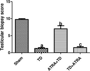
Figure 22. Effect of ATRA on testicular biopsy score in the ipsilateral Testis following TDData were presented as mean ± SEM (n=6). arepresents a significant decrease at (p<0.001) when compared with the sham group. brepresents a significant increase at (p<0.001) compared with the TD group. crepresents a sig-nificant decrease at (p<0.001) compared with the ATRA +TD group
DISCUSSION
Infertility is the major complication of TIRI. To reduce the risk of testicular damage, surgical detorsion is essential to restore blood flow and prevent necrosis (Patoulias et al., 2017). Despite restoration of blood flow to ischemic tissue, testicular reperfusion injury may occur, causing significant testicular injury and dysfunction of cellular components (Kalogeris et al., 2014). Reperfusion injury is characterized by ROS production, which triggers pathophysiological events in the testes (Afolabi et al., 2023). Therefore, interventions aimed at minimizing oxidative stress may help prevent TIRI. All-trans retinoic acid (ATRA) has been reported to influence the transcription of antioxidant enzymes and exhibit cytoprotective properties (Rao et al., 2010; Sakamoto et al., 2010; Sapiro et al., 2017). It also indirectly prevents NF-κB-mediated transcription of inflammatory cytokines, which can increase ROS production and exacerbate tissue damage (El-Sisi et al., 2017). Furthermore, ATRA plays a key role in spermatogenesis by regulating gene transcription, supporting germ-cell differentiation, and contributing to the formation of the blood-testis barrier (Teletin et al., 2017). Although pre-torsion administration does not strictly reflect clinical practice, it was included to clarify the mechanistic potential of ATRA in mitigating ischemia-induced oxidative injury, whereas post-torsion administration reflects a more realistic clinical scenario.
ROS-induced oxidative stress is one of the major pathophysiological events during TIRI (Kalogeris et al., 2012; Afolabi et al., 2022a). In this study, the observed increase in testicular MDA and nitrite (NO) levels indicates lipid peroxidation and nitrosative stress in the TD group, suggesting oxidative stress-induced testicular dysfunction. Reperfusion injury accelerates lipid peroxidation, which in turn initiates pro-inflammatory pathways in ipsilateral testicular tissue (Ayala et al., 2014; Bei et al., 2019). At high concentrations, nitrite can interact with other radicals to form peroxynitrite, which may disrupt mitochondrial respiratory-chain components in the testes, causing cellular energy deficits and, ultimately, cell death (Zhang et al., 2013).
Testicular antioxidant enzymes neutralize free radicals, thereby limiting oxidative modifications of lipids, proteins, and nucleic acids and maintaining proper testicular-cell function under stress (Li, 2022). In testicular torsion, antioxidant enzymes such as GPx, GSH, GST, SOD, and CAT are severely depleted, as shown in this study. This creates an imbalance between pro-oxidants and antioxidants, making the testicular milieu vulnerable to oxidative damage. This finding agrees with previous studies reporting depletion of the antioxidant defense system following TIRI (Afolabi et al., 2022b; Li, 2022; Ajike et al., 2025). However, ATRA administration reversed oxidative damage, as shown by increased CAT, SOD, GPx, GST, GSH, and total protein levels in both the ATRA-pretreated and ATRA-post-treated groups. Because the antioxidant defense system improved in both ATRA-treated groups after reperfusion injury, the oxidative stress markers MDA and NO were also reduced to near-baseline levels. These findings agree with those of Njar et al. (2006) and le Maire et al. (2019), who reported high antioxidant transcriptional activity of ATRA. In this study, ATRA reduced ROS production during TIRI, possibly by promoting transcription of antioxidant enzymes through binding to retinoid X receptors (RXRs) in the nucleus, thereby supporting upregulation or stabilization of antioxidant machinery against reperfusion injury.
Activation of inflammatory cascades is another mechanism by which TIRI causes testicular damage and compromises testicular function (Kalogeris et al., 2012; Granger & Kvietys, 2015; Ajike et al., 2025). During testicular injury, inflammatory cascades are activated, resulting in the release of pro-inflammatory mediators such as TNF-α, IL-1β, and IL-6 from macrophages. These cytokines increase leukocyte mobilization and recruitment to the injured testicular tissue (Shimizu et al., 2016; Li, 2022; Kölükçü et al., 2024). This process results in ROS and reactive nitrogen species (RNS) production and the release of proteolytic enzymes (Cheng et al., 2020). These pro-inflammatory cytokines activate NF-κB, a major transcription factor that regulates the expression of pro-inflammatory genes (Courtois et al., 2016; Anilkumar & Wright-Jin, 2024). During TIRI, as observed in this study, NF-κB is overactivated by pro-inflammatory cytokines, amplifying the inflammatory response (Courtois et al., 2016). Excessive inflammation following TIRI may result in testicular atrophy and infertility (Sharp et al., 2013). In the present study, administration of ATRA before and after TIRI reduced TNF-α, IL-6, MPO, and NF-κB levels, suggesting suppression of inflammation. ATRA may exert its anti-inflammatory effect by decreasing TNF-α secretion from macrophages and interfering with NF-κB upregulation in the testicular nucleus. This is followed by reduced transcription of pro-inflammatory cytokines and decreased secretion of TNF-α, IL-6, and MPO in the ipsilateral testis. This finding is consistent with other studies reporting the anti-inflammatory properties of ATRA (Mehta et al., 1994; Na et al., 1999; Dolin et al., 2023). Other studies have reported that ATRA reduces phosphorylation of NF-κB signaling intermediates, such as p65 and IκB subunits, and binds directly to retinoid X receptor (RXR), which may disrupt the NF-κB signaling cascade and suppress downstream inflammatory damage in testicular tissue (Karkeni et al., 2017; Franco & Pan, 2023). The observed reduction in IL-6 levels in both the ATRA-pretreated and ATRA-post-treated groups may be due to activation of RARE pathways. Previous studies have shown that ATRA can activate the retinoic acid response element (RARE) pathway, associated with anti-inflammatory gene regulation, and downregulate IL-6 expression (Ogawa et al., 2018). The observed reduction in MPO levels may also be attributed to the ability of ATRA to upregulate anti-inflammatory signaling in immune cells. This action may reduce not only TNF-α release from macrophages but also leukocyte recruitment to the injury site. Previous studies have also documented the anti-inflammatory action of ATRA on macrophages as a possible mechanism for halting the progression of various diseases (Rafieian-Kopaei et al., 2014; Deng & Chen, 2022).
Physiologically, apoptosis regulates the germ-cell population and supports effective spermatogenesis. However, during TIRI, massive ROS production increases activation of apoptotic pathways, which may result in germ-cell loss, testicular-cell disruption, and eventually infertility (Kostakis et al., 2017; Xu et al., 2022). TIRI can trigger intrinsic apoptotic pathways in the mitochondria (Hu et al., 2023). This occurs through activation of MAPKs, which can disrupt mitochondrial function and structure, reduce calcium storage capacity, and cause calcium accumulation. Increased calcium levels stimulate the mitochondrial permeability transition pore and activate calcium/calmodulin-dependent proteases, which disrupt cellular structures and eventually trigger cell death (Kalogeris et al., 2012). Another possible mechanism is stimulation of mitochondrial Bax secretion, causing release of cytochrome C from the mitochondria into the cytoplasm, where it binds to Apaf-1 (apoptotic protease activating factor-1) and triggers caspase-3-mediated apoptosis (Shoaib et al., 2021; Khbouz et al., 2022; Wang et al., 2022). In this study, increased testicular Bax and caspase-3 levels and reduced Bcl-2 levels indicate that TIRI triggers apoptosis in the ipsilateral testis, consistent with other studies that reported elevated Bax and caspase-3 levels as evidence of apoptosis in testicular cells following TIRI (Afolabi et al., 2022a). The elevated Bax and caspase-3 levels and reduced Bcl-2 levels observed after TIRI suggest compromised sperm integrity and possibly altered spermatogenesis. ATRA-pretreated and ATRA-post-treated groups showed reduced levels of the pro-apoptotic proteins Bax and caspase-3 and increased levels of Bcl-2, suggesting a possible anti-apoptotic effect of ATRA. ATRA may exert this effect indirectly by downregulating MAPK phosphorylation, thereby reducing activation of pro-apoptotic proteins in the mitochondria. Previous studies have also reported that ATRA can reduce cardiac apoptosis following myocardial ischemia-reperfusion injury (Zhu et al., 2015).
Reproductive hormones such as LH, FSH, testosterone, and inhibin are key indicators of spermatogenesis and male fertility (Zhao et al., 2024). Massive free-radical production in testicular-cell mitochondria during TIRI can injure Leydig and Sertoli cells, disrupting testosterone production and spermatogenesis in the seminiferous tubules and resulting in defective sperm production and infertility (Shimizu et al., 2016; Elmimehr et al., 2021). The observed decrease in testosterone and inhibin levels in the TD group suggests disruption of spermatogenesis in the testes. Specifically, reduced inhibin and testosterone levels indicate loss of Sertoli-cell and Leydig-cell hormonal function, respectively. This abnormality may disrupt FSH secretion and culminate in abnormal or incomplete spermatogenesis. The increased LH and FSH levels in the TD group may be attributed to activation of compensatory mechanisms in response to reduced testosterone and inhibin levels, as reported in other studies (Benny et al., 2017; Balci et al., 2021; Ajike et al., 2025). The observed increase in testosterone levels in the ATRA-treated group suggests improved Leydig-cell function. This improvement may be related to the ability of ATRA to increase transcription of steroidogenic enzymes in Leydig cells, which may help restore testosterone levels following TIRI. Restored testosterone levels not only help establish an environment that supports the initiation of spermatogenesis, but also contribute to completion of the spermatogenic process, thereby restoring normal sperm function after ATRA administration (Munetsuna et al., 2009). Improved testosterone levels also promote Sertoli-cell function, accompanied by increased inhibin levels that regulate FSH activity. These coordinated effects mediated by ATRA help restore spermatogenic and steroidogenic processes during TIRI.
Moreover, the findings of this study showed that ipsilateral TIRI induced a marked increase in testicular DNA damage, as evidenced by significant elevations in testicular DNA fragmentation index (DFI) and 8-hydroxy-2’-deoxyguanosine (8-OHdG) concentrations in the TD group compared with the sham group. Elevated DFI indicates strand breaks and compromised chromatin integrity (Li & Lloyd, 2020), whereas elevated 8-OHdG indicates oxidative DNA damage and strand breaks (Omari Shekaftik & Nasirzadeh, 2021). Elevation of these markers is detrimental to germ-cell viability and spermatogenesis and ultimately leads to germ-cell apoptosis and impaired spermatogenesis (Fadel et al., 2020). Notably, pretreatment and post-treatment with ATRA significantly reduced 8-OHdG levels, suggesting that ATRA effectively mitigates oxidative DNA damage. By lowering oxidative damage, ATRA helps preserve the structural and functional integrity of the testis, which is critical for spermatogenesis and fertility maintenance following TIRI (Zhao et al., 2024). ATRA is known to modulate cell differentiation, immune function, and redox homeostasis through direct ROS scavenging, indirect upregulation of endogenous antioxidant enzymes, and inhibition of pro-inflammatory signaling pathways (Behairi et al., 2016; Brown, 2023). ATRA also markedly decreased DFI, with pretreatment achieving a greater reduction than post-treatment. This suggests that prophylactic administration provides superior genomic protection by priming the testicular antioxidant defense system before reperfusion occurs (Hasbullah et al., 2022). This preemptive effect likely reduces the initial ROS surge and limits downstream apoptotic signaling cascades.
Sperm count, motility, and viability were significantly decreased in the TD group compared with the sham group, demonstrating that TIRI impairs spermatogenesis and compromises sperm quality. These findings are consistent with evidence that ROS attack sperm plasma membranes, disrupt mitochondrial function, and induce sperm DNA fragmentation, ultimately reducing sperm fertilizing capacity. Following ATRA administration, sperm parameters improved significantly, with ATRA pretreatment showing a more pronounced effect than post-treatment (Malivindi et al., 2018; Havel & Griswold, 2025). This improvement further supports the role of ATRA in reducing oxidative damage and preserving germ-cell function (Teletin et al., 2017). The number of sperm cells with abnormal morphology was significantly increased in the TD group compared with the sham group, indicating that TIRI also disrupts spermiogenesis and results in morphological defects, such as head, midpiece, or tail abnormalities, that compromise fertilizing potential (Li, 2022). Notably, ATRA treatment preserved the structural integrity of sperm cells, as shown by the significantly reduced proportion of abnormally shaped sperm, likely due to prevention of ROS-mediated damage (Drevet et al., 2022).
The spermatogenesis indices and histological evaluation of testes following unilateral TIRI allow visualization of testicular cells and specific changes in the seminiferous tubules. Careful observation of the seminiferous tubules of TD rats showed reduced Leydig-cell, Sertoli-cell, spermatogonia, spermatocyte, spermatid, and testicular biopsy score counts, indicating disruption of spermatogenesis caused by oxidative cellular damage. Histological assessment of the testes in the TD group also showed distortion of testicular histoarchitecture and a reduced number of testicular cells. This confirms that TD alters spermatogenesis and is consistent with previous studies (Yíldíz et al., 2011; Parlaktas et al., 2014). It is also plausible that oxidative stress, inflammation, and apoptosis contribute to cellular damage within the seminiferous tubules, as noted in other studies (Yíldíz et al., 2011; Kalogeris et al., 2012). However, ATRA treatment improved testicular and epididymal structures, likely because of its ability to activate endogenous antioxidants, neutralize ROS during oxidative stress, promote germ-cell differentiation, and exert cytoprotective effects, as confirmed by previous studies (Rao et al., 2010; Sakamoto et al., 2010; Sapiro et al., 2017; Teletin et al., 2017).
Clinical Implications and Translational Relevance
Although both preand post-torsion administration of ATRA were evaluated in this study, post-detorsion administration has greater clinical relevance because pharmacological intervention in patients with testicular torsion would usually occur after surgical correction. The protective effects observed with post-treatment suggest that ATRA may be used as an adjunct to surgical detorsion to attenuate reperfusion-induced oxidative stress, inflammation, and apoptotic injury. However, translation to clinical practice requires caution because differences in dosing, timing, and pharmacokinetics exist between experimental models and humans. Therefore, further preclinical optimization and controlled clinical trials are necessary to establish the safety, efficacy, and therapeutic applicability of ATRA in human testicular torsion.
CONCLUSION
The present study demonstrates that ATRA effectively protects against TIRI by enhancing antioxidant defenses, suppressing inflammatory and apoptotic pathways, and preserving spermatogenic and hormonal functions. These findings highlight the potential of ATRA as a therapeutic adjunct in the management of testicular torsion. However, this study was limited to short-term evaluation and did not assess molecular signaling or fertility outcomes; therefore, further investigations of long-term reproductive recovery and mechanistic pathways are warranted. Future studies extending these findings to human clinical settings are also needed to evaluate the safety, optimal dosing, and therapeutic potential of ATRA as an adjunct treatment following surgical detorsion.
ETHICAL APPROVAL
This study was approved by the Ethical Research Committee of the Faculty of Basic Medical Sciences, Ladoke Akintola University of Technology, Ogbomoso, with reference number ERCFBMSLAUTECH: 033/05/2024.
REFERENCES
Adamczewska D, Słowikowska-Hilczer J, Walczak-Jędrzejowska R. The Fate of Leydig Cells in Men with Spermatogenic Failure. Life (Basel). 2022;12:570. PMID: 35455061 DOI: 10.3390/life12040570
Medline
Adegunlola JG, Afolabi OK, Akhigbe RE, Adegunlola GA, Adewumi OM, Oyeyipo IP, Ige SF, Afolabi AO. Lipid peroxidation in brain tissue following administration of low and high doses of arsenite and L-ascorbate in wistar strain rats. Toxicol Int. 2012;19:47-50. PMID: 22736903 DOI: 10.4103/0971-6580.94516
Medline
Afolabi O, Alabi B, Omobowale T, Oluranti O, Iwalewa O. Cysteamine mitigates torsion/detorsion-induced reperfusion injury via inhibition of apoptosis, oxidative stress and inflammatory responses in experimental rat model. Andrologia. 2022a;54:e14243. PMID: 34498746 DOI: 10.1111/and.14243
Medline
Afolabi OA, Anyogu DC, Hamed MA, Odetayo AF, Adeyemi DH, Akhigbe RE. Glutamine prevents upregulation of NF-kB signaling and caspase 3 activation in ischaemia/ reperfusion-induced testicular damage: An animal model. Biomed Pharmacother. 2022b;150:113056. PMID: 35658227 DOI: 10.1016/j.biopha.2022.113056
Medline
Afolabi OA, Adebola Alabi B, Adedamola Ajike R, Simeon Oyekunle O, Adegoke W, Adebayo Ojetola A. Evaluation of testicular torsion management in Ogbomoso, South-Western Nigeria and surgical detorsion-augmented treatment with phytochemical fractions of Corchorus olitorius leaf in experimental rats. Saudi J Biol Sci. 2023;30:103495. PMID: 36439959 DOI: 10.1016/j. sjbs.2022.103495
Medline
Ajike RA, Afolabi OA, Alabi BA, Ajayi AF, Oyekunle OS, Lawal SK, Olojede SO, Nku-Ekpang OA, Hezekiah OS, Hammed OS. Sequential administration of febuxostat and vitamin E protects against testicular ischemia/reperfusion injury via inhibition of sperm DNA damage in Wistar rats. Naunyn Schmiedebergs Arch Pharmacol. 2025;398:14025-39. PMID: 40261353 DOI: 10.1007/s00210-025-04095-x
Medline
Anilkumar S, Wright-Jin E. NF-κB as an Inducible Regulator of Inflammation in the Central Nervous System. Cells. 2024;13:485. PMID: 38534329 DOI: 10.3390/cells13060485
Medline
Avwioro G, Iyiola S, Aghoghovwia B. Histological and biochemical markers of the liver of Wistar rats on subchronic oral administration of green tea. N Am J Med Sci. 2010;2:376-80. PMID: 22737676 DOI: 10.4297/najms.2010.2376
Medline
Ayala A, Muñoz MF, Argüelles S. Lipid peroxidation: production, metabolism, and signaling mechanisms of malondialdehyde and 4-hydroxy-2-nonenal. Oxid Med Cell Longev. 2014;2014:360438. PMID: 24999379 DOI: 10.1155/2014/360438
Medline
Balci CN, Firat T, Acar N, Kukner A. Carvacrol treatment opens Kir6.2 ATP-dependent potassium channels and prevents apoptosis on rat testis following ischemia-reperfusion injury model. Rom J Morphol Embryol. 2021;62:179-90. PMID: 34609420 DOI: 10.47162/ RJME.62.1.17
Medline
Behairi N, Belkhelfa M, Rafa H, Labsi M, Deghbar N, Bouzid N, Mesbah-Amroun H, Touil-Boukoffa C. All-trans retinoic acid (ATRA) prevents lipopolysaccharide-induced neuroinflammation, amyloidogenesis and memory impairment in aged rats. J Neuroimmunol. 2016;300:21-9. PMID: 27806872 DOI: 10.1016/j.jneuroim.2016.10.004
Medline
Bei Y, Pan LL, Zhou Q, Zhao C, Xie Y, Wu C, Meng X, Gu H, Xu J, Zhou L, Sluijter JPG, Das S, Agerberth B, Sun J, Xiao J. Cathelicidin-related antimicrobial peptide protects against myocardial ischemia/reperfusion injury. BMC Med. 2019;17:42. PMID: 30782145 DOI: 10.1186/s12916-019-1268-y
Medline
Benny B, George Priya Doss C, Thirumal Kumar D, Asha Devi S. Assessing reproductive toxicity and antioxidant enzymes on beta asarone induced male Wistar albino rats: In vivo and computational analysis. Life Sci. 2017;173:150-60. PMID: 27569590 DOI: 10.1016/j.lfs.2016.08.021
Medline
Brown G. Retinoic acid receptor regulation of decision-making for cell differentiation. Front Cell Dev Biol. 2023;11:1182204. PMID: 37082619 DOI: 10.3389/ fcell.2023.1182204
Medline
Cheng Z, Qian S, Qingtao M, Zhongyuan X, Yeda X. Effects of ATRA on diabetic rats with renal ischemia-reperfusion injury. Acta Cir Bras. 2020;35:e202000106. PMID: 32236320 DOI: 10.1590/s0102-865020200010000006
Medline
Chovanec M, Lauritsen J, Bandak M, Oing C, Kier GG, Kreiberg M, Rosenvilde J, Wagner T, Bokemeyer C, Daugaard G. Late adverse effects and quality of life in survivors of testicular germ cell tumour. Nat Rev Urol. 2021;18:227-45. PMID: 33686290 DOI: 10.1038/s41585-021-00440-w
Medline
Deng Q, Chen J. Potential Therapeutic Effect of All-Trans Retinoic Acid on Atherosclerosis. Biomolecules. 2022;12:869. PMID: 35883425 DOI: 10.3390/biom12070869
Medline
Desser RK, Himmelhoch SR, Evans WH, Januska M, Mage M, Shelton E. Guinea pig heterophil and eosinophil peroxidase. Arch Biochem Biophys. 1972;148:452-65. PMID: 4623114 DOI: 10.1016/0003-9861(72)90164-6
Medline
Dolin HH, Franco JH, Chen X, Pan ZK. Retinoic Acid-Induced Regulation of Inflammatory Pathways Is a Potential Sepsis Treatment. Infect Immun. 2023;91:e0045722. PMID: 36877073 DOI: 10.1128/iai.00457-22
Medline
Drevet JR, Hallak J, Nasr-Esfahani MH, Aitken RJ. Reactive Oxygen Species and Their Consequences on the Structure and Function of Mammalian Spermatozoa. Antioxid Redox Signal. 2022;37:481-500. PMID: 34913729 DOI: 10.1089/ ars.2021.0235
Medline
Elmimehr R, Motamed-Sanaye A, Brazvan B, Abtahi-Eivary SH, Moghimian M, Fani M. Effects of hypothermia and pentoxifylline on the adnexal torsion/detorsion injuries in a rat testis model. Andrologia. 2021;53:e14143. PMID: 34115392 DOI: 10.1111/and.14143
Medline
El-Sisi AE, El-Sayad ME, Abdelsalam NM. Protective effects of mirtazapine and chrysin on experimentally induced testicular damage in rats. Biomed Pharmacother. 2017;95:1059-66. PMID: 28922723 DOI: 10.1016/j.biopha.2017.09.022
Medline
Fadel F, Al-Kandari N, Khashab F, Al-Saleh F, Al-Maghrebi M. JNK inhibition alleviates oxidative DNA damage, germ cell apoptosis, and mitochondrial dysfunction in testicular ischemia reperfusion injury. Acta Biochim Biophys Sin (Shanghai). 2020;52:891-900. PMID: 32662511 DOI: 10.1093/abbs/gmaa074
Medline
Gornall AG, Bardawill CJ, David MM. Determination of serum proteins by means of the biuret reaction. J Biol Chem. 1949;177:751-66. PMID: 18110453
Medline
Granger DN, Kvietys PR. Reperfusion injury and reactive oxygen species: The evolution of a concept. Redox Biol. 2015;6:524-51. PMID: 26484802 DOI: 10.1016/j. redox.2015.08.020
Medline
Hasbullah JS, Scott EN, Bhavsar AP, Gunaretnam EP, Miao F, Soliman H, Carleton BC, Ross CJD. All-trans retinoic acid (ATRA) regulates key genes in the RARG-TOP2B pathway and reduces anthracycline-induced cardiotoxicity. PLoS One. 2022;17:e0276541. PMID: 36331922 DOI: 10.1371/journal.pone.0276541
Medline
Havel SL, Griswold MD. The action of retinoic acid on spermatogonia in the testis. Curr Top Dev Biol. 2025;161:143-66. PMID: 39870432 DOI: 10.1016/bs.ctdb.2024.10.005
Medline
Hu T, Zou HX, Le SY, Wang YR, Qiao YM, Yuan Y, Liu JC, Lai SQ, Huang H. Tanshinone IIA confers protection against myocardial ischemia/reperfusion injury by inhibiting ferroptosis and apoptosis via VDAC1. Int J Mol Med. 2023;52:109. PMID: 37800609 DOI: 10.3892/ ijmm.2023.5312
Medline
Johnsen SG. Testicular biopsy score count--a method for registration of spermatogenesis in human testes: normal values and results in 335 hypogonadal males. Hormones. 1970;1:2-25. PMID: 5527187 DOI: 10.1159/000178170
Medline
Juan CA, Pérez de la Lastra JM, Plou FJ, Pérez-Lebeña E. The Chemistry of Reactive Oxygen Species (ROS) Revisited: Outlining Their Role in Biological Macromolecules (DNA, Lipids and Proteins) and Induced Pathologies. Int J Mol Sci. 2021;22:4642. PMID: 33924958 DOI: 10.3390/ijms22094642
Medline
Kalogeris T, Baines CP, Krenz M, Korthuis RJ. Cell biology of ischemia/reperfusion injury. Int Rev Cell Mol Biol. 2012;298:229-317. PMID: 22878108 DOI: 10.1016/ B978-0-12-394309-5.00006-7
Medline
Kalogeris T, Bao Y, Korthuis RJ. Mitochondrial reactive oxygen species: a double edged sword in ischemia/ reperfusion vs preconditioning. Redox Biol. 2014;2:702-14. PMID: 24944913 DOI: 10.1016/j.redox.2014.05.006
Medline
Kang JB, Son HK, Shah MA, Koh PO. Retinoic acid attenuates ischemic injury-induced activation of glial cells and inflammatory factors in a rat stroke model. PLoS One. 2024;19:e0300072. PMID: 38527023 DOI: 10.1371/journal.pone.0300072
Medline
Karkeni E, Bonnet L, Astier J, Couturier C, Dalifard J, Tourniaire F, Landrier JF. All-trans-retinoic acid represses chemokine expression in adipocytes and adipose tissue by inhibiting NF-κB signaling. J Nutr Biochem. 2017;42:101-7. PMID: 28157617 DOI: 10.1016/j.jnutbio.2017.01.004
Medline
Kaur G, Wright K, Verma S, Haynes A, Dufour JM. The Good, the Bad and the Ugly of Testicular Immune Regulation: A Delicate Balance Between Immune Function and Immune Privilege. Adv Exp Med Biol. 2021;1288:21-47. PMID: 34453730. DOI: 10.1007/978-3-030-77779-1_2
Medline
Khbouz B, Lallemand F, Cirillo A, Rowart P, Legouis D, Sounni NE, Noël A, De Tullio P, de Seigneux S, Jouret F. Kidney-targeted irradiation triggers renal ischemic preconditioning in mice. Am J Physiol Renal Physiol. 2022;323:F198-F211. PMID: 35796462 DOI: 10.1152/ajprenal.00005.2022
Medline
Korkes F, Cabral PR, Alves CD, Savioli ML, Pompeo AC. Testicular torsion and weather conditions: analysis of 21,289 cases in Brazil. Int Braz J Urol. 2012;38:222-8. PMID: 22555045 DOI: 10.1590/s1677-55382012000200010
Medline
Kostakis ID, Zavras N, Damaskos C, Sakellariou S, Korkolopoulou P, Misiakos EP, Tsaparas P, Vaos G, Karatzas T. Erythropoietin and sildenafil protect against ischemia/ reperfusion injury following testicular torsion in adult rats. Exp Ther Med. 2017;13:3341-7. PMID: 28587411 DOI: 10.3892/etm.2017.4441
Medline
Kostenko V, Akimov O, Gutnik O, Kostenko H, Kostenko V, Romantseva T, Morhun Y, Nazarenko S, Taran O. Modulation of redox-sensitive transcription factors with polyphenols as pathogenetically grounded approach in therapy of systemic inflammatory response. Heliyon. 2023;9:e15551. PMID: 37180884 DOI: 10.1016/j.heliyon.2023.e15551
Medline
Laher A, Ragavan S, Mehta P, Adam A. Testicular Torsion in the Emergency Room: A Review of Detection and Management Strategies. Open Access Emerg Med. 2020;12:237-46. PMID: 33116959 DOI: 10.2147/OAEM.S236767
Medline
le Maire A, Teyssier C, Balaguer P, Bourguet W, Germain P. Regulation of RXR-RAR Heterodimers by RXR-and RAR-Specific Ligands and Their Combinations. Cells. 2019;8:1392. PMID: 31694317 DOI: 10.3390/cells8111392
Medline
Li MW, Lloyd KCK. DNA fragmentation index (DFI) as a measure of sperm quality and fertility in mice. Sci Rep. 2020;10:3833. PMID: 32123279 DOI: 10.1038/s41598-020-60876-9
Medline
Li ZM. Role of antioxidants in preventing testicular ischemiareperfusion injury: a narrative review. Eur Rev Med Pharmacol Sci. 2022;26:9126-43. PMID: 36591825 DOI: 10.26355/eurrev_202212_30663
Medline
Malivindi R, Rago V, De Rose D, Gervasi MC, Cione E, Russo G, Santoro M, Aquila S. Influence of all-trans retinoic acid on sperm metabolism and oxidative stress: Its involvement in the physiopathology of varicocele-associated male infertility. J Cell Physiol. 2018;233:9526-37. PMID: 29943815 DOI: 10.1002/jcp.26872
Medline
Mehta K, McQueen T, Tucker S, Pandita R, Aggarwal BB. Inhibition by all-trans-retinoic acid of tumor necrosis factor and nitric oxide production by peritoneal macrophages. J Leukoc Biol. 1994;55:336-42. PMID: 8120450 DOI: 10.1002/jlb.55.3.336
Medline
Misra HP, Fridovich I. The role of superoxide anion in the autoxidation of epinephrine and a simple assay for superoxide dismutase. J Biol Chem. 1972;247:3170-5. PMID: 4623845
Medline
Munetsuna E, Hojo Y, Hattori M, Ishii H, Kawato S, Ishida A, Kominami SA, Yamazaki T. Retinoic acid stimulates 17beta-estradiol and testosterone synthesis in rat hippocampal slice cultures. Endocrinology. 2009;150:4260-9. PMID: 19497980 DOI: 10.1210/en.2008-1644
Medline
Na SY, Kang BY, Chung SW, Han SJ, Ma X, Trinchieri G, Im SY, Lee JW, Kim TS. Retinoids inhibit interleukin-12 production in macrophages through physical associations of retinoid X receptor and NFkappaB. J Biol Chem. 1999;274:7674-80. PMID: 10075655. DOI: 10.1074/jbc.274.12.7674
Medline
Njar VC, Gediya L, Purushottamachar P, Chopra P, Vasaitis TS, Khandelwal A, Mehta J, Huynh C, Belosay A, Patel J. Retinoic acid metabolism blocking agents (RAMBAs) for treatment of cancer and dermatological diseases. Bioorg Med Chem. 2006;14:4323-40. PMID: 16530416 DOI: 10.1016/j.bmc.2006.02.041
Medline
Ogawa E, Sato Y, Minagawa A, Okuyama R. Pathogenesis of psoriasis and development of treatment. J Dermatol. 2018;45:264-72. PMID: 29226422 DOI: 10.1111/1346-8138.14139
Medline
Omari Shekaftik S, Nasirzadeh N. 8-Hydroxy-2’-deoxyguanosine (8-OHdG) as a biomarker of oxidative DNA damage induced by occupational exposure to nanomaterials: a systematic review. Nanotoxicology. 2021;15:850-64. PMID: 34171202 DOI: 10.1080/17435390.2021.1936254
Medline
Paglia DE, Valentine WN. Studies on the quantitative and qualitative characterization of erythrocyte glutathione peroxidase. J Lab Clin Med. 1967;70:158-69. PMID: 6066618.
Medline
Parlaktas BS, Atilgan D, Gencten Y, Akbas A, Markoc F, Erdemir F, Ozyurt H, Uluocak N. The effects of carvedilol on ischemia-reperfusion injury in the rat testis. Int Braz J Urol. 2014;40:109-17. PMID: 24642157 DOI: 10.1590/ S1677-5538.IBJU.2014.01.16
Medline
Patoulias D, Farmakis K, Kalogirou M, Patoulias I. Transient testicular torsion: from early diagnosis to appropriate therapeutic intervention (a prospective clinical study). Folia Med Cracov. 2017;57:53-62. PMID: 29121037.
Medline
Rafieian-Kopaei M, Setorki M, Doudi M, Baradaran A, Nasri H. Atherosclerosis: process, indicators, risk factors and new hopes. Int J Prev Med. 2014;5:927-46. PMID: 25489440
Medline
Rahman I, Kode A, Biswas SK. Assay for quantitative determination of glutathione and glutathione disulfide levels using enzymatic recycling method. Nat Protoc. 2006;1:3159-65. PMID: 17406579 DOI: 10.1038/nprot.2006.378
Medline
Raji Y, Hart VO. Influence of prolonged exposure to Nigerian Bonny light crude oil on fertility indices in rats. Niger J Physiol Sci. 2012;27:55-63. PMID: 23235309
Medline
Rao J, Zhang C, Wang P, Lu L, Zhang F. All-trans retinoic acid alleviates hepatic ischemia/reperfusion injury by enhancing manganese superoxide dismutase in rats. Biol Pharm Bull. 2010;33:869-75. PMID: 20460768 DOI: 10.1248/bpb.33.869
Medline
Ridnour LA, Sim JE, Hayward MA, Wink DA, Martin SM, Buettner GR, Spitz DR. A spectrophotometric method for the direct detection and quantitation of nitric oxide, nitrite, and nitrate in cell culture media. Anal Biochem. 2000;281:223-9. PMID: 10870839 DOI: 10.1006/ abio.2000.4583
Medline
Sakamoto K, Hiraiwa M, Saito M, Nakahara T, Sato Y, Nagao T, Ishii K. Protective effect of all-trans retinoic acid on NMDA-induced neuronal cell death in rat retina. Eur J Pharmacol. 2010;635:56-61. PMID: 20303946 DOI: 10.1016/j.ejphar.2010.03.001
Medline
Sapiro JM, Monks TJ, Lau SS. All-trans-retinoic acid-mediated cytoprotection in LLC-PK1 renal epithelial cells is coupled to p-ERK activation in a ROS-independent manner. Am J Physiol Renal Physiol. 2017;313:F1200-8. PMID: 28768661 DOI: 10.1152/ajprenal.00085.2017
Medline
Sharma A, Minhas S, Dhillo WS, Jayasena CN. Male infertility due to testicular disorders. J Clin Endocrinol Metab. 2021;106:e442-e59. PMID: 33295608 DOI: 10.1210/clinem/dgaa781
Medline
Sharp VJ, Kieran K, Arlen AM. Testicular torsion: diagnosis, evaluation, and management. Am Fam Physician. 2013;88:835-40. PMID: 24364548
Medline
Shimizu S, Tsounapi P, Dimitriadis F, Higashi Y, Shimizu T, Saito M. Testicular torsion-detorsion and potential therapeutic treatments: A possible role for ischemic postconditioning. Int J Urol. 2016;23:454-63. PMID: 27217335 DOI: 10.1111/iju.13110
Medline
Shoaib S, Tufail S, Sherwani MA, Yusuf N, Islam N. Phenethyl Isothiocyanate Induces Apoptosis Through ROS Generation and Caspase-3 Activation in Cervical Cancer Cells. Front Pharmacol. 2021;12:673103. PMID: 34393773 DOI: 10.3389/fphar.2021.673103
Medline
Siddikuzzaman, Grace VM. Antioxidant potential of all-trans retinoic acid (ATRA) and enhanced activity of liposome encapsulated ATRA against inflammation and tumor-directed angiogenesis. Immunopharmacol Immunotoxicol. 2013;35:164-73. PMID: 23116338. DOI: 10.3109/08923973.2012.736520
Medline
Sies H, Jones DP. Reactive oxygen species (ROS) as pleiotropic physiological signalling agents. Nat Rev Mol Cell Biol. 2020;21:363-83. PMID: 32231263 DOI: 10.1038/ s41580-020-0230-3
Medline
Singhal A, Kumar S. Neutrophil and remnant clearance in immunity and inflammation. Immunology. 2022;165:22-43. PMID: 34704249 DOI: 10.1111/imm.13423
Medline
Sinha AK. Colorimetric assay of catalase. Anal Biochem. 1972;47:389-94. PMID: 4556490 DOI: 10.1016/0003-2697(72)90132-7
Medline
Sodhi RK, Singh N. All-trans retinoic acid rescues memory deficits and neuropathological changes in mouse model of streptozotocin-induced dementia of Alzheimer’s type. Prog Neuropsychopharmacol Biol Psychiatry. 2013;40:38-46. PMID: 23044340 DOI: 10.1016/j.pnpbp.2012.09.012
Medline
Sultana S, Jabeen F, Sultana T, Al-Ghanim KA, Al-Misned F, Mahboob S. Assessment of heavy metals and its impact on DNA fragmentation in different fish species. Braz J Biol. 2020;80:823-8. PMID: 31778486 DOI: 10.1590/1519-6984.221849.
Medline
Teletin M, Vernet N, Ghyselinck NB, Mark M. Roles of Retinoic Acid in Germ Cell Differentiation. Curr Top Dev Biol. 2017;125:191-225. PMID: 28527572 DOI: 10.1016/bs.ctdb.2016.11.013
Medline
Törzsök P, Steiner C, Pallauf M, Abenhardt M, Milinovic L, Plank B, Rückl A, Sieberer M, Lusuardi L, Deininger S. Long-Term Follow-Up after Testicular Torsion: Prospective Evaluation of Endocrine and Exocrine Testicular Function, Fertility, Oxidative Stress and Erectile Function. J Clin Med. 2022;11:6507. PMID: 36362741 DOI: 10.3390/jcm11216507
Medline
Wang YC, Wang LT, Hung TI, Hong YR, Chen CH, Ho CJ, Wang C. Severe cellular stress drives apoptosis through a dual control mechanism independently of p53. Cell Death Discov. 2022;8:282. PMID: 35680784 DOI: 10.1038/ s41420-022-01078-2
Medline
Xu LZ, He KX, Ning JZ, Cheng F. Oleuropein attenuates testicular ischemia-reperfusion by inhibiting apoptosis and inflammation. Tissue Cell. 2022;78:101876. PMID: 35914339 DOI: 10.1016/j.tice.2022.101876
Medline
Yíldíz H, Durmus AS, Simşek H, Yaman M. Protective effect of sildenafil citrate on contralateral testis injury after unilateral testicular torsion/detorsion. Clinics. 2011;66:137-42. PMID: 21437450 DOI: 10.1590/s1807-59322011000100024
Medline
Yucel C, Arslan FD, Ekmekci S, Ulker V, Kisa E, Erdogan Yucel E, Ucar M, Ilbey YO, Celik O, Basok BI, Kozacioglu Z. Protective Effect of All-Trans Retinoic Acid in Cisplatin-Induced Testicular Damage in Rats. World J Mens Health. 2019;37:249-56. PMID: 30799561 DOI: 10.5534/wjmh.180105
Medline
Zhang J, Deng Z, Liao J, Song C, Liang C, Xue H, Wang L, Zhang K, Yan G. Leptin attenuates cerebral ischemia injury through the promotion of energy metabolism via the PI3K/ Akt pathway. J Cereb Blood Flow Metab. 2013;33:567-74. PMID: 23299243 DOI: 10.1038/jcbfm.2012.202
Medline
Zhao Y, Deng S, Li C, Cao J, Wu A, Chen M, Ma X, Wu S, Lian Z. The Role of Retinoic Acid in Spermatogenesis and Its Application in Male Reproduction. Cells. 2024;13:1092. PMID: 38994945 DOI: 10.3390/cells13131092
Medline
Zhu Y, Liu Y, Yang F, Chen W, Jiang J, He P, Jiang S, Li M, Xu R. All-Trans Retinoic Acid Exerts Neuroprotective Effects in Amyotrophic Lateral Sclerosis-Like Tg (SOD1*G93A)1Gur Mice. Mol Neurobiol. 2020;57:3603-15. PMID: 32548665 DOI: 10.1007/s12035-020-01973-8
Medline
Zhu Z, Zhu J, Zhao X, Yang K, Lu L, Zhang F, Shen W, Zhang R. All-Trans Retinoic Acid Ameliorates Myocardial Ischemia/ Reperfusion Injury by Reducing Cardiomyocyte Apoptosis. PLoS One. 2015;10:e0133414. PMID: 26186635 DOI: 10.1371/journal.pone.0133414
Medline