JBRA Assist. Reprod. 2024;28(1):78-89
REVIEW
doi: 10.5935/1518-0557.20230044
Immune system cells modulation in patients with reproductive issues: A systematic review approach
1Post-Graduate Program in Pharmaceutical Sciences (PPGCF) Federal University of Ceará (UFC), Fortaleza, CE, Brazil
2Oswaldo Cruz Foundation (FIOCRUZ CEARÁ), Eusébio, CE, Brazil
3Ceará Hematology and Hemotherapy Center (HEMOCE), Fortaleza, CE, Brazil
4North Northeast Biotechnology Network (RENORBIO), State University of Ceará (UECE), Fortaleza, CE, Brazil
CONFLICT OF INTERESTS
The authors declare no conflict of interest.
ABSTRACT
The aim of this study was to carry out a systematic literature review to investigate the main immune cells responsible for implantation failures. We selected papers from PubMed, Embase and Virtual Health Library databases. Eligible articles included publications between January 1, 2010 and April 24, 2022. Inclusion criteria were: observational and case-control studies; and the exclusion criteria were: review papers, letters to the editor, abstracts, animal studies and case reports. We extracted the following information: day of collection, number of patients, control group, age of patients, type of sample used, immune cells and cytokines. As main findings in our mapping, we found that in peripheral blood, CD3+, CD4+, CD8+, CD16+, CD56+, CD57+, CD69+, CD154+, CD158a+, NKp46 cells were increased and the CD4+, CD45+, Foxp3 and NKp46 markers were reduced. From the endometrial biopsies, there was an increase in CD3+, CD4+, CD5+, CD8+, CD16+, CD25+, CD45+, CD56+, CD57+, CD68+, CD127+ and a reduction in CD45+, CD56+, NKp46 and FoxP3 cells. Cytokines found increased in peripheral blood included IL-6, IL-10, IL-17, INF-γ, TGF-ß, TNF-α; while IL-4, IL-6, IL-10, IL-35, FoxP3, TGF-ß, SOCS3 were reduced. As for the biopsies, there was an increase in IL-2, IL-6, IL-17, IL-22, IL-23, INF-A1, INF-B1, INF-γ, TNF-R and a reduction in IL-6, IL-10, INF-γ, TGFß, TNF-α. We concluded that immune cells can be modulated during pregnancy failure, but further studies are needed to elucidate the modulating effect of the immune system on the endometrium of these patients.
Keywords: abortion, embryo implantation, assisted reproductive techniques, cytokines
INTRODUCTION
Assisted Reproductive Techniques (ART) have evolved exponentially to improve the rates of obtaining clinical pregnancy in couples with infertility. Despite these advances, embryo implantation is still a limiting step in the success rate of these ARTs (van Hoogenhuijze et al., 2017). Globally, the in vitro fertilization (IVF) technique enables a rate of 25 to 30% live births per initiated cycle, differing from rodents or rabbits, which have a 95% implantation rates. This fact may be directly associated with endometrial and decidual control in embryo implantation events in different mammalian species (Simon et al., 2020). Several factors can interfere with the embryo implantation process, such as endometrium, embryo, anatomical structure, lifestyle, thrombophilic conditions, and immunological factors (Kalem et al., 2020).
Among these mechanisms, the immunological factor stands out. During the communication between the endometrium and the embryo, a pattern of pro-inflammatory cytokines is established to help regulate the endometrium receptivity (Madkour et al., 2016). We assume that the uterus local immune system (IS) significantly assists in tolerance to the semi-allogeneic graft of the conceptum (Sho et al., 2017). Furthermore, it has been proposed that IS dysfunction in the endometrial milieu is related to mechanisms that compromise embryo implantation, potentially causing problems such as Recurrent Implantation Failure (RIF) and increased Recurrent Abortion Rates (RAR) in ART procedures (Singh et al., 2019). Given the potential relevance of the IS in embryo implantation rates, it may become an excellent reliable, non-invasive biomarker for verifying gestational success rates (Qasemi et al., 2021).
Despite multiple improvements in ARTs, implantation failure is still considered a relevant issue (van Hoogenhuijze et al., 2017). Such a fact is mainly due to the emotional burden that unsuccessful ART cycles can generate in patients undergoing these procedures (Bashiri et al., 2018). Implementing biomarkers in reproductive medicine may be a viable alternative for reducing implantation problems (Palmer & Barnhat, 2013). As an element distributed throughout the body, the IS may become an effective biomarker for specific implantation events (Qasemi et al., 2021). Thus, our study aims to investigate, through a systematic literature review, the main immune cells present in the process of embryo implantation failure.
MATERIALS AND METHODS
Study
This study is a systematic literature review based on papers from PubMed, Embase, and Virtual Health Library (VHL) databases. To this end, the study was prepared following the guidelines for Reporting Items for Systematic Reviews and Meta-Analysis (PRISMA). It is worth noting that the entire protocol of this review was submitted to the National Institute for Health Research database (CRD42022343288).
Eligibility Criteria
For study selection, we adapted the PICOS methodological model used by Rocha et al. (2021). The full summary description of the criteria is presented in Table 1. The eligibility criteria adopted were:
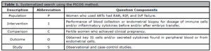
Table 1. Systematized search using the PICOS method.
ART: Assisted Reproductive Techniques; IVF: In vitro fertilization; RAR: Recurrent Pregnancy Loss; RIF: Recurrent Implantation Failure; IS: Immune System.
1. Studies Included: Clinical and case-control trials that addressed the proposed topic;
2. Publication Period: The selected studies were published between January 1, 2010, and April 24, 2022;
3. Language of the Studies: we analyzed papers in English, Portuguese (Brazil), and Spanish;
4. Intervention Used: Patients with RAR, RIF, and fertilization failures submitted to ART, who underwent before or after endometrial biopsy, treatments, or blood collection to study their immune profile; and
5. Study outcome: Obtaining the major IS cells and/or secreted cytokines found in peripheral blood or endometrial cells.
Database Search
We searched the “PubMed”, “Embase” and VHL databases. The search was done manually using a combination of previously selected keywords from the Medical Subjects Headings (MESH) database in the advanced search. The following keywords were used: (immune system) AND (embryo implantation) AND (women).
Inclusion and Exclusion Criteria
To filter the best results within the suggested scope of the review, we used the following inclusion and exclusion criteria:
Inclusion Criteria
1. Observational Studies; and
2. Case-Control Studies.
Exclusion Criteria
1. Articles out of scope;
2. Literature reviews;
3. Letter to the editor;
4. Abstracts;
5. Animal Studies; and
6. Case Reports.
Methodological Quality Assessment of Studies
To reduce the chance of bias among the selected papers we assessed the risk of methodological bias. To do so, we used the Newcastle Ottawa scale for bias assessment of observational studies. Studies considered to have a low risk of bias entered our review assessment. All data from the methodological evaluation are presented in Table 2.
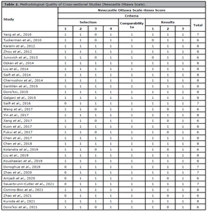
Table 2. Methodological Quality of Cross-sectional Studies (Newcastle Ottawa Scale).
Selection 1: Representativeness of the exposed cohort; Selection 2: Selection of the unexposed cohort; Selection 3: Exposure determination; Selection 4: Demonstration that the result of interest was not present in the baseline; Comparability 1a and 1b: Comparability of cohorts based on design or analysis; Results 1: Results assessment; Results 2: Follow-up of cohorts; Results 3: Adequacy of cohort follow-up.
Data Collection and Extraction
After screening the articles mentioned above, they were evaluated by four authors (MGA; RYM; FLDM; JOF). In case of divergence, a fifth author (NR) specialized in the field would reevaluate the paper and the results were tabulated. The data extracted from each paper were:
1. Day on which the sample was collected;
2. Number of patients;
3. Control group;
4. Age of patients;
5. Type of sample used; and
6. Immune cell mediators.
RESULTS
Methodological Screening
According to the methodological model used to formulate the review, we found 467 papers. Of these, only 31 were selected for the review. Overall, 1,800 patients with a mean age between 20 to 39.9 years were evaluated. The data are available in Table 1. It is worth pointing out that the primary sampling methodologies to identify immunological profiles of these patients were acquired from endometrial biopsies and peripheral blood sampling. Based on this, we extracted the data extraction from these two different methodologies. Figure 1 depicts the entire methodological screening.
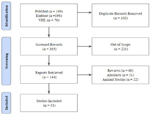
Figure 1. Methodological screening.
Immune Cell Mapping in Peripheral Blood
We found that in the peripheral blood of patients with implantation problems, there was an increase in cells compared to controls. The main increased cells were CD3+, CD4+, CD8+, CD16+, CD56+, CD57+, CD69+, CD154+, CD158a+, NKp46 (Yang et al., 2010; Karami et al., 2012; Chernyshov et al., 2014; Santillán et al., 2015; Dons’koi, 2015; Jiang et al., 2017; Dons’koi et al., 2021; Comins-Boo et al., 2021). On the other hand, CD4+, CD45+, Foxp3, and NKp46 cells were reduced in patients with reproductive issues, compared to control groups (Zhou et al., 2012; Fukui et al., 2017; Liu et al., 2019; Comins-Boo et al., 2021). Th1 type immune responses were also increased in two studies (Saifi et al., 2014; Kuroda et al., 2021).
Immune Cell Assessment in Endometrial Biopsy Samples
The literature also infers that implantation issues may modulate the immune response in the endometrium. There was a marked increase of cells in samples from these patients when compared to control groups. Among these, CD3+, CD4+, CD5+, CD8+, CD16+, CD25+, CD45+, CD56+, CD57+, CD68+, CD127+ (Tuckerman et al., 2010; Junovich et al., 2013; Santillán et al., 2015; Galgani et al., 2015; Wang et al., 2017; Kuon et al., 2017; Jiang et al., 2017; Chen et al., 2017; 2018; Zhao et al., 2020; 2021; Sauerbrunn-Cutler et al., 2021). In contrast, cells such as CD45+, CD56+, NKp46 and Foxp3 had reduced levels in endometrial biopsies from patients with RIF, RAR, or who resorted to ART (Jiang et al., 2017; Fukui et al., 2017; Sauerbrunn-Cutler et al., 2021).
Inflammatory cytokines in the peripheral blood of patients
In our review, there was an increase in inflammatory cytokines in the peripheral blood of patients with gestational complications, among which we list IL-6, IL-10, IL-17, INF-γ, TGF-ß, TNF-α (Ozkan et al., 2014; Saifi et al., 2014; Yin et al., 2017). In contrast, a group of inflammatory cytokines was reduced in patients with gestational complications in six studies, such as IL-4, IL-6, IL-10, IL- 35, FoxP3, TGF-ß, SOCS3 (Junovich et al., 2013; Zhou et al., 2012; Ozkan et al., 2014; Saifi et al., 2014; 2016; Koushaeian et al., 2019). All data are detailed in Table 3.
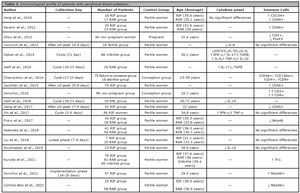
Table 3. Immunological profile of patients with peripheral blood collection.
CD: Cluster of Differentiation; RIF: Recurrent Implantation Failure; RAR: Recurrent Abortion Rates; LH: Luteinizing Hormone; Th: Helper T Cells; NKp: Natural Cytotoxicity Receptor Protein; and - : not available.
Inflammatory cytokines from endometrial biopsies
Two studies reported an increase in inflammatory cytokines in endometrial biopsies, such as IL-2, IL-6, IL-17, IL-22, IL-23, INF-A1, INF-B1, INF-γ, TNF- R (Galgani et al., 2015; Amjadi et al., 2020). In endometrial tissues, some inflammatory cytokines had their levels reduced in four studies, such as IL-6, IL-10, INF-γ, TGFß, TNF-α (Junovich et al., 2013; Galgani et al., 2015; Wang et al., 2017; Fukui et al., 2017). All data are detailed in Table 4.
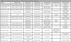
Table 4. Immunological profile of patients who underwent endometrial biopsy procedures.
CD: Cluster of Differentiation; RIF: Recurrent Implantation Failures; PRIF: Potential Recurrent Implantation Failures; RAR: Recurrent Abortion Rates; ET: Embryo transfer; LH: Luteinizing Hormone; NKp: Natural Cytotoxicity Receptor Protein; FoxP3+: Forkhead box P3 protein; and -- : not available.
Common Points Between Collections
To better plot, the commonalities between both collections, the main immune cells found in the studies were selected and placed in a Venn diagram, as shown in Figures 2 and 3. By plotting the main immune cells, we noticed that there was a common immune enhancement in both techniques in CD3 +, CD8 +, CD16 +, and CD57 + cells. In the cells found, there was an intersection point of the FoxP3+ phenotype. Interestingly, among the inflammatory cytokines in the endometrial biopsies used in the review, the only intersection point found in the study was the increase in IL-17 levels.
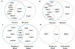
Figure 2. Venn diagram containing peripheral blood immune cells and embryo biopsy in RIF, RAR, and infertile patients. Caption: Immune cells from different types of collection. In blue: increase in cell quantity. In pink: Reduction of cell quantity. A: Peripheral blood immune cells. B: Immune cells derived from embryonic biopsies. C: Just enlarged cells between the two collection types. D: Just reduced cells between the two collection types.
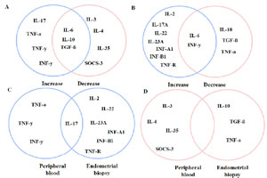
Figure 3. Venn diagram containing peripheral blood inflammatory cytokines and endometrial biopsies from RIF, RAR and infertile patients. Caption: Cytokines from different types of collection. In blue: increase in the number of cytokines. In pink: Reduction in the number of cytokines. A: peripheral blood immunological cytokines. B: Immune cytokines derived from embryonic biopsies. C: Only amplified cytokine pattern between the two types of collection. D: Only reduced cytokine pattern between the two types of collection.
DISCUSSION
In a successful pregnancy, the body must undergo many physiological processes that act directly on proper endometrial function and dynamic interaction between the endometrium and the blastocyst (Mrozikiewicz et al., 2021). This implantation process is somewhat inefficient in human reproduction, since about 25% of conceptual matings generate live births, and 5-15% can be identified clinically (Nowak et al., 2017). In this context, investigating biomarkers and clinical trials to determine or prospect possible treatment and/or immune abnormalities in these patients could effectively improve conception rates (Parhizkar et al., 2021).
One of the methodologies found in this search was the study of immune components from the peripheral blood of patients. Peripheral blood cell analyses consist mainly of evaluating lymphocytes and monocytes that are enabled in a wide range of assays, from the most straight forward, such as cytotoxicity assessments, to the most complex, such as functional and phenotypic evaluation of immune cells (Navas et al., 2019). Among the significant disadvantages of peripheral blood cell analyses is the similarity between immune responses in the body, which can lead to erroneous analysis and interpretation of data. However, non-generic-specific biomarkers can improve tissue specificity (Jogia et al., 2021). However, one of the main advantages of peripheral blood collection over other collection types is its better accessibility (Medrone Junior, 2009).
Another well-used methodology for the identification of immunological biomarkers was an endometrial biopsy. The primary purpose of this technique is to obtain a sampling of endometrial material to evaluate lesions of the endometrial lining and investigate other benign pathologies (Nicholls-Dempsey et al., 2018). One of the main disadvantages of endometrial biopsy is still the invasiveness of the technique. Although it is less harmful compared to other methodologies, such as laparoscopy, it still requires anesthesia or strong analgesics in some cases (Al-Jefout et al., 2007). However, the endometrial tissue structure has many distinct molecules that may contribute to adhesion, cell distribution, transit, and more specific signaling processes involved in implantation disorders (Marron et al., 2019).
RIF, RAR, and infertile patients, had higher T lymphocyte counts (CD3+, CD4+, CD8+, CD69+, CD154+) in their peripheral blood and (CD3+, CD4+, CD8+, CD25+, and CD127+) in endometrial biopsies (Yang et al., 2010; Chernyshov et al., 2014; Dons’koi, 2015; Galgani et al., 2015; Wang et al., 2017; Zhao et al., 2020). The literature has already described that T-type lymphocytes are located in the tissue stroma and glandular epithelium. These cells control the Th1 response involved in immune surveillance, preventing excessive trophoblast invasion and assisting in allograft immune tolerance (Zolfaghari et al., 2021). However, the literature also reports that in many cases of maternal anti-fetal rejection and gestational problems, there is an increased infiltration of cytotoxic T-lymphocytes into the fetal tissues (Kim et al., 2015). This fact was previously observed in the study by Quinn et al. (2011) who found an increased infiltration of cytotoxic CD8+ cells in women who had severe late preeclampsia.
Cells of the innate immune response have also been shown to be affected in cases of conception failure. The amount of CD68+ macrophages in endometrial biopsies increased in patients who intended to achieve clinical pregnancy from ART (Zhao et al., 2020; 2021). Under normal conditions, peripheral blood macrophages can reduce the invasiveness of trophoblast implantation. However, when these macrophages are dysregulated, there is no complete explanation for their effects on implantation (Han et al., 2021). In a previous study by Kim et al. (2008) there was an increase of macrophages in chorionic villi of patients with villitis of unknown cause, in addition to the macrophage infiltrate increase in the presence of CD8+ T cells in these patients.
One of the main unknowns in the literature is the impact of NK cells on embryo implantation. The attack of cytotoxic NK cells is immediate and does not require prior antigen preparation. Instead, these cells orchestrate the attack uniquely from interactions with receptors or inhibitory functions (Becker et al., 2016). An increase of NK cells in peripheral blood and endometrial biopsies (CD8+, CD16+, CD56+, CD57+, and CD158a+) in most studies (Tuckerman et al., 2010; Karami et al., 2012; Junovich et al., 2013; Chernyshov et al., 2014; Dons’koi, 2015; Santillán et al., 2015; Jiang et al., 2017; Kuon et al., 2017; Chen et al., 2017; 2018; Sauerbrunn-Cutler et al., 2021; Comins-Boo et al., 2021; Zhao et al., 2021). In a previous retrospective study evaluating the ovarian reserve of women undergoing cycles of ART, they found that the high rate of NK cells led to a reduction in ovarian reserve, which confirms the reproductive impacts shown previously (Hur et al., 2020). However, NK cells in the peripheral blood of healthy individuals can vary around 5%-29% depending on gender, stress, ethnicity, and age. Based on this, confirming their actual activity on fecundity it is still a controversial issue in the literature (Canella et al., 2021).
A curious finding was that some immune system cells adopted both a growth and a declining pattern in the different studies analyzed. These patterns occurred in dendritic cell lineages (CD86+) in the peripheral blood and NK cells expressing NKp46 receptors. In addition, regulatory T (Foxp3) and (CD4+) cells were also expressed in a lower amount in endometrial biopsies (Jiang et al., 2017; Fukui et al., 2017; Liu et al., 2019; Comins-Boo et al., 2021; Sauerbrunn-Cutler et al., 2021). As for the lower expression in dendritic cells, the literature already reports that this low amount of cells can lead to a reduction in IL-10 that controls tissue levels of effector T-cells that consequently infiltrate the endometrium (Kong et al., 2018). Recent studies have reported that reducing NK cells expressing NKp46 receptors on endometrial cells increases the high risk of gestational loss. However, their correlation with this loss is still unknown in the literature (Fukui et al., 2006; Takeyama et al., 2021).
Regulatory T-cells are defined by their expression of CD4+, CD25+, and the Foxp3 domain, which control the developmental function of naturally occurring T-reg populations (Heitmann et al., 2017). In this review, we found increased CD4+ cells in peripheral blood and a reduction in FoxP3. The primary function of these regulatory cells is endometrium modulation to receive the semi-allograft without complete immunosuppression during implantation, in addition to its protective function against opportunistic infections, which may risk the mother’s health (Royster et al., 2019). In a previous study, Teles et al. (2013) used mouse models with depletion for regulatory T-lymphocytes and reported that they had potential problems in embryo implantation, which is in tune with the findings of this review.
From the studies analyzed, we found a panel of cytokines sharply increased in the peripheral blood of the patients, including cytokines IL-6, IL-10, IL-17, INF-γ, TGF-ß, TNF-α (Ozkan et al., 2014; Saifi et al., 2014; Yin et al., 2017). However, according to a review by Chaouat et al. (2003), after the coitus phase until the apposition phase, the uterus is subjected to an inflammatory reaction with a transient cellular influx of lymphocytes and macrophages near the uterine lumen, leading to a high production of interleukins such as IL-6 and TNF-a, which could explain this change in peripheral blood cytokines, which also showed a reduction in IL-4, IL-6, IL-10, IL-35, FoxP3, TGF-ß, SOCS3 cytokines (Junovich et al., 2013; Zhou et al., 2012; Ozkan et al., 2014; Saifi et al., 2014; 2016; Koushaeian et al., 2019). On the other hand, when we checked the expression patterns of inflammatory cytokines in endometrial biopsies, IL-2, IL-6, IL-17, IL-22, IL-23, INF-A1, INF-B1, INF-γ, TNF-R were relatively increased in patients with reproductive complications (Galgani et al., 2015; Amjadi et al., 2020). This fact was reported by Saini et al. (2011) saying that Th1- cells, especially IL-2 and gamma interferon producers, play a crucial role in allograft rejection during fetal implantation. On the other hand, the levels of IL-6, IL-10, INF-γ, TGFß, TNF-α had their levels reduced (Junovich et al., 2013; Galgani et al., 2015; Wang et al., 2017; Fukui et al., 2017). This fact can also be explained by Chaouat et al. (2003) as previously mentioned, in which the reduction of cytokines by defense cells can lead to an unfavorable environment for embryo development, resulting in development imbalance and pregnancy loss. One of the main limitations of that study is the unspecificity of immune factors collected mainly from peripheral blood, as mentioned before, which may account for the increase and decrease in some immune cells (Jogia et al., 2021). Another factor that can lead to failures in these methodologies is the collections performed at different times, which can modulate the proliferation of immune cells and make it impossible to collect more homogeneous data and responses. Hormonal balance may be an interesting parameter to be addressed in further studies to investigate its correlation with the immune system and its implantation impacts on ART outcomes (Kim et al., 2015).
In conclusion, identifying immunological biomarkers may be an effective way to address fertility issues. Increased cytotoxic T-lymphocytes such as CD3+, CD25+, CD69+, CD127+, and CD154+ have been shown to correlate with fertility in patients. Innate immune response cells CD68+ were also increased in patients with problems to achieve pregnancy. CD8+, CD16+, CD56+, CD57+, and CD158a+ NK cells may exhibit a possible increase in the rates of reproductive problems. However, this finding is still uncertain in the literature. Regulatory T-cells such as Foxp3 have been shown to have the potential to aid in implantation immune tolerance, and their reduction has been seen in patients with reproductive disorders. In this context, further studies are needed to verify these cells’ main activity in the IS and identify functional biomarkers. In addition, the data presented in this review can be of great value for the design of studies aimed at developing methodologies, such as rapid tests that could predict the implantation rate of patients who resort to ARTs, enhancing the chances of clinical pregnancy success.
ABBREVIATIONS
• Recurrent implantation failure (RIF);
• In vitro fertilization (IVF);
• Medical Subjects Headings (MESH);
• Recurrent Abortion Rates (RAR);
• Assisted Reproductive Techniques (ART);
• Reporting Items for Systematic Reviews and Meta-Analysis (PRISMA) and
• Immune system (IS)
REFERENCES
Al-Jefout M, Andreadis N, Tokushige N, Markham R, Fraser I. A pilot study to evaluate the relative efficacy of endometrial biopsy and full curettage in making a diagnosis of endometriosis by the detection of endometrial nerve fibers. Am J Obstet Gynecol. 2007;197:578.e1-4. PMID: 18060940 DOI: 10.1016/j.ajog.2007.04.032 Medline
Amjadi F, Zandieh Z, Mehdizadeh M, Aghajanpour S, Raoufi E, Aghamajidi A, Aflatoonian R. The uterine immunological changes may be responsible for repeated implantation failure. J Reprod Immunol. 2020;138:103080. PMID: 32120158 DOI: 10.1016/j.jri.2020.103080 Medline
Bashiri A, Halper KI, Orvieto R. Recurrent Implantation Failure-update overview on etiology, diagnosis, treatment and future directions. Reprod Biol Endocrinol. 2018;16:121. PMID: 30518389 DOI: 10.1186/s12958-018-0414-2 Medline
Becker PS, Suck G, Nowakowska P, Ullrich E, Seifried E, Bader P, Tonn T, Seidl C. Selection and expansion of natural killer cells for NK cell-based immunotherapy. Cancer Immunol Immunother. 2016;65:477-84. PMID: 26810567 DOI: 10.1007/s00262-016-1792-y Medline
Canella PRBC, Barini R, Carvalho PO, Razolli DS. Lipid emulsion therapy in women with recurrent pregnancy loss and repeated implantation failure: The role of abnormal natural killer cell activity. J Cell Mol Med. 2021;25:2290-6. PMID: 33544456 DOI: 10.1111/jcmm.16257 Medline
Chaouat G, Lédée-Bataille N, Zourbas S, Ostojic S, Dubanchet S, Martal J, Frydman R. Cytokines, implantation and early abortion: re-examining the Th1/Th2 paradigm leads to question the single pathway, single therapy concept. Am J Reprod Immunol. 2003;50:177-86. PMID: 14629021 DOI: 10.1034/j.1600-0897.2003.00080.x Medline
Chen X, Mariee N, Jiang L, Liu Y, Wang CC, Li TC, Laird S. Measurement of uterine natural killer cell percentage in the periimplantation endometrium from fertile women and women with recurrent reproductive failure: establishment of a reference range. Am J Obstet Gynecol. 2017;217:680.e1-e6. PMID: 28935491 DOI: 10.1016/j.ajog.2017.09.010 Medline
Chen X, Liu Y, Cheung WC, Zhao Y, Huang J, Chung JPW, Wang CC, Li TC. Increased expression of angiogenic cytokines in CD56+ uterine natural killer cells from women with recurrent miscarriage. Cytokine. 2018;110:272-6. PMID: 29396049 DOI: 10.1016/j.cyto.2018.01.013 Medline
Chernyshov VP, Dons’koi BV, Sudoma IO, Goncharova YO. Favorable immune phenotype predicts successful implantation and pregnancy. Immunol Lett. 2014;162:217-21. PMID: 25445617 DOI: 10.1016/j.imlet.2014.10.022 Medline
Comins-Boo A, Cristóbal I, Fernández-Arquero M, Rodríguez de Frías E, Calvo Urrutia M, Pilar Suárez L, Gasca Escorial P, Ángel Herráiz M, Sánchez-Ramón S. Functional NK surrogate biomarkers for inflammatory recurrent pregnancy loss and recurrent implantation failure. Am J Reprod Immunol. 2021;86:e13426. PMID: 33811416 DOI: 10.1111/aji.13426 Medline
Donoghue JF, Paiva P, Teh WT, Cann LM, Nowell C, Rees H, Bittinger S, Obers V, Bulmer JN, Stern C, McBain J, Rogers PAW. Endometrial uNK cell counts do not predict successful implantation in an IVF population. Hum Reprod. 2019;34:2456-66. PMID: 31825483 DOI: 10.1093/humrep/dez194 Medline
Dons’koi BV. Accentuated hypoand hyper-NK lymphocyte CD8 expression is a marker of NK subsets’ misbalance and is predictive for reproductive failures. Immunobiology. 2015;220:649-55. PMID: 25576428 DOI: 10.1016/j.imbio.2014.11.015 Medline
Dons’koi BV, Osypchuk DV, Chernyshov VP, Khazhylenko KG. Expression of natural cytotoxicity receptor NKp46 on peripheral blood natural killer cells in women with a history of recurrent implantation failures. J Obstet Gynaecol Res. 2021;47:1009-15. PMID: 33368832 DOI: 10.1111/jog.14631 Medline
Fukui A, Ntrivalas E, Gilman-Sachs A, Kwak-Kim J, Lee SK, Levine R, Beaman K. Expression of natural cytotoxicity receptors and a2V-ATPase on peripheral blood NK cell subsets in women with recurrent spontaneous abortions and implantation failures. Am J Reprod Immunol. 2006;56:312-20. PMID: 17076675 DOI: 10.1111/j.1600-0897.2006.00431.x Medline
Fukui A, Funamizu A, Fukuhara R, Shibahara H. Expression of natural cytotoxicity receptors and cytokine production on endometrial natural killer cells in women with recurrent pregnancy loss or implantation failure, and the expression of natural cytotoxicity receptors on peripheral blood natural killer cells in pregnant women with a history of recurrent pregnancy loss. J Obstet Gynaecol Res. 2017;43:1678-86. PMID: 28815854 DOI: 10.1111/jog.13448 Medline
Galgani M, Insabato L, Calì G, Della Gatta AN, Mirra P, Papaccio F, Santopaolo M, Alviggi C, Mollo A, Strina I, Matarese G, Beguinot F, De Placido G, Ulianich L. Regulatory T cells, inflammation, and endoplasmic reticulum stress in women with defective endometrial receptivity. Fertil Steril. 2015;103:1579-86.e1. PMID: 25935494 DOI: 10.1016/j.fertnstert.2015.03.014 Medline
Han X, Li W, Li P, Zheng Z, Lin B, Zhou B, Guo K, He P, Yang J. Stimulation of α7 Nicotinic Acetylcholine Receptor by Nicotine Suppresses Decidual M1 Macrophage Polarization Against Inflammation in Lipopolysaccharide-Induced Preeclampsia-Like Mouse Model. Front Immunol. 2021;12:642071. PMID: 33995360 DOI: 10.3389/fimmu.2021.642071 Medline
Heitmann RJ, Weitzel RP, Feng Y, Segars JH, Tisdale JF, Wolff EF. Maternal T Regulatory Cell Depletion Impairs Embryo Implantation Which Can Be Corrected With Adoptive T Regulatory Cell Transfer. Reprod Sci. 2017;24:1014-24. PMID: 27834288 DOI: 10.1177/1933719116675054 Medline
Hur YJ, Yu EJ, Choe SA, Paek J, Kim YS. Peripheral blood natural killer cell proportion and ovarian function in women with recurrent implantation failure. Gynecol Endocrinol. 2020;36:922-5. PMID: 32162570 DOI: 10.1080/09513590.2020.1725969 Medline
Jiang R, Yan G, Xing J, Wu H, Liu Y, Wu H, Fan X, Zhou J, Ding L, Sun H. Abnormal ratio of CD57+ cells to CD56+ cells in women with recurrent implantation failure. Am J Reprod Immunol. 2017;78:e12708. PMID: 28543977 DOI: 10.1111/aji.12708 Medline
Jogia T, Kopp MA, Schwab JM, Ruitenberg MJ. Peripheral white blood cell responses as emerging biomarkers for patient stratification and prognosis in acute spinal cord injury. Curr Opin Neurol. 2021;34:796-803. PMID: 34608075 DOI: 10.1097/WCO.0000000000000995 Medline
Junovich G, Azpiroz A, Incera E, Ferrer C, Pasqualini A, Gutierrez G. Endometrial CD16(+) and CD16(-) NK cell count in fertility and unexplained infertility. Am J Reprod Immunol. 2013;70:182-9. PMID: 23648010 DOI: 10.1111/aji.12132 Medline
Kalem Z, Namli Kalem M, Bakirarar B, Kent E, Makrigiannakis A, Gurgan T. Intrauterine G-CSF Administration in Recurrent Implantation Failure (RIF): An Rct. Sci Rep. 2020;10:5139. PMID: 32198409 DOI: 10.1038/s41598-020-61955-7 Medline
Karami N, Boroujerdnia MG, Nikbakht R, Khodadadi A. Enhancement of peripheral blood CD56(dim) cell and NK cell cytotoxicity in women with recurrent spontaneous abortion or in vitro fertilization failure. J Reprod Immunol. 2012;95:87-92. PMID: 22854126 DOI: 10.1016/j.jri.2012.06.005 Medline
Kim JS, Romero R, Kim MR, Kim YM, Friel L, Espinoza J, Kim CJ. Involvement of Hofbauer cells and maternal T cells in villitis of unknown aetiology. Histopathology. 2008;52:457-64. PMID: 18315598 DOI: 10.1111/j.1365-2559.2008.02964.x Medline
Kim CJ, Romero R, Chaemsaithong P, Kim JS. Chronic inflammation of the placenta: definition, classification, pathogenesis, and clinical significance. Am J Obstet Gynecol. 2015;213:S53-69. PMID: 26428503 DOI: 10.1016/j.ajog.2015.08.041 Medline
Kolanska K, Suner L, Cohen J, Ben Kraiem Y, Placais L, Fain O, Bornes M, Selleret L, Delhommeau F, Feger F, Mathieu d’Argent E, Darai E, Chabbert-Buffet N, Antoine JM, Kayem G, Mekinian A. Proportion of Cytotoxic Peripheral Blood Natural Killer Cells and T-Cell Large Granular Lymphocytes in Recurrent Miscarriage and Repeated Implantation Failure: Case-Control Study and Meta-analysis. Arch Immunol Ther Exp. 2019;67:225-36. PMID: 31147723 DOI: 10.1007/s00005-019-00546-5 Medline
Kong X, Kong Y, Zhang F, Wang T, Zhu X. Expression and significance of dendritic cells and Th17/Treg in serum and placental tissues of patients with intrahepatic cholestasis of pregnancy. J Matern Fetal Neonatal Med. 2018;31:901-6. PMID: 28298162 DOI: 10.1080/14767058.2017.1300652 Medline
Koushaeian L, Ghorbani F, Ahmadi M, Eghbal-Fard S, Zamani M, Danaii S, Yousefi B, Jadidi-Niaragh F, Hamdi K, Yousefi M. The role of IL-10-producing B cells in repeated implantation failure patients with cellular immune abnormalities. Immunol Lett. 2019;214:16-22. PMID: 31442543 DOI: 10.1016/j.imlet.2019.08.002 Medline
Kuon R, Weber M, Heger J, Santillán I, Vomstein K, Bär C, Strowitzki T, Markert UR, Toth B. Uterine natural killer cells in patients with idiopathic recurrent miscarriage. Am J Reprod Immunol. 2017;78:e12721. PMID: 28639334 DOI: 10.1111/aji.12721 Medline
Kuroda K, Nakagawa K, Horikawa T, Moriyama A, Ojiro Y, Takamizawa S, Ochiai A, Matsumura Y, Ikemoto Y, Yamaguchi K, Sugiyama R. Increasing number of implantation failures and pregnancy losses associated with elevated Th1/Th2 cell ratio. Am J Reprod Immunol. 2021;86:e13429. PMID: 33835626 DOI: 10.1111/aji.13429 Medline
Liu B, Mariee N, Laird S, Smith J, Li J, Li TC. The prognostic value of uNK cell count and histological dating in the mid-luteal phase of women with reproductive failure. Eur J Obstet Gynecol Reprod Biol. 2014;181:171-5. PMID: 25150956 DOI: 10.1016/j.ejogrb.2014.07.010 Medline
Liu S, Wei H, Li Y, Diao L, Lian R, Zhang X, Zeng Y. Characterization of dendritic cell (DC)-10 in recurrent miscarriage and recurrent implantation failure. Reproduction. 2019;158:247-55. PMID: 31284267 DOI: 10.1530/REP-19-0172 Medline
Madkour A, Bouamoud N, Louanjli N, Kaarouch I, Copin H, Benkhalifa M, Sefrioui O. Intrauterine insemination of cultured peripheral blood mononuclear cells prior to embryo transfer improves clinical outcome for patients with repeated implantation failures. Zygote. 2016;24:58-69. PMID: 25613318 DOI: 10.1017/S0967199414000719 Medline
Marron K, Walsh D, Harrity C. Detailed endometrial immune assessment of both normal and adverse reproductive outcome populations. J Assist Reprod Genet. 2019;36:199-210. PMID: 30194617 DOI: 10.1007/s10815-018-1300-8 Medline
Mrozikiewicz AE, Ożarowski M, Jędrzejczak P. Biomolecular Markers of Recurrent Implantation Failure-A Review. Int J Mol Sci. 2021;22:10082. PMID: 34576245 DOI: 10.3390/ijms221810082 Medline
Navas A, Giraldo-Parra L, Prieto MD, Cabrera J, Gómez MA. Phenotypic and functional stability of leukocytes from human peripheral blood samples: considerations for the design of immunological studies. BMC Immunol. 2019;20:5. PMID: 30658588 DOI: 10.1186/s12865-019-0286-z Medline
Nicholls-Dempsey L, Kamga-Ngande C, Bélisle S, Lapensée L, Roy G, Tremblay C, Simard-Émond L. Endometrial Biopsy in an Outpatient Gynaecological Setting: Overinvestigation. J Obstet Gynaecol Can. 2018;40:1309-14. PMID: 29937135 DOI: 10.1016/j.jogc.2018.01.020 Medline
Nowak I, Wilczyńska K, Wilczyński JR, Malinowski A, Radwan P, Radwan M, Kuśnierczyk P. KIR, LILRB and their Ligands’ Genes as Potential Biomarkers in Recurrent Implantation Failure. Arch Immunol Ther Exp (Warsz). 2017;65:391-9. PMID: 28523429 DOI: 10.1007/s00005-017-0474-6 Medline
Ozkan ZS, Deveci D, Kumbak B, Simsek M, Ilhan F, Sekercioglu S, Sapmaz E. What is the impact of Th1/Th2 ratio, SOCS3, IL17, and IL35 levels in unexplained infertility? J Reprod Immunol. 2014;103:53-8. PMID: 24368037 DOI: 10.1016/j.jri.2013.11.002 Medline
Palmer SS, Barnhart KT. Biomarkers in reproductive medicine: the promise, and can it be fulfilled? Fertil Steril. 2013;99:954-62. PMID: 23246448 DOI: 10.1016/j.fertnstert.2012.11.019 Medline
Parhizkar F, Motavalli-Khiavi R, Aghebati-Maleki L, Parhizkar Z, Pourakbari R, Kafil HS, Danaii S, Yousefi M. The Impact of New Immunological Therapeutic Strategies on Recurrent Miscarriage and Recurrent Implantation Failure. Immunol Lett. 2021;236:20-30. PMID: 34090942 DOI: 10.1016/j.imlet.2021.05.008 Medline
PRISMA guide. Available at: <http://www.prisma-statement.org/PRISMAStatement>. Access in: May 25, 2022.
Qasemi M, Mahdian R, Amidi F. Cell-free DNA discoveries in human reproductive medicine: providing a new tool for biomarker and genetic assays in ART. J Assist Reprod Genet. 2021;38:277-88. PMID: 33421023 DOI: 10.1007/s10815-020-02038-4 Medline
Quinn KH, Lacoursiere DY, Cui L, Bui J, Parast MM. The unique pathophysiology of early-onset severe preeclampsia: role of decidual T regulatory cells. J Reprod Immunol. 2011;91:76-82. PMID: 21782252 DOI: 10.1016/j.jri.2011.05.006 Medline
Rocha YM, de Moura GA, Desidério GA, de Oliveira CH, Lourenço FD, de Figueiredo Nicolete LD. The impact of fake news on social media and its influence on health during the COVID-19 pandemic: a systematic review. Z Gesundh Wiss. 2021:1-10. PMID: 34660175 DOI: 10.1007/s10389-021-01658-z Medline
Royster GD, Harris JC, Nelson A, Castro Y, Weitzel RP, Tisdale J, Heitmann RJ, DeCherney AH, Wolff EF. Rapamycin Corrects T Regulatory Cell Depletion and Improves Embryo Implantation and Live Birth Rates in a Murine Model. Reprod Sci. 2019;26:1545-56. PMID: 30782087 DOI: 10.1177/1933719119828110 Medline
Saifi B, Rezaae SA, Tajik N, Ahmadpour ME, Ashrafi M, Vakill R, SoleimaniAsl S, Aflatoonian R, Mehdizadeh M. Th17 cells and related cytokines in unexplained recurrent spontaneous miscarriage at the implantation window. Reprod Biomed Online. 2014;29:481-9. PMID: 25154016 DOI: 10.1016/j.rbmo.2014.06.008 Medline
Saifi B, Aflatoonian R, Tajik N, Erfanian Ahmadpour M, Valiki R, Amjadi F, Valizade N, Ahmadi S, Rezaae SA, Mehdizadeh M. T regulatory markers expression in unexplained recurrent spontaneous abortion. J Matern Fetal Neonatal Med. 2016;29:1175-80. PMID: 26037627 DOI: 10.3109/14767058.2015.1039507 Medline
Saini V, Arora S, Yadav A, Bhattacharjee J. Cytokines in recurrent pregnancy loss. Clin Chim Acta. 2011;412:702-8. PMID: 21236247 DOI: 10.1016/j.cca.2011.01.002 Medline
Santillán I, Lozano I, Illán J, Verdú V, Coca S, Bajo-Arenas JM, Martinez F. Where and when should natural killer cells be tested in women with repeated implantation failure? J Reprod Immunol. 2015;108:142-8. PMID: 25708533 DOI: 10.1016/j.jri.2014.12.009 Medline
Sauerbrunn-Cutler MT, Huber WJ, Krueger PM, Sung CJ, Has P, Sharma S. Do endometrial natural killer and regulatory T cells differ in infertile and clinical pregnancy patients? An analysis in patients undergoing frozen embryo transfer cycles. Am J Reprod Immunol. 2021;85:e13393. PMID: 33501767 DOI: 10.1111/aji.13393 Medline
Sho T, Hachisuga T, Koi C, Kurita T, Kagami S, Kawagoe T, Matsuura Y, Yoshimura K, Hisaoka M. 17β-Estradiol induces proliferation of endometrial NK cells (CD56+) in postmenopausal women. Climacteric. 2017;20:571-6. PMID: 28933961 DOI: 10.1080/13697137.2017.1377173 Medline
Simon C, Gómez C, Cabanillas S, Vladimirov I, Castillón G, Giles J, Boynukalin K, Findikli N, Bahçeci M, Ortega I, Vidal C, Funabiki M, Izquierdo A, López L, Portela S, Frantz N, Kulmann M, Taguchi S, Labarta E, Colucci F, et al.; ERA-RCT Study Consortium Group. A 5-year multicentre randomized controlled trial comparing personalized, frozen and fresh blastocyst transfer in IVF. Reprod Biomed Online. 2020;41:402-15. PMID: 32723696 DOI: 10.1016/j.rbmo.2020.06.002 Medline
Singh N, Davis AA, Kumar S, Kriplani A. The effect of administration of intravenous intralipid on pregnancy outcomes in women with implantation failure after IVF/ICSI with non-donor oocytes: A randomised controlled trial. Eur J Obstet Gynecol Reprod Biol. 2019;240:45-51. PMID: 31228675 DOI: 10.1016/j.ejogrb.2019.06.007 Medline
Takeyama R, Fukui A, Mai C, Yamamoto M, Saeki S, Yamaya A, Shibahara H. Co-expression of NKp46 with activating or inhibitory receptors on, and cytokine production by, uterine endometrial NK cells in recurrent pregnancy loss. J Reprod Immunol. 2021;145:103324. PMID: 33930666 DOI: 10.1016/j.jri.2021.103324 Medline
Teles A, Schumacher A, Kühnle MC, Linzke N, Thuere C, Reichardt P, Tadokoro CE, Hämmerling GJ, Zenclussen AC. Control of uterine microenvironment by foxp3(+) cells facilitates embryo implantation. Front Immunol. 2013;4:158. PMID: 23801995 DOI: 10.3389/fimmu.2013.00158 Medline
Tuckerman E, Mariee N, Prakash A, Li TC, Laird S. Uterine natural killer cells in peri-implantation endometrium from women with repeated implantation failure after IVF. J Reprod Immunol. 2010;87:60-6. PMID: 20800899 DOI: 10.1016/j.jri.2010.07.001 Medline
van Hoogenhuijze NE, Torrance HL, Mol F, Laven JSE, Scheenjes E, Traas MAF, Janssen C, Cohlen B, Teklenburg G, de Bruin JP, van Oppenraaij R, Maas JWM, Moll E, Fleischer K, van Hooff MH, de Koning C, Cantineau A, Lambalk CB, Verberg M, Nijs M, et al. Endometrial scratching in women with implantation failure after a first IVF/ICSI cycle; does it lead to a higher live birth rate? The SCRaTCH study: a randomized controlled trial (NTR 5342). BMC Womens Health. 2017;17:47. PMID: 28732531 DOI: 10.1186/s12905-017-0378-y Medline
Wang WJ, Liu FJ, Zhang X, Liu XM, Qu QL, Li FH, Zhuang LL, Li XX, Hao CF. Periodic elevation of regulatory T cells on the day of embryo transfer is associated with better in vitro fertilization outcome. J Reprod Immunol. 2017;119:49-53. PMID: 28122284 DOI: 10.1016/j.jri.2017.01.002 Medline
Yang KM, Ntrivalas E, Cho HJ, Kim NY, Beaman K, Gilman-Sachs A, Kwak-Kim J. Women with multiple implantation failures and recurrent pregnancy losses have increased peripheral blood T cell activation. Am J Reprod Immunol. 2010;63:370-8. PMID: 20236264 DOI: 10.1111/j.1600-0897.2010.00811.x Medline
Yin B, Zeng Y, Wu T, Yu S, Xu J, Liu S, Diao L, Zhao Z, Liang D, Li Y. Functional properties of peripheral CD8+ T cells in patients with repeated implantation failure. Am J Reprod Immunol. 2017;78:e12704. PMID: 28508475 DOI: 10.1111/aji.12704 Medline
Zhao Y, Chen X, Zhang T, Chan LKY, Liu Y, Chung JP, Kwong J, Li TC. The use of multiplex staining to measure the density and clustering of four endometrial immune cells around the implantation period in women with recurrent miscarriage: comparison with fertile controls. J Mol Histol. 2020;51:593-603. PMID: 32857228 DOI: 10.1007/s10735-020-09908-2 Medline
Zhao Y, Man GCW, Wang J, Liu Y, Kwong J, Zhang T, Chung JPW, Wang CC, Chen X, Li TC. The identification of endometrial immune cell densities and clustering analysis in the mid-luteal phase as predictor for pregnancy outcomes after IVF-ET treatment. J Reprod Immunol. 2021;148:103431. PMID: 34627075 DOI: 10.1016/j.jri.2021.103431 Medline
Zhou J, Wang Z, Zhao X, Wang J, Sun H, Hu Y. An increase of Treg cells in the peripheral blood is associated with a better in vitro fertilization treatment outcome. Am J Reprod Immunol. 2012;68:100-6. PMID: 22687138 DOI: 10.1111/j.1600-0897.2012.01153.x Medline
Zolfaghari MA, Arefnezhad R, Parhizkar F, Hejazi MS, Motavalli Khiavi F, Mahmoodpoor A, Yousefi M. T lymphocytes and preeclampsia: The potential role of T-cell subsets and related MicroRNAs in the pathogenesis of preeclampsia. Am J Reprod Immunol. 2021;86:e13475. PMID: 34043850 DOI: 10.1111/aji.13475 Medline