JBRA Assist. Reprod. 2001;05(01):7-13
ARTIGO ORIGINAL
doi: 10.5935/1518-0557.2001.5.1.02
Apoptosis-related changes of fractionated human spermatozoa: effects of cryopreservation-thawing and ineubation conditions
Abstract
The objectives of these studies were: (1) to evaluate the effect of cryopreservation-thawing of human spermatozoa on DNA fragmentation and membrane integrity; and (2) to examine time dependent effects on membrane integrity and motion parameters of sperm incubated under capacitating conditions. This was a prospective, controlled cohort study. In experiment one, ejaculates from 16 men undergoing infertility evaluation (patients) and from 5 donors were examined. Purified sperm populations with high motility were prepared by gradient centrifugation, cryopreserved using a manual method and TEST-yolk buffer and glycerol (TYB-G), followed by quick thaw. Annexin V binding was used for assessing membrane translocation of phosphatidylserine (PS) and terminal deoxynucleotidyl transferase-mediated dUTP nick end labeling (TUNEL) was utilized for the evaluation ofDNA fragmentation. In experiment two, 11 ejaculates from patients and 5 from donors were examined. Purified fractions with high and low sperm motility (90% and 40% layers) were studied in order to assess PS translocation. In experiment three, we studied ejaculates from 16 patients and 5 donors; populations of sperm with high motility were prepared and incubated up to 24 hours under capacitating conditions with temporal assessment of motion parameters and annexin V binding. The results were as follows: Experiment 1: the percentage oflive cells with intact membranes (annexin V, Iive) was significantly reduced after cryopreservation-thawing, and cells with PS translocation. (annexin V, Iive) and necrosis increased significantly. TUNEL revealed percentages of cells with DNA fragmentation in the pre-freeze and post-thaw samples that were not significantly different. In experiment 2: the percentages oflive cells with PS translocation and of necrotic cells increased significantly after thawing in both fractions; however, such induction of PS translocation was significantly higher in the fractions with high sperm motility. In experiment 3: the percentage of live cells with intact membranes was significantly reduced mainly at 6-8 hours of incubation and cells with PS translocation and necrotic increased signi ficantly. We concl uded that cryopreservation-thawing of human sperm from patients and donors was associated with membrane change as revealed by membrane translocation of PS while having no major impact on DNA fragmentation. Prolonged incubation ofhuman sperm was associated with membrane PS translocation and a significant time-dependent motility loss.
Key Words: apoptosis, cryopreservation, DNA fragmentation, human spermatozoa, incubation, phosphatidylserine translocation.
Resumo
Os objetivos desse estudo foram avaliar (1) o efeito da criopreservação de esperma humano, a fragmentação do DNA e a integridade da membrana, (2) examinar os efeitos do tempo sobre a integridade da membrana e os parâmetros de motilidade do esperma incubado preparado em condições capacitantes.Este foi um estudo prospectivo e controlado. No experimento 1 foram avaliados a ejaculação de 16 homens que sofrem de infertilidade (pacientes) e 5 doadores. A população do esperma purificado com alta motilidade foram preparado por gradiente de centrifugação, criopreservado e usado no método manual e no Test-Yolk buffer e glicerol. (TYB-G), seguido de um rápido descongelamento. Annexin V binding foi usado para avaliar a translocação da membrana de phosphstidylsenine (PS) e terminal deoxynucleotidyl transferase-mediated dUTP nick end labeling ( cortar e rotular) (TUNEL) era utilizado para a validação do fragmento de DNA No experimento 2 foram avaliados ejaculação de 11 pacientes e de 5 doadores. Purificados e fracionados com alta e baixa motilidade (90% e 40% camada) foram estudados em ordem de avaliação PS translocação. No experimento 3 foram estudados a ejaculação de 16 pacientes e 5 doadores, população com esperma de alta motilidade foram preparados e incubados por 24 horas de acordo com as condições de capacitação com avaliação temporal de parâmetros de motilidade e Annexim V binding. Os resultados foram: experimento 1 - a porcentagem de células vivas com membrana intacta (annexin V, binding) foi significantemente reduzida após a criopreservação e as células com PS translocação e as células mortas aumentaram significantemente após. TUNEL revelou porcentagens de células com fragmentação de DNA em amostra pré congelamento e pós descongelamento que não foram significantemente diferentes. No experimento 2 as porcentagens de vida das células com PS translocação e células mortas aumentaram significantemente mais em frações com esperma de alta motilidade. No experimento 3 a porcentagem de células vivas com membranas intactas foram significantemente reduzidas principalmente entre 6-8 horas de incubação e células com PS translocação e mortas cresceram significantemente. Concluímos que a criopreservação de sêmen humano vindos de pacientes e doadores estavam associados com a mudança da membrana como revelado pela membrana da translocação de PS enquanto não tinham o principal impacto com fragmentação do DNA. Incubação prolongada de sêmen humano esta associada 'a membrana PS translocação e a um tempo significantemente dependente de motilidade menor.
Introduction
Evidence has been presented that membrane stress, phase transitions encountered by the plasma membrane during freezing-thawing making it more prone to fracture, and to a lesser extent lipid peroxidation, contribute to sub-lethal cryodamage in human spermatozoa (Alvarez & Storey 1992, 1993). Lipid diffusion through the plasma membrane is significantly compromised in frozen-thawed compared to fresh human sperm (James et al., 1999). The membrane phospholipid content decreases after cryopreservation with loss of phosphatidylcholine and phosphatidylethanolamine being the more pronounced (Alvarez & Storey, 1992). During cryopreservation-thawing, phospholipids that are normally located on the inner layer of plasma membrane have been reported to move to the outer layer. Frozen-thawed ram spermatozoa depicted translocation of phosphatidylglycerol to the outer layer (Hinkovska-Galcheva et al., 1989).
It has been demonstrated that PS is externalized in cells with membrane disturbance (Vermes et al., 1995). Plasma membrane PS externalization is widely accepted as an early marker of programmed cell death (Martin et al., 1995). Using annexin V (which can selectively bind translocated PS) and the vital dye propidium iodide, Glander and Schaller (1999) reported on the effects of cryopreservationthaw in human spermatozoa and Anzar et al. (2000) reported on bovine spermatozoa, and suggested that this assay can be used to detect membrane integrity of frozen-thawed spermatozoa.
Sperm cryopreservation may also affect DNA,nuclear protein interactions. Although cryopreservation had no effect on the chromosomal content of human spermatozoa (Martin et al., 1991), others have reported on the effects of freezing leading to changes of sperrn chromatin condensation using acridine orange, Feulgen-DNA cytophotometric analysis and aniline blue staining (Royere et al., 1988, 1991; Hamadeh et al., 1999). Pre-lytic DNA fragmentation is commonly accepted as a marker of programmed cell death. Various assays including the terminal deoxynucleotidyl transferase-mediated dUTP nick end labeling (TUNEL) have been used to evaluate DNA integrity of human spermatozoa (Manicardi et al., 1995; Sun et al., 1997; Aitken et al., 1998; Lopes et al., 1998; Hughes et al., 1999).
As released from the male reproductive tract, spermatozoa are incapable of fertilizing the oocyte. In vivo, sperrn require a period of residence in the female reproductive tract during which cellular changes take place that enable a successful gamete interaction. The changes are known collectively as capacitation (reviewed in Yanagimachi, 1994). Since the timing of ovulation and concomitant availability of fully capacitated sperrn cannot easily be orchestrated, efforts to establish conditions that improve sperm longevity and functional status in vivo are desirable. During capacitation, sperrn undergo a variety of metabolic and functional changes as well as membrane structure modifications that lead to a decrease in membrane stability (Nolan et al., 1995). Prolonged incubation of stallion and mouse sperm under different in vitro (capacitating and non-capacitating) conditions leads to loss of motility and viability (Weil et al., 1998; Bedford et al., 1999). This phenomenon is also observed when incubating human spermatozoa (Aitken et al., 1996). Such changes may be the consequence of metabolic alterations due to the culture conditions; however, it has also been suggested that they may be secondary to the triggering of an internally controlled, programmed cell death mechanism (Weil et al., 1998).
In the present study, we aimed to evaluate the effects of cryopreservation-thawing on membrane and DNA integrity, as well as the temporal effects of sperm incubation at body temperature on plasma membrane integrity and motion characteristics of human spennatozoa. We used the annexin V binding assay for assessing plasma membrane externalization of PS and the TUNEL assay for evaluation of DNA integrity, and monitored sperm motion parameters with a computer-assisted semen analyzer (CASA).
Materials and methods
Patients Inclusion Criteria and Study Design
These studies were performed under approval of the Institutional Review Board at Eastem Virginia Medical School. In all experiments semen specimens were collected after a 2-4 day sexual abstinence period. All patients suffered from primary infertility more than a one-year duration, had a normal physical examination and were nonsmokers. All ejaculates had less than 1 x 10 leukocytes/mL of semen (peroxidase staining) (World Health Organization - WHO, 1999). In order to provide sufficient number of cells for all tested, subjects with a concentration of motile spermatozoa >20x 106 /mL in the original sample were included. After semen liquefaction for 30 min at room temperature, a basic semen analysis (concentration, motility and morphology) was performed.In experiment 1 ejaculates from men undergoing evaluation for infertility (n= 16 patients) at our andrology laboratory (study group) and ejaculates from 5 fertile men participating in our artificial insemination donor (AIO) program (control group) were examined independently. This was immediately followed by discontinuous Percoll gradient centrifugation in order to isolate the purified populations with high sperm motility (90% layer). Pre-freeze evaluation of motion parameters, annexin V binding assay and lUNEL assay were performed. In experiment 2,11 ejaculates from patients and 5 fTOm donors were examined independently. Pre-freeze evaluation of motion parameters, annexin V binding assay were performed in the fractions with high and low sperm motility (90% and 40% layers fractions). In experiment 3, sixteen patients and 6 donors were examined and followed by discontinuous gradient centrifugation isolating high motility sperm (90% layer). Annexin V binding assay and motion parameters were analyzed at 0, 1, 3, 6 and 24 hours while incubating at body temperature under capacitating conditions.
Preparation ofspermatozoa and basic semen analysis
Sperrn concentration and motion parameters (progressive motility, curvilinear velocity [μm/sec],percentage of rapid cells [i e., % sperrnatozoa with > 50μm/sec] and linearity) were objectively evaluated using the HTM-IVOS semen analyzer (Hamilton 1borne Research, Beverly, MA, USA) with fixed parameter settings (Oehninger et al., 1990). Sperm concentration and motility readings were manually monitored and corrections were made as appropriate. Sperm concentration and motility were assessed according to the WHO criteria (1999), and sperm morphology was examined according to strict criteria after Diff-Quik staining (Kruger et al., 1988).The sperm fractions were isolated using discontinuous Percoll (Sigma Chemical Co,St. Louis, MO,USA) gradient separation (90% and 40% layers). Up to 2 mL of semen was carefully placed on Percoll layers, centrifuged at 380xg for 20 minutes and the fractions from the 40% and 90% layers were carefully washed with human tubal fluid (HTF; Irvine Scientific) supplemented with 0.3% human serul11 albumin (HSA; Irvine Scientific) at 380xg for 10 mino The supernatant was discarded and the pellet was resuspended in 0.5 mL ofHTF supplemented with 0.3% HSA.
Detection of Membrane Phosphatidylserine Translocation
During early stages of membrane disturbance PS is translocated to the outer portion of plasma membrane, eventually becoming available for binding to annexin V. In the present experiments we used annexin V Cy3. 18 (Annexin V.Cy3; Sigma, USA) for detection of PS externalization with simultaneous assessment of cell viability. In order to differentiate between live cells with and without PS translocation and necrotic cells, we used 6-carboxyfluorescein diacetate (6-CFDA) in combination with Ann V Cy3. The non-fluorescent 6-CFDA enters the cell and is converted to the fluorescent compound 6- carboxyfluorescein (6-CF). This conversion is a function of the esterases present only in the living cells. Thus, no fluorescence can be observed in the necrotic (dead) cells.By fluorescence microscopy, 6-CF is observed as green fluorescence and Ann V.Cy3 as red. Three patterns of fluorescence are observed: 1. Live, normal cells that stain only with 6-CF (green); 2. Live cells with translocation of membrane phosphatidylserine that stain with both 6-CF (green) and Ann V Cy3 (red); 3. Necrotic or dead cells that stain only with Ann VCy3 (red).Sperm suspensions were washed twice with phosphate buffered saline (PBS; Irvine) and adjusted to 10 X 106/mL. A fifty μL aliquot of sperm suspension was placed on a poly-L-Iysine-coated slide and stained with 6-CFDNAnn V.Cy3 solution in the presence of calcium. After incubation in the dark for 10 min, the slide was covered with a 24 x50 mm cover slip and immediately read blindly by two observers using a magnification of 600x by epifluorescence microscope with an ultraviolet filter. At least one hundred spermatozoa were counted per slide. Our laboratory has reported an intraobserver variability of < 6% and an inter-observer vaniability of< 3% for the technique (Barroso et al., 2000; Duru et al., 2001 a; Schuffner et al., 2001 a).
Detection ofDNA Fragmentation
DNA cleavage may yield doublestranded, low molecular weight DNA fragments as well as single strand breaks in high molecular weight DNA. Such DNA strand breaks can be detected by TUNEL. Here, we used the In Situ Cell Death Detection kit (Boehringer Mannheim, Indianapolis, IN). Briefly, the purified motile sperm fractions were washed two times in PBS supplemented with 0.3% HSA, and the concentration was adjusted to 20x 106 cells/mL. One hundred f.iL of sperm suspension was fixed with 100 μL of 4% paraformaldehyde for 30 minutes at room temperature and washed in PBS supplemented with 0.3% HSA. Cells were permeabilized using 0. 1 % Triton X- 100 in 0. 1 % sodium citrate for 2 minutes at room temperature and washed two times in PBS supplemented with 0.3% HSA. Then, spermatozoa were incubated with the TUNEL reaction mixture (calf thymus TdT plus dUTP label) in the dark at 37°C for 1 hour and washed two times in PBS supplemented with 0.3% HSA. The final pellet was transferred to a final volume of 250 μL in PBS. Fifteen μL of sperm suspension was put on a slide, mixed with 5 μL of anti-fade solution, covered with a slip and analyzed immediately using epifluorescent microscopy as described for PS translocation detection.Each cell was assigned as having DNA fragmentation (intense green fluorescence) or normal (no fluorescence). For each test, one aliquot of fixed and permeabilized spermatozoa were incubated either with only dUTP label solution (negative controls) or using only DNAse 1,1 mg/mL, for 10 min at room temperature (positive controls). At least 100 cells were analyzed by two investigators in duplicate slides and results averaged. Our laboratory has reported intra-observer variability < 8% and an inter-observer variability < 7% for the technique (Barroso et al., 2000).
Sperm Cryopreservation
The techniques for freezing-thawing have been previously described (Morshedi 1996; Srisombut et al., 1998). Briefly, sperm suspensions were mixed slowly with freezing medium containing TES-TRIS citrate and 20% egg yolk, with 12% glycerol as cryoprotectant (TYB-G) at a final concentration of 4.5% (Irvine Scientific, USA) in a drop-wise fashion until a i : 1/2 volume ratio of sperm suspension to freezing medium was attained in 10 mino Aliquots were then placed in 2 mL cryovials at a volume of 0.4 mL/vial, refrigerated for 1 h. Subsequently, the vials were immersed in liquid nitrogen vapor for 30 min before finally being plunged in liquid nitrogen at -196°C. The specimens were thawed in a water bath at 40°C for 3 min after 24 h of cryostorage.
Statistical Analysis
Parametric and non-parametric paired and unpaired t-tests, Spearman rank correlations and analysis ofvariance (ANOVA) were used as appropriate. Data are presented as mean ± standard error of the mean. The level of significance was set at<0.05.
Results
The mean basic semen parameters ofpatients (n= 16) included in experiment 1 were as follows: concentration, 76±14 (x 106mL); motility, 70±6 (%); concentration motile, 49± 13 (x 106/mL); curvilinear velocitv, 42±2 (flm/s); linearity, 62±2; and normal morphology, 8±2 (%). The mean basic semen parameters of donors (n=5) included in the study were as follows: concentration, 116±21 (x 106/mL); motility, 74±4 (%); concentration motile, 88±18 (x 106 /mL), curvilinear velocity, 66±6 (μm/s); linearity, 62±2; and normal morphology, 18±5 (%). Results ofpre-freeze and post-thaw motion parameters as well as cryosurvival rate of experiment 1 are depicted in Table I. In both groups (patients and controls), and after separation of the fractions with high sperm motility (90% Percoll layers), all postthaw motion parameters (except linearity) were significantly lower than the corresponding pre-freezing values. The cryosurvival rate (expressed as post-thaw/prefreeze ratio of percentage of motility) was lower in the patient group but the difference did not attain significance.Table II presents annexin V binding and TUNEL results of the cohorts studied comparing pre-freezing versus post-thaw using the standard TYB-G protocol in experiment 1. The percentage oflive cells with intact membranes (Ann V, live, normal) was significantly reduced after cryopreservation-thawing in the study and control groups (P<0.0001 .and P=0.0005, respectively). The percentage oflive cells with PS externalization (Ann V+, live) increased significantly in the post-thaw samples of patients (P=0.002) and controls (P= 0.001 ). There was also a significant increase in the percentage of necrotic (dead) cells in post-thaw samples (P< 0.0004 and P= 0.007, respectively, for the patient and the control groups).
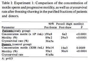
Table I. Experiment 1: Comparison of the concentration of motile sperm and progressive motility, as well as cryosurvival rate after freeziilg-thawing in the purified fractions of patients and donors.
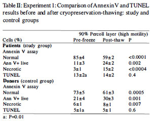
Table II. Experiment 1: Comparison of Annexin V and TUNEL results before and after cryopreservation-thawing: study and control groups
TUNEL results (Table II) revealed no significant differences in the percentages of cells with DNA fragmentation in the pre-freeze and post-thaw samples of both patient and donor groups. Of note, the percentage of TUNEL positive cells was significantly higher in the pre-freeze motile sperm fractions of patients than donors (P= 0.01).
There was no significant correlation between PS translocation or TUNEL results (pre-freeze or post-thaw) and motility or loss of progressive motility following cryopreservation thawing.
Table III shows experiment 2 results; before freezing, the percentage of annexin V+ live cells and necrotic cells were significantly higher in the 40% fractions when compared with those in the 90% fractions (P<0.05). For both fractions with those in the 90% fractions (P<0.05). For both fractions with high and low sperm motility, the post-thaw percentage of normal cells decreased significantly, whereas the percentage of cells with membrane PS translocation and necrosis increased significantly when compared with pre-freeze values. Similar results were observed n the control group.
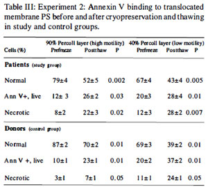
Table III. Experiment 2: Annexin V binding to translocated membrane PS before and after cryopreservation and thawing in study and control groups.
In experiment 3, as can be observed in Table IV considering the results of the annexin V binding assay, in both patients and donors there was a similar pattern of change over time. In the patientgroup the changes in annexin V binding were more evident in the first 6 hours as follows: decrease in the percentage of normal cells (P<0.001), increase in the percentage of cells with membrane PS translocation (annexin V+, live) and necrotic cells (P<0.001). In the donor group the decrease in the percentage ofnormal cells was significant only at 24 hours (P=0.002) and the increase in the percentage of cells with membrane PS translocation (annexin V, tive) (P=0.007) was marked by 6 hours of incubation. Overall, patients had higher percentages of PS translocation, particularly at 24 hours, than donors (P=0.01). T here was no difference in the percentage of necrotic cells between donors and patients (P=0.9).
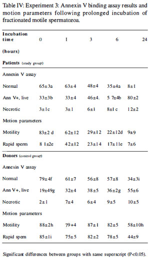
Table IV. Experiment 3: Annexin V binding assay results and motion parameters following prolonged incubation of fractionated motile spermatozoa.
In regard to motion parameters (Table IV), in donors and patients, motility and rapid cells decreased over time. In the patient group, motility and rapid cells decreased more pronouncedly by 6 hours (P=O.OO5 and P=0.OO4, respectively); on the other hand, in the donor group this decrease in motility and rapid cells was observed only by 24 hours (P=0.05 and P=0.02, respectively). In patients, there was a significant negative correlation between the decrease of motility and the increase of membrane PS translocation over time (r=0.47, P=0.02).
Significant differences between groups with sarne superscript (P<0.05).
Discussion
In the present study, we first evaluated the effects of cryopreservation-thawing (using a standard protocol of manual quick freezing with TYB-G and quick thawing) on both membrane and DNA integrity of fractionated, motile fractions of human spermatozoa. The clinical preparation of cryopreserved semen using intra-uterine insemination-ready processed samples by washing-centrifugation or gradient separation has recently been advocated (Sharma & Agarwal, 1996; Larson et al., 1997). Our results indicated that cryopreservation-thawing altered plasma membrane symmetry and was associated with translocation of PS while DNA integrity was maintained. In regard to the prolonged incubation under capacitating conditions we observed an increase on PS translocation associated with motility loss.
It has been reported that annexin V binding combined with propidium iodide staining is more sensitive in detecting a deterioration of membrane functions than the vital stain propidium iodide alone (Glander & Schaller, 1999). Propidium iodide is normally a membrane-impermeable dye. However, dead cells lose their resistance to the influx of propidium resulting in intracellular staining. In our study, we used annexin V in combination with 6-CFDA; this compound is converted to 6-CF by esterases in the living cells, which allows for a clear distinction under epifluorescent microscopy of live-normal cells, live cells with altered membranes (here depicting PS translocation) and necrotic (dead) éells. We observed that annexin V stained the entire spermatozoon (head, midpiece and flagellum) in more than 90% of the annexin V+ live cells on a siide (data not shown). We postulate that PS is therefore translocated to the outer leaflet at different domains of the cell membrane.
We showed that the percentage of annexin V+ live cells increased significantly fo llowing freezi ng-thawing as compared to prefreeze values (Duru et al., 2001 a,b; Schuffner et al., 2001 b). These cells are alive, but they have lost membrane integrity as demonstrated by translocation of PS. Additionally, the cryopreservation-thawing process also induced necrosis. Therefore, freezing-thawing significantly reduced the percentage of normal cells. Whether this is true also for samples from infertile men with more severe degrees of oligoasthenoteratozoospermia needs to be evaluated.
It has been suggested that an aminophospholipjd translocase mediates specifically a rapid ATP-dependent translocation of aminophospholipids from the exoplasmic to the cytoplasmic leaflet (Muller et al., 1999). In cryopreserved ram spermatozoa, the sequestering of endogenous PS to the cytoplasmic leaflet is maintained in intact cells, but not in impaired cells. Furthermore, in this system, post-thaw activity of the putative aminophospholipid translocase was significantly reduced in intact cells (Muller et al., 1999). If the activity of this putative enzyme is inhibited in cryopreserved human spermatozoa, an increased extemalization of PS is to be expected.
The present experiments indicated no significant impact on DNA fragmentation (as measured by TUNEL) after freezing-thawing using the standard TYB-G protocol . Although the cohort of patients studied herein did not differ from the donor group in terms of the basic sperm parameters (except morphology), this group of men had significantly higher basal levels of DNA fragmentation than controls. Our . studies focused on examination of samples readily following thawing. More studies are needed to address delayed (time-dependent) effects on chromatin-DNA integrity.
In both donors and patients, the percentages of motility and rapid cells decreased significantly over time when incubation was prolonged for 24 hours under capacitating conditions. Greater detrimental changes were observed in patients as compared to donors. Motility loss was associated with membrane translocation of PS. Such spermatozoa depicting viability yet PS extemalization. may represent cells undergoing apoptotic changes. The question remains, consequently, whether ejaculated human spermatozoa undergo programmed cell death, and if so, which mechanisms are operati ve. Loss of viability under culture conditions could also be secondary to a lethal cell dysfunction resulting from metabolic disturbances due to the culture environment. Alternatively, such membrane changes may be associated with events that render spermatozoa ready for fertilization.
In conclusion, cryopreservation-thawing of motile sperm from patients and donors using standard methodology (manual freezing with TYB-G) was associated with plasma membrane translocation of PS, an expression of membrane alteration and perhaps a damage. Conversely, freezing with TYB-G had no obvious impact on DNA fragmentation as assessed by TUNEL. Prolonged incubation of human sperm was associated with membrane PS translocation and a significant time-dependent motility loss.