JBRA Assist. Reprod. 2023;27(3):436-441
ORIGINAL ARTICLE
doi: 10.5935/1518-0557.20230004
Effect of progesterone administration on tissue mast cell population and histamine content in mice uterus after ovulation induction
1Razi Herbal Medicines Research Center, School of Medicine, Lorestan University of Medical Sciences, Khorramabad, Iran
2Social Determinants of Health Research Center, School of Medicine, Lorestan University of Medical Sciences, Khorramabad, Iran
CONFLICTS OF INTEREST
The authors declare that they have conflict of interest.
ABSTRACT
Objective: Mast cell population and histamine affect on blastocyst implantation. This study aimed to evaluate the effects of progesterone administration after induction of ovulation on the uterine tissue mast cell population and histamine content in mice.
Methods: We ran an experimental study on three groups of mice; control group, ovulation induction (induction group), and ovulation induction along with progesterone administration (progesterone group). Mast cells were counted using toluidine blue staining, and the histamine level was measured through spectrophotometry.
Results: According to the analysis of variance (ANOVA), there was no difference in mast cell population in endometrium (p=0.138) nor in myometrium (p=0.611). The ratio of mast cells in the myometrium per endometrium increased in the progesterone group in comparison to the control group based on a generalized linear model (p=0.041). The uterine histamine level was different between the groups, based on the ANOVA (p=0.039), in which the progesterone group had lower amounts of histamine.
Conclusions: Progesterone administration after ovulation induction did not decrease the number of endometrial mast cells and could have increased the ratio of myometrium mast cells per endometrium mast cell. The histamine level in uterus decreased by the administration of progesterone in the ovulation-induced mice.
Keywords: progesterone, histamine, ovulation, assisted reproduction
INTRODUCTION
Ovarian stimulation is used in infertility treatments and results in simultaneous growth of several follicles during a one month cycle (Gad et al., 2011). The uterus is made up of endometrium, myometrium and perimetrium. The endometrium is a unique tissue that undergoes proliferation, differentiation, destruction and repair. Such processes are necessary for embryo implantation. These cycles are affected by sexual steroid hormones (King & Critchley, 2010).
Mast cells (mastocytes) are a part of the innate immune system, stemming from the bone marrow. The major source of tissue histamine is the mast cell and they mature in their targeted tissues (Witkowski et al., 2015). The role histamine released from the uterine mast cells is notable upon implantation, pregnancy and delivery (King & Critchley, 2010). Histamine derived from the uterus is known as a major regulatory agent playing a role in implantation through the induction of vascular permeability and decidualization (Jensen et al., 2010). Female sexual hormones affect mast cell function. Estradiol and progesterone result in mast cell maturation, especially maturation of uterine mast cells (King & Critchley, 2010). These hormones also affect mast cell activation (Zierau et al., 2012). Reduction of mast cells upon implantation is associated with increased estrogen, believing that implantation time decidualization is regulated by the histamine released from mast cells (Harvey, 1964). It has been reported that mast cells in humans and rodents are necessary for ovulation, implantation of blastocysts and placentation. In addition, mast cells support decidual repair and uterine contractions (Meyer et al., 2018). Woidacki et al. (2013) mentioned that mast cells had receptors for estradiol and progesterone. Hamouzova et al. (2016) mentioned that the most number of mast cells was seen when estradiol levels were at their upper limit (Hamouzova et al., 2016). Noor et al. (2010) mentioned that histamine affects implantation, ovulation, regulation of secretions of placental blood and myometrium contraction.
Sexual hormones balance change during ovulation induction, and considering the roles of mast cells and histamine in the implantation process, the present study aimed to investigate the effects of progesterone administration after ovulation induction on the uterine tissue mast cell population and histamine content in mice.
MATERIALS AND METHODS
Study design and groups
We ran this experimental study on 15 NMRI mice aged 6-8 weeks. The animals were kept for two estrus cycles under standard condition. Then, they were broken down into three groups, including the control group, ovulation induction group (called induction group), and a group for progesterone administration after ovulation induction (called progesterone group). Concerning the control group (n=5), pseudo pregnancy was performed through vaginal swabbing, and after 3.5 days the animals were slaughtered for study. For induction group (n=5), a single dose of 10 IU human menopausal gonadotropin (hMG) hormone was injected intraperitoneally (IP), followed by a single dose of 10 IU human chorionic gonadotropin (hCG) hormone IP injection after 48 hours to induce ovulation. Then pseudo pregnancy was performed and after 3.5 days the animals were slaughtered for the study. Concerning the progesterone group (n=5), the process in the induction group was performed followed by daily administration of 1 mg subcutaneous (SC) progesterone for up to 3.5 days (Narimani et al., 2020). Ethical guidelines for working with laboratory animals were followed and the protocol of this study was approved in the ethics committee of Lorestan University of Medical Sciences under registration number IR.LUMS.REC.1399.300.
Tissue study and mast cell count
In order to take samples, the animals were slaughtered by neck dislocation after general anesthesia. One uterus horn was used for tissue study and the other horn was used for histamine analysis. For tissue study, the mid one-third of uterus horns were separated and fixed in formalin solution for 24 to 48 hours. The paraffin processing was performed manually. After tissue processing, the samples were embedded horizontally in paraffin in order to make cross-sections from paraffin blocks. Then 5μm serial sections were prepared in which each 5 consecutive sections were used for one staining method as sections 1 - 5 for hematoxylin and eosin (H&E), sections 6 - 10 for periodic acid Schiff (PAS), sections 11 - 15 for toluidine blue staining and this sequence was repeated.Toluidine blue staining was performed to stain mast cells; H&E and PAS staining were used for qualitative morphological study of decidualization. After dewaxing, the samples were stained, and then they were dehydrated in ascending alcohol concentrations, clear in xylene and mounted with a cover glass. For H&E staining, the samples were put in hematoxylin Harris that we prepared in house according to Suvarna et al. (2018) from hematoxylin crystal (No. 1.04302.0025, Merck, Germany), for 5 min, and after we washed in water, acid alcohol, water again, lithium carbonate 1% (No. 1.05680.0520, Merck, Germany); water again; then the samples were put in eosin - prepared from dry eosin (No. 1.15935.0025, Merck, Germany) according to Suvarna et al. (2018) for 2 min. PAS staining was performed in house according to Suvarna et al. (2013). For toluidine blue staining, 1 g toluidine powder (No. 1.15930.0025, Merck, Germany) was mixed with 20 ml alcohol 95%, 80 ml distilled water and 1 ml glacial acetic acid. The concentration of toluidine blue was 1% with Ph 1.0 - according to the literature (Karaca et al., 2008; Liu et al., 2012).To count mast cells per area, 10 sections were studied in different fields as the morphometric method of using a square frame 8.5×8.5 cm with magnification ×400 a under LEICA microscope (DM500, ICC50 HD, Germany), using Leica Applications Suit LAS ES Version 3.4.0 (Leica microsystems limited, 2016) software. The count process was performed in the endometrium and the myometrium. The count was done per area (later in the paper this variable is called mast cell count instead of mast cell count per area). The morphometric protocol used was that discussed by Gundersen et al. (1988).
Tissue histamine level
After separating the samples and clearing fat and extra tissues, they were kept temporarily at -70ºC until histamine analysis. The samples were thawed and homogenized as follows: the samples were added four folds (volume wise) of sodium chloride 0.85% (1:4 v/v) in homogenizer (ULTRA TURRAX, IKA-T18 basic, Germany). Then the samples were passed through a filter. Thereafter, the samples were centrifuged at 12000 rpm for 10 min. We used a cold reagent: 1.5 ml of sulphanilic acid (0.9% w/v) (No. 8.22338.1000, Merck, Germany) in 4% hydrochloric acid (No. 1.00317.2500, Merck, Germany), plus 1.5 ml of sodium nitrite 5% w/v poured into a 50 ml volumetric container and kept in an ice bath for 5 minutes. Some 6 ml of 5% sodium nitrite was added and then cooled to 25 ml with cooled distilled water and kept in an ice bath for 15 minutes. Measurement method: 5 ml of sodium carbonate 1.1% (NO.1.06398.1000, Merck, Germany) was slowly mixed with 2 ml of cold reagent and then 1 ml of the homogenized sample was added to it, after 5 minutes of absorption at 496 wavelength, it was read by a spectrophotometer. Distilled water was used as a blank (histamine was prepared as standard from 0-100μg/ml and measured). We used a colorimetric method to evaluate the histamine levels using a spectrophotometer (Cecil instrument CE2020, Serial Number 93183) at a wavelength of 496 nm. Histamine was used as the standard from 0 - 100 µg. Then the histamine level was reported at the unit of µg/g-tissue.
Statistical analysis
The sample size was calculated based on the “law of diminishing returns”, proposed by Charan & Kantharia (2013) for animal studies. Accordingly, we had five samples for each of the three groups, which resulted in the acceptable range of 10 to 20 degrees of freedom for variance analysis (ANOVA) (Arifin & Zahiruddin, 2017; Charan & Kantharia, 2013). Therefore, we used the one way ANOVA with Tukey post hoc test to compare the variability of the means between the groups of study. When non-significant, we used the generalized linear modeling with Poisson family for such cases. Non-parametric tests did not have enough power for this sample size. P=0.05 was considered as the significance level. We used the software packages SPSS24 (IBM, US) and Stata14 (Stata Corp. LLC, US) to analyze and plot the data.
RESULTS
Qualitative study of the tissues showed successful decidualization in progesterone group (Figure 1, A and B). The mast cells were counted in endometrium and myometrium (Figure 1, C and D). The means of mast cell count in the groups of study were compared. According to the ANOVA, the differences of the means were not significantly greater than the differences existed within the groups neither in endometrium (p=0.138) nor in myometrium (p=0.611) (Table 1, Figure 2).
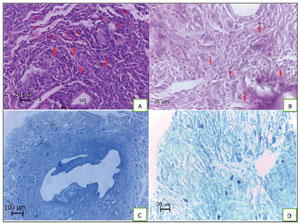
Figure 1. Morphology of uterus samples. Micrographs A: UG indicates uterine glands, S indicates sinusoid and the arrows indicate decidual cells (H&E, ×400). Micrographs B: decidualization of endometrium and the arrowed cells indicate large granular inflammatory cells (PAS, ×400). Micrographs C and D: The dark blue cells indicate mast cells (toluidine blue, ×100 [C] and ×400 [D]). Distribution of mast cells is seen that most of them are in myometrium.
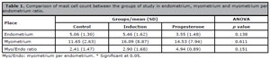
Table 1. Comparison of mast cell count between the groups of study in endometrium, myometrium and myometrium per endometrium ratio.
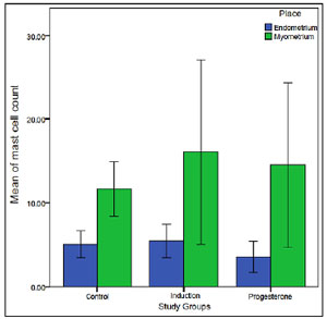
Figure 2. The mean of mast cell count per area among the groups and places. No significant difference was observed (ANOVA). The error bars indicate 95% CI.
According to the increasing mast cell count in myometrium (against control group) along with decreasing the mast cell count in endometrium (against control group), the new variable “myo/endo ratio” was generated indicating the ratio of mast cell count in myometrium per mast cell count in endometrium. Nevertheless, the means of this ratio were not significantly different between the groups of study (p=0.151) (Table 1). Since general linear model (i.e. ANOVA) could not predict the outcome, generalized linear model (here Poisson regression) was conducted. Accordingly, progesterone group showed a significant higher myo/endo ratio in comparison to the control group (incidence rate ratio [IRR] =2.051, p=0.041) (Table 2, Figure 3). Poisson regression was also performed for mast cell count in endometrium and myometrium, but no significant result was observed (not shown).
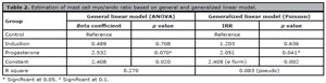
Table 2. Estimation of mast cell myo/endo ratio based on general and generalized linear model.
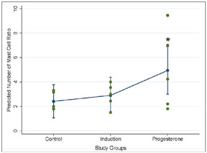
Figure 3. Poisson regression for prediction of mast cell count ratio in myometrium per endometrium (the plot is marginal post estimation output of the regression, Stata14). The error bars indicate 95% CI. * Significant difference considering the control group as the base.
Uterine histamine level was compared between the groups. According to the ANOVA, the differences of the means were significantly greater than the differences existed within the groups (p=0.039) in which progesterone group showed lower amounts of histamine in comparison to other groups based on Tukey post hoc test (Table 3).
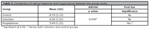
Table 3. Comparison of uterus histamine level (µg/g-tissue) between the groups study.
DISCUSSION
Induction of ovulation is a process commonly used in management of infertility. Progesterone administration is a way to improve success rate of implantation. Animal studies are performed to find the mechanisms of effect for progesterone. The present study was performed to show the effect of progesterone on endometrium and myometrium mast cell population and uterus histamine level after induction of ovulation in a mice model.
In the present study, decidualization process was observed dominantly in progesterone group. De Clercq et al. (2017) showed that deciualization was dependent to progesterone. This finding was aligned with the present study. Dursun et al. (2004) showed that exogenous gonadotropins could affect the morphology of endometrium and mitotic index during implantation, and these changes were more dominant in higher doses of exogenous gonadotropins.
The means of mast cell count were not significantly different between the groups neither in endometrium nor in myometrium. It means that the groups of study could not predict the mast cell count linearly. Tissue histamine level - as the clinical outcome of mast cell population - decreased in progesterone group in comparison to both control and induction groups. The mentioned findings mean that the effect of progesterone is via reduction in mast cell activity (i.e. histamine secretion).
According to the literature, the role of mast cells in implantation process and pregnancy is controversial. Although it has been known that estrogen and progesterone affect the population of mast cells (Noor et al., 2010; Wang et al., 2008), it seems that its increase may have different clinical outcomes in different conditions. In the present study, the authors found a non-significant increase in mast cell count of myometrium in induction and progesterone groups, and a non-significant decrease in mast cell count of endometrium in progesterone group. Hence, the authors intended to analyze the mast cell count ratio of myometrium per endometrium. Accordingly, this ratio was increased in progesterone group in comparison to control group based on Poisson regression. It seems that endometrial mast cells may play role against implantation while myometrium mast cells are necessary for better blood supply and uterine contractions. This interpretation should be studied specifically in future.
Historically, Harvey (1964) studied the distribution of mast cells in different days of estrous cycle in hamster. They found reduction of mast cells in endometrium and myometrium at the time of implantation. However, the percentage of myometrium mast cells per total mast cells increased (from 66% at day 1 to 96% at day 4) (Harvey, 1964). This result supported the hypothesis and result of the present study. Maraspin & Bo (1971) performed an animal study on a rat model. In their progesterone group, the mast cell count in myometrium per endometrium was 291 versus (vs.) 20 while in control group it was 255 vs. 25 and in estrogen group it was 150 vs. 31. They believed that mast cells play role in decidualization after pseudo pregnancy and therefore had considered positive effects for both endometrium and myometrium mast cell count (Maraspin & Bo, 1971). In the present study, there was no difference in this ratio between control and induction groups. The authors believe that after induction of ovulation decidualization is needed and progesterone help us to reach this aim. Mori et al. (1997) performed a human study. In their results, the mast cell count in inner myometrium per endometrium was 40 vs. 6 in proliferative phase while 30 vs 8 in secretory phase. Considering secretory phase as the time of decidualitation, the reduction of this ratio cannot be justified. However, it should be regarded that secretory phase has different physiology in pregnancy and non-pregnancy conditions. Bytautiene et al. (2004) found that myometrium mast cells and their degranulation could modulate uterine contractility during pregnancy. Szukiewicz et al. (2007) hypothesized that mast cell-derived interleukin-8 (IL-8) was associated with follicular growth and ovulation. They studied in vitro administration of IL-8 and found this association. It showed that the role of mast cells in female reproduction system was not limited to histamine secretion (Szukiewicz et al., 2007). Karaca et al. (2008) investigated the mast cells in female reproductive tract of goats. They found that distribution of the mast cells were different among the different parts of uterus in different days of the cycle (Karaca et al., 2008). Jensen et al. (2010) investigated the role of estradiol and progesterone in migration, maturation and degranulation of the mast cells in mice. They demonstrated that the hormones resulted in mast cell migration from periphery to uterus, maturation and degranulation as well as improve in angiogenesis (Jensen et al., 2010). Zierau et al. (2012) reviewed the role of estradiol and progesterone in mast cell behavior. They mentioned that female sex hormones induced mast cell maturation and degranulation (Zierau et al., 2012). Hamouzova et al. (2020) studied changes of mast cell distribution in canine uterus at early follicular and luteal phases of oestrous cycle. A significant decrease in mast cell count was observed at luteal phase in comparison to early follicular phase in both endometrium and myometrium. According to our calculation on their results, myo/endo ratio was 1.328 and 1.181 for luteal and early follicular phases respectively (Hamouzova et al., 2020).
The limitation of this study was lack of evaluating different parts of uterus separately (including horns, corpus and cervix), and releasing factors other than histamine. Although the authors had different results in comparison to the literature in terms of decrease in histamine level, it should be regarded that the present study had different design including pseudo pregnancy and induction of ovulation followed by progesterone administration. This result (reduction in histamine level based on the design of our study) may have beneficial effect for fertility of mice and possibly in human.
CONCLUSIONS
Progesterone administration after induction of ovulation could not change the number of endometrial mast cells (ANOVA) and could increase the ratio of myometrium mast cells per endometrium mast cell (generalized linear model). Histamine level of uterus was decreased by administration of progesterone in ovulation induced mice. The ratio of mast cell in myometrium per endometrium is introduced to researchers for further study.
ACKNOWLEDGEMENTS
This manuscript was the MSc thesis of NA and we acknowledge the deputy of research.
Funding
Lorestan University of Medical Sciences with grant number 1562-99-1-1397.
REFERENCES
Arifin WN, Zahiruddin WM. Sample Size Calculation in Animal Studies Using Resource Equation Approach. Malays J Med Sci. 2017;24:101-5. PMID: 29386977 DOI: 10.21315/mjms2017.24.5.11
Medline
Bytautiene E, Vedernikov YP, Saade GR, Romero R, Garfield RE. Degranulation of uterine mast cell modifies contractility of isolated myometrium from pregnant women. Am J Obstet Gynecol. 2004;191:1705-10. PMID: 15547545 DOI: 10.1016/j.ajog.2004.04.008
Medline
Charan J, Kantharia ND. How to calculate sample size in animal studies? J Pharmacol Pharmacother. 2013;4:303-6. PMID: 24250214 DOI: 10.4103/0976-500X.119726
Medline
De Clercq K, Hennes A, Vriens J. Isolation of Mouse Endometrial Epithelial and Stromal Cells for In Vitro Decidualization. J Vis Exp. 2017;121:55168. PMID: 28287563 DOI: 10.3791/55168
Medline
Dursun A, Sendag F, Terek MC, Yilmaz H, Oztekin K, Baka M, Tanyalcin T. Morphometric changes in the endometrium and serum leptin levels during the implantation period of the embryo in the rat in response to exogenous ovarian stimulation. Fertil Steril. 2004;82:1121-6. PMID: 15474084 DOI: 10.1016/j.fertnstert.2004.04.039
Medline
Gad A, Besenfelder U, Rings F, Ghanem N, Salilew-Wondim D, Hossain MM, Tesfaye D, Lonergan P, Becker A, Cinar U, Schellander K, Havlicek V, Hölker M. Effect of reproductive tract environment following controlled ovarian hyperstimulation treatment on embryo development and global transcriptome profile of blastocysts: implications for animal breeding and human assisted reproduction. Hum Reprod. 2011;26:1693-707. PMID: 21531990 DOI: 10.1093/humrep/der110
Medline
Gundersen HJ, Bendtsen TF, Korbo L, Marcussen N, Møller A, Nielsen K, Nyengaard JR, Pakkenberg B, Sørensen FB, Vesterby A, et al. Some new, simple and efficient stereological methods and their use in pathological research and diagnosis. APMIS. 1988;96:379-94. PMID: 3288247 DOI: 10.1111/j.1699-0463.1988.tb05320.x
Medline
Hamouzova P, Cizek P, Bartoskova A, Vitasek R, Tichy F. Changes in the mast cell distribution in the canine ovary and uterus throughout the oestrous cycle. Reprod Domest Anim. 2020;55:479-85. PMID: 31961006 DOI: 10.1111/rda.13641
Medline
Harvey EB. Mast cell distribution in the uterus of cycling and pregnant hamsters. Anat Rec. 1964;148:507-16. PMID: 14153310 DOI: 10.1002/ar.1091480309
Medline
Jensen F, Woudwyk M, Teles A, Woidacki K, Taran F, Costa S, Malfertheiner SF, Zenclussen AC. Estradiol and progesterone regulate the migration of mast cells from the periphery to the uterus and induce their maturation and degranulation. PLoS One. 2010;5:e14409. PMID: 21203555 DOI: 10.1371/journal.pone.0014409
Medline
Karaca T, Arikan S, Kalender H, Yoruk M. Distribution and heterogeneity of mast cells in female reproductive tract and ovary on different days of the oestrus cycle in Angora goats. Reprod Domest Anim. 2008;43:451-6. PMID: 18298406 DOI: 10.1111/j.1439-0531.2007.00934.x
Medline
King AE, Critchley HO. Oestrogen and progesterone regulation of inflammatory processes in the human endometrium. J Steroid Biochem Mol Biol. 2010;120:116-26. PMID: 20067835 DOI: 10.1016/j.jsbmb.2010.01.003
Medline
Liu YX, Yu M, Wang C, Peng KM, Liu HZ. Distribution patterns of mast cells in the uterus of pregnant Meishan pigs. Reprod Domest Anim. 2012;47:574-7. PMID: 22066801 DOI: 10.1111/j.1439-0531.2011.01920.x
Medline
Maraspin LE, Bo WJ. Effects of hormones, pregnancy and pseudopregnancy on the mast cell count in the rat uterus. Life Sci I. 1971;10:111-20. PMID: 5575403 DOI: 10.1016/0024-3205(71)90084-1
Medline
Meyer N, Schüler T, Zenclussen AC. Simultaneous Ablation of Uterine Natural Killer Cells and Uterine Mast Cells in Mice Leads to Poor Vascularization and Abnormal Doppler Measurements That Compromise Fetal Well-being. Front Immunol. 2018;8:1913. PMID: 29375562 DOI: 10.3389/fimmu.2017.01913
Medline
Mori A, Zhai YL, Toki T, Nikaido T, Fujii S. Distribution and heterogeneity of mast cells in the human uterus. Hum Reprod. 1997;12:368-72. PMID: 9070727 DOI: 10.1093/humrep/12.2.368
Medline
Narimani L, Boroujeni NB, Gholami M, Anbari K, Alavi SER, Ahmadi SAY, Boroujeni MB. Pre-Implantation Effects of Progesterone Administration on Ovarian Angiogenesis after Ovarian Stimulation: A Histological, Hormonal, and Molecular Analysis. JBRA Assist Reprod. 2020;24:289-95. PMID: 32155017 DOI: 10.5935/1518-0557.20190076
Medline
Szukiewicz D, Pyzlak M, Klimkiewicz J, Szewczyk G, Maslinska D. Mast cell-derived interleukin-8 may be involved in the ovarian mechanisms of follicle growth and ovulation. Inflamm Res. 2007;56:S35-6. PMID: 17806170 DOI: 10.1007/s00011-006-0517-7
Medline
Woidacki K, Jensen F, Zenclussen AC. Mast cells as novel mediators of reproductive processes. Front Immunol. 2013;4:29. PMID: 23419968 DOI: 10.3389/fimmu.2013.00029
Medline
Zierau O, Zenclussen AC, Jensen F. Role of female sex hormones, estradiol and progesterone, in mast cell behavior. Front Immunol. 2012;3:169. PMID: 22723800 DOI: 10.3389/fimmu.2012.00169
Medline