JBRA Assist. Reprod. 2025;29(2):289-297
ORIGINAL ARTICLE
doi: 10.5935/1518-0557.20240108
Prednisolone at a therapeutic dose is not detrimental to mouse oocyte competence
1Centre of Excellence in Clinical Embryology, Department of Reproductive Science, Kasturba Medical College, Manipal, Manipal Academy of Higher Education, Manipal-576 104, India
2Division of Reproductive Genetics, Department of Reproductive Science, Kasturba Medical College, Manipal. Manipal Academy of Higher Education, Manipal-576 104, India
CONFLICTS OF INTEREST
The authors have no conflict of interest to declare
*Equally contributed to the work.
ABSTRACT
Objective: To examine the impact of PRDL on mouse oocyte developmental competence.
Methods: This study was conducted on 6-8-week-old female Swiss albino mice in which different doses of PRDL were administered for three days during superovulation. Oocytes were assessed for quality, and subsequently, embryos derived from these oocytes were evaluated at the blastocyst stage. Simultaneously, oocytes were also exposed to PRDL during in vitro maturation and examined for their spindle and activation potential.
Results: Compared to control group, the total cell number of blastocysts in the PRDL group increased. Nevertheless, the incidence of apoptosis was comparable between the groups. However, direct exposure at 10 µM significantly increased spindle abnormalities compared to those in the control group (p<0.05).
Conclusions: Short-term PRDL exposure at a therapeutic dose is not detrimental to oocyte developmental potential in vitro. Although the current study confirmed the safety of PRDL in a mouse model, further studies involving long-term exposure are warranted, as patients receive PRDL treatment for extended periods of time.
Keywords: assisted reproductive technology, glucocorticoids, oocytes, prednisolone, preimplantation embryo development
INTRODUCTION
The off-label use of glucocorticoids (GCs) as an adjuvant therapy during ovarian stimulation has attracted increased interest in recent years (Beltrán et al., 2020; Conforti et al., 2021; Farzaneh & Afshar, 2020). During the peri-implantation phase, both natural and synthetic GCs are believed to improve the uterine environment as immunomodulators (Boomsma et al., 2022; Robertson et al., 2016), whereas during the follicular phase, GCs ameliorate follicular development by suppressing androgen levels and increasing the production of growth factors, which are known to amplify the action of gonadotropins (Langford & Miell, 1993; Miell et al., 1993). Low doses of GCs (10 mg/day) have been shown to have a positive impact on pregnancy, while women experiencing recurrent miscarriages and other conditions of immunological infertility have also been treated with higher doses of GCs (up to 60 mg/day) (Boomsma et al., 2007; Kalampokas et al., 2017). Nevertheless, there is scarce evidence regarding the efficacy of administering GCs during ovarian stimulation to increase live birth rates in women undergoing assisted reproductive technology (ART) treatment (Kalampokas et al., 2017; Kim, 2021; Lin et al., 2023).
Animal studies on the effect of GC exposure on oocyte maturation and developmental competence have shown conflicting results. Low-dose corticosterone administration in mice caused no adverse effects on oocyte maturation; however, the administration of higher concentrations of corticosterone caused alterations in oocyte ultrastructure and increased mitochondrial mutations (Li et al., 2018). Cortisol administration impaired mouse oocyte developmental competence by inducing apoptosis (Yuan et al., 2016; 2020). In contrast, supplementation of culture media supplemented with dexamethasone and cortisol improved oocyte maturation and development competence in bovine oocytes (Barroso et al., 2020; da Costa et al., 2016), but a negative effect of both cortisol and dexamethasone was observed on lamb and pig oocyte maturation (González et al., 2010a; Yang et al., 1999). In addition, high concentrations of corticosterone significantly affect oocyte maturation, fertilization and embryo development in vitro in a mouse model (Andersen, 2003; Cikoš et al., 2019; González et al., 2010b).
Prednisolone (PRDL) is a synthetic glucocorticoid given to patients with infertility conditions to ameliorate the ovarian response and pregnancy outcome in ART treatment (Revelli et al., 2008); it is either given alone or in combination with other drugs (Duvan et al., 2006; Ubaldi et al., 2002). PRDL is widely prescribed for women with recurrent miscarriages or immunological infertility conditions to improve implantation outcomes (Robertson et al., 2016). PRDL treatment may start a month before controlled ovarian stimulation (COS) (Geva et al., 2000), during COS (Duvan et al., 2006) or just after oocyte retrieval (Motteram et al., 2015). PRDL is additionally employed during the COS protocol for patients with polycystic ovarian syndrome (PCOS) (Fridström et al., 1999; Mohammadi Yeganeh et al., 2018) and ovarian hyperstimulation syndrome (OHSS) (Fang et al., 2021; Weigert et al., 2002). Importantly, when used during the peri-implantation phase, PRDL treatment has specific adverse effects on preimplantation embryos, fetuses, and placentas in mice (Kieffer et al., 2020; Uppangala et al., 2021). However, no experimental studies have explored the effect of PRDL on oocyte maturation and developmental competence.
Recent meta-analyses have suggested that there is no added value in the use of PRDL in IVF cycles (Lin et al., 2023). The clinical benefits of PRDL are debatable, and further studies are warranted to understand the impact of PRDL on oocytes exposed during the follicular phase. The main research question is whether there is any negative impact of PRDL on oocyte functional and genetic competence influencing its developmental competence and genetic integrity. Therefore, to bridge the knowledge gap on the impact of PRDL on oocyte maturation and developmental competence, we investigated the effect of PRDL administration during ovarian stimulation on oocyte maturation and the genetic integrity of embryos derived from oocytes exposed to PRDL. In our study, mice were administered different concentrations of PRDL (0.01, 0.1 and 1mg/kg body weight) for three days during the superovulation regimen to mimic the clinical scenario, and the oocyte number, maturation, mitochondrial potential, developmental competence and genetic integrity of the blastocysts were assessed. Additionally, to understand the direct effects of PRDL on oocyte maturation, immature mouse oocytes were exposed to PRDL at various concentrations (0.1, 1 and 10µM) for 24 h during in vitro maturation.
MATERIAL AND METHODS
Animals and ethics declaration
Healthy Swiss albino mice aged 6-8 weeks (female, n=85; male, n=25) maintained at 25±2°C and 45-55% humidity with a 12:12 h photoperiod and fed a standard diet and water ad libitum were used for the experiments. Animal handling and procedures were carried out as per the institutional guidelines, and the study was approved by the institutional animal ethics committee (IAEC/KMC/14/2021 and IAEC/KMC/16/2021).
Preparation of prednisolone (PRDL)
A stock solution of 3 mM prednisolone (PRDL, Cat No. P6004, Sigma Aldrich, USA) was prepared using absolute ethanol (Cat No. F204325, Hayman, UK) as previously described (Uppangala et al., 2021). For the in vivo approach, working concentrations of PRDL (0.01, 0.1 and 1 mg/kg body weight) were prepared by diluting the stock solution with phosphate-buffered saline (PBS). According to Kieffer et al. (2020), 1 mg/kg of PRDL in mice corresponds to the highest dose used in ART (60 mg/day). Hence, we used concentrations of 0.01, 0.1 and 1 mg/kg, where 0.01 mg/kg corresponds to the physiological concentration of GCs and 0.1 corresponds to the therapeutic dose (5-10 mg/day). For the in vitro approach, the stock solution was further diluted to 0.1, 1 and 10 μM PRDL using in vitro maturation (IVM) medium. A concentration of 0.1 μM corresponds to the physiological level of GCs (Burkuš et al., 2013; Gong et al., 2015), 1 μM is close to the therapeutic dose (5-6 mg/day), and 10 μM is ten times the therapeutic dose and is hence considered the supraphysiological concentration.
PRDL administration and Cumulus oocyte complex (COC) collection
To mimic the clinical scenario, PRDL administration (intraperitoneal) started on the day of pregnant mare serum gonadotropin (PMSG) injection. Furthermore, PRDL was given for 3 days, with a 24 h time interval between each administration during the superovulation regimen. For superovulation, 5 IU PMSG (Cat No. HOR-272, ProSpecTany TechnoGene Ltd., Israel) followed by 10 IU human chorionic gonadotropin (hCG) (Cat No. Lupi-HCG 2000, Lupin, India) was administered to female mice at 48 h intervals. An approximately 8 h time interval was maintained between PRDL and PMSG injection as well as between PRDL and hCG administration. The animals were sacrificed 12 h after hCG injection, and COC was collected. COCs were either denuded to assess oocyte number, nuclear maturity, fragmentation and mitochondrial potential or subjected to IVF to assess oocyte developmental competence.
Oocyte denudation and morphological assessment
The COCs were enzymatically denuded by treatment with 1 mg/mL hyaluronidase (Cat No. H4272, Sigma Aldrich, USA) for 30 seconds, and the denuded oocytes were assessed morphologically under an inverted phase contrast microscope (IX73, Olympus, Japan) to determine nuclear maturity and morphological abnormalities.
Measurement of oocyte mitochondrial potential by JC-1 staining
To determine whether PRDL alters oocyte mitochondrial potential, PRDL-exposed oocytes were subjected to JC1 staining as described previously (Uppangala et al., 2015). Briefly, oocytes were incubated with prewarmed 1μg/mL 5,5´,6,6´-tetrachloro-1,1´,3,3´-tetraethyl-imidacarbocyanine iodide (JC-1, Cat No. T3168, Molecular Probes, Life Technologies, USA) for 30 min at 37°C and 5% CO2. After 30 min, the excess probe was removed by washing with M16 medium supplemented with 0.1% BSA, and the plates were mounted on glass slides. The oocytes were observed under a fluorescence microscope (Imager-A1; Zeiss, Gottingen, Germany). Oocytes with low mitochondrial potential fluoresce green, while oocytes with high mitochondrial potential fluoresce orange owing to the aggregation of JC-1. The mitochondrial membrane potential is represented as the ratio of orange to green fluorescence, which was determined by ImageJ software (National Institute of Health, Bethesda, Maryland, USA).
In vitro fertilization (IVF) and embryo culture
The COCs retrieved from the oviduct were incubated in potassium simplex optimization medium (KSOM) supplemented with 0.1% BSA (Cat No. A3311, Sigma Aldrich, USA) at 37°C and 5% CO2. Caudal sperm retrieved from male mice were subjected to swim up for 45 min at 37°C and 5% CO2 in KSOM supplemented with 1% BSA. The motile sperm fraction postswim-up was coincubated with COC. Oocytes were denuded and assessed for fertilization 10 h post insemination (hereafter referred to as hpi). Normally fertilized oocytes were cultured at 37°C and 5% CO2 in 20 µL of KSOM supplemented with 0.1% BSA and overlaid with paraffin liquid light oil. Preimplantation embryo development was assessed at regular intervals until 96 hpi. The total cell number and DNA integrity of the blastocysts were assessed.
DNA damage assessment by terminal deoxynucleotidyl transferase (TdT) dUTP nick end labeling (TUNEL) assay
The TUNEL assay was performed as described previously (D’Souza et al., 2016) with slight modifications. Briefly, at 96 h, blastocysts were fixed using 4% PFA overnight at 4°C in 60-well plates (Cat No. 163118, Nunc, India). The fixed blastocysts were permeabilized for 1 h using 0.1% Triton-100, 0.1% sodium citrate and 0.5% BSA in PBS. Later, the embryos were incubated for 1 h with a TUNEL reaction mixture (Cat No. 12156792910, Roche Diagnostics, Germany) in the dark at 37°C. After washing, the slides were counterstained with 4μg/mL DAPI and mounted onto a clean slide using mounting medium (Cat No. S3023, DAKO, Denmark). The embryos were observed under a fluorescence microscope (x400 magnification; Imager-A1; Zeiss, Gottingen, Germany) and imaged using Q-Capture software (Media Cybernetics Inc., USA). TUNEL-labeled cells displayed red fluorescence, whereas the nuclei of the cells displayed blue fluorescence. The TUNEL index was calculated by calculating the percentage of TUNEL-labeled cells relative to the total number of cells present in each embryo.
Germinal vesicle (GV) oocyte isolation and PRDL treatment in vitro
To study the direct effects of PRDL on mouse oocytes, GV oocytes were collected from ovaries excised from female mice in prewarmed homemade M2 media. The cumulus-free GV oocytes isolated from the five study groups were randomly divided and cultured in vitro for 24 h in IVM medium supplemented with (0.1, 1 or 10 µM) or without PRDL.
Briefly, in vitro maturation of GV oocytes was carried out in 20 µL droplets of IVM medium with oil overlay for 24 h under growth conditions (37°C and 5% CO2). IVM medium was prepared by adding 0.05% pyruvate (Cat No. P3662, Sigma Aldrich, USA), 1% nonessential α-amino acids (Cat No. 11140-050, Gibco, USA), 1% ITS (Cat No. 51500-056, Gibco, USA), 0.05% penicillin‒streptomycin (Cat No. 15140-122, Gibco, USA) and 0.3% bovine serum albumin (BSA) to Dulbecco’s modified Eagle’s medium (DMEM). After IVM, oocytes were assessed for the presence of an extruded polar body, which indicates the maturity of the oocyte.
Spindle integrity assessment
Mature oocytes obtained post IVM were examined for spindle morphology as per an earlier protocol (Daddangadi et al., 2020). Briefly, oocytes were incubated with extraction buffer containing 50mM potassium chloride, 5 mM ethylenediaminetetraacetic acid disodium salt, 0.5mM magnesium chloride, 25% glycerol, 25mM HEPES, 20 μM phenyl methane sulfonyl fluoride and 2% Triton X-100, adjusted to pH 6.75 for 1 h under growth conditions. Oocyte fixation was performed at -20°C by exposing the oocytes to ice-cold ethanol for 12 minutes. Furthermore, the cells were treated with 0.25% Triton-X and 5% knockout serum (Cat No. 10828-010, Gibco, India) for 1 h at 37°C. Furthermore, the oocytes were incubated with a primary anti-α-tubulin antibody (1:150) (Cat. No. T9026, Sigma Aldrich, USA) overnight at 4°C. After washing, the oocytes were incubated with a secondary goat anti-mouse IgG antibody (1:500) (Cat No. NB7535, Novus Biologicals, USA) for 1 h at 37°C. After counterstaining with 4 μg/mL DAPI (40,6-diamidino-2-phenylindole, Cat. no. D9542, Sigma Aldrich, USA), the spindle was observed under a 40X objective of fluorescence microscope (Imager-A1, Zeiss, Gottingen, Germany). The spindle images were captured using Q capture software (Media Cybernetics, Inc., USA), and the spindle morphology was analyzed.
Parthenogenetic oocyte activation
Parthenogenetic activation of in vitro matured oocytes was performed as previously described with slight modifications (Ma et al., 2005). Oocytes were incubated with freshly prepared 10 mM strontium chloride (StCl2, Cat No. 107865, Merck, India) in Ca2+- and Mg2+-free M16 media supplemented with 0.1% BSA for 1 h. After incubation at 37°C in 5% CO2, the washed oocytes were cultured for 2 h in 30 µL of prewarmed M16 medium. After 2 h of culture, the oocytes were evaluated under an inverted phase contrast microscope (40X, IX 73, Olympus, Japan) for the presence of one pro nucleus (PN) and two extruded polar bodies in the perivitelline space, which confirmed oocyte activation.
Statistical analysis
The data obtained in this study are from a minimum of 3 independent trials and are represented either as the mean±standard error of the mean (SEM) or as a percentage (%). Differences between the groups were tested either by one-way analysis of variance (ANOVA) or the Kruskal‒Wallis test depending on the distribution of the data. The chi-square test was applied when the data were in percentage form. Differences were considered significant when p<0.05. GraphPAD InStat software (GraphPad Inc., La Jolla, CA, USA) was used for statistical evaluation, and Microcal Origin 6.0 software (Origin Lab Corporation, Northampton, MA, USA) was used to prepare the graphs.
RESULTS
PRDL administration resulted in comparable oocyte maturation and improved the mitochondrial potential
To mimic the clinical scenario, PRDL was administered for a total of three days, beginning on the day of PMSG administration and ending on the day of hCG injection. The retrieved COC was denuded and assessed for maturity. We observed a decrease in the average number of oocytes retrieved in the treated groups as the dose of PRDL increased. However, the difference was not statistically significant (Table 1). Although the 1 mg/kg PRDL group had the lowest number of oocytes (29.14±5.94), the highest maturation rate was also observed in the same group (83.57±7.87), which was comparable to that of the control group (77.11±9.79). These oocytes were further subjected to JC1 staining to determine the effect of PRDL administration on the mitochondrial potential of the oocytes. The JC1 ratio was significantly lower in oocytes from the 1 mg/kg PRDL group (0.75±0.02) than in those from the 0.01 mg/kg PRDL group (0.81±0.01; p<0.05; Fig. 1A) and 0.1 mg/kg PRDL group (0.91±0.01; p<0.001). The 0.1 mg/kg PRDL group even had a significantly greater JC1 ratio than the VC group (0.77±0.01; p<0.01). However, none of the PRDL-exposed groups showed significantly altered mitochondrial potential compared to that of the control group. Representative images of oocytes displaying various levels of mitochondrial potential are provided in Fig. 1B.
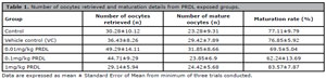
Table 1. Number of oocytes retrieved and maturation details from PRDL exposed groups.
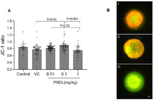
Figure 1. Mitochondrial membrane potential of oocytes after PRDL administration. A. Comparison of mitochondrial potential by JC-1 staining in control (n=25), VC (n=65), 0.01 (n=79), 0.1 (n=66) and 1mg/kg (n=42) PRDL-treated oocytes. B i-iii. Representative fluorescence microscopy images (40×) of JC-1-stained oocytes in decreasing order of mitochondrial potential. Scale bar =20 µm.
PRDL administration did not alter oocyte developmental competence or apoptotic index in the blastocyst
To understand the developmental competence of PRDL-exposed oocytes, COCs isolated post superovulation were subjected to IVF. Complete details regarding fertilization and embryo development are presented in Table 2. However, after 10 hpi, the control group demonstrated the highest fertilization rate (91.07±4.98) among the study groups, but the difference was not statistically significant. Further progression of fertilized oocytes was monitored up to the blastocyst stage (96 hpi), and embryo progression was evaluated. Embryos derived from the PRDL group showed a comparable embryo development rate to that of control embryos. Even the blastocyst rate and hatching rate were comparable at 96 hpi. Although the rates of embryo progression in the PRDL group were similar to those in the control group, the total cell number (TCN) increased in blastocysts derived from oocytes in the 0.01 (p<0.001) and 1 mg/kg PRDL groups (p<0.05) compared to that in the control group (Fig. 2A). Furthermore, blastocysts obtained from each group were subjected to a TUNEL assay to assess DNA integrity. Interestingly, blastocysts across various study groups displayed comparable TUNEL indices, indicating that PRDL did not affect oocyte developmental competence or blastocyst quality (Fig. 2B). Representative images of blastocysts with TUNEL-positive cells are shown in Fig. 2C.
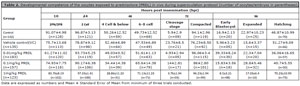
Table 2. Developmental competence of the oocytes exposed to prednisolone (PRDL) in vivo during superovulation protocol (number of oocytes/embryos in parentheses).
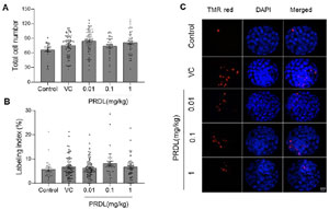
Figure 2. Impact of PRDL on blastocyst cell number and DNA integrity. A. Comparison of total cell numbers and B. Labeling indices of blastocysts from the control (n=27), VC (n=70), 0.01 (n=80), 0.1 (n=38) and 1 mg/kg (n=45) PRDL groups. C. Representative fluorescence microscopy (40×) images showing TUNEL-positive cells (red) and total cells (blue). Scale bar =20 µm.
Direct exposure to PRDL did not affect the maturation or mitochondrial potential of oocytes matured in vitro but affected spindle morphology
To assess the direct effect of PRDL, GV oocytes were subjected to IVM in the presence or absence of PRDL. PRDL at various concentrations (0.1, 1 and 10 µM) did not affect the oocyte maturation rate compared to that of control oocytes. The maturation rates between the groups were comparable. When these oocytes were subjected to JC1 staining to determine the effect of PRDL on oocyte mitochondria, the JCI ratio was comparable between the groups, indicating that PRDL did not affect the mitochondrial potential of the oocytes (Table 3). However, when the spindle morphology of these oocytes was assessed, the oocytes exposed to the highest PRDL concentration had a significantly greater incidence of abnormal spindle morphology than did the control oocytes (Fig. 3A). Representative images displaying normal and abnormal spindle morphology are shown in Fig. 3B. Furthermore, the activation rate of the oocytes exposed to PRDL was also comparable between the study groups.
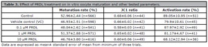
Table 3. Effect of PRDL treatment on in vitro oocyte maturation and other tested parameters.
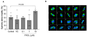
Figure 3. Spindle integrity of oocytes exposed to PRDL in vitro. A. Comparison of the percentage of spindle abnormalities in control (n=29), vehicle control (n=28), 0.1 µM (n=34), 1 µM (n=21), and 10 µM (n=36) PRDL-exposed MII oocytes. C. Representative fluorescence microscopy images (40×) of the spindle. (a-j) normal spindle arrangement; (k-t) damaged spindles and misaligned chromosomes. Scale bar =20 µm.
DISCUSSION
Although PRDL is used in routine clinical practice, there are limited experimental studies that have elucidated the effect of PRDL exposure on oocytes and embryos. This study demonstrated that short-term PRDL exposure to mouse oocytes at a therapeutic dose does not have a deleterious effect on oocyte competence when it is administered in vivo. However, direct exposure to high concentrations of PRDL can affect oocyte spindle morphology. This is the first experimental study to assess the impact of PRDL on mouse oocyte functional, genetic and developmental competence.
Initial reports on the administration of GCs alone or in combination with other adjuvants suggested that GCs improve the ovarian response during COS (Keay et al., 1997; Trott et al., 1996). It has been proposed that GC therapy might improve follicular development by suppressing androgen levels and increasing the production of growth factors, which are known to amplify the action of gonadotropins (Langford & Miell, 1993). Clinical studies involving PRDL administration during COS have demonstrated conflicting results with respect to oocyte yield and maturation potential (Revelli et al., 2008; Ubaldi et al., 2002). Notably, these studies included patients with good prognosis. In this study, a marginal reduction in oocyte yield was observed with an increasing PRDL dose. However, comparable maturation potential was observed between the PRDL-exposed oocytes and the control group. In mice, the administration of glucocorticoids such as cortisol and corticosterone did not affect oocyte maturation potential (Li et al., 2018; Yuan et al., 2016). However, a positive correlation was observed between the follicular fluid cortisol concentration and the morphological maturity of the follicles in women undergoing IVF (Fateh et al., 1989).
Mitochondrial potential is an important indicator of oocyte quality because mitochondria are important organelles involved in various embryological processes, from fertilization to embryonic development (Tan et al., 2017). Despite having comparable maturation rates, the 1mg/kg PRDL group showed significantly reduced mitochondrial potential compared to the other PRDL groups used in this study. This finding aligns with an earlier study in which the oocyte mitochondrial potential decreased with a simultaneous increase in oxidative stress when female mice were exposed to 50 mg/kg cortisol (Yuan et al., 2020). However, a low concentration of PRDL had a positive effect on the oocyte mitochondrial potential. These differences could be due to variations in the chemical nature of the drug, the concentrations used (0.01, 0.1 and 1 mg/kg PRDL) and differences in study design.
To understand the developmental potential of oocytes treated with PRDL in vivo, we performed IVF. PRDL treatment (0.01-1 mg/kg) did not affect the embryo development rate. PRDL-treated oocytes had comparable blastocyst formation rates and hatching rates at 96 hpi. Similarly, the rate of blastocyst formation in oocytes treated with corticosterone did not differ from that in untreated oocytes (Li et al., 2018). However, the developmental potential of oocytes decreased significantly when cortisol (50 mg/kg) was administered to female mice (Yuan et al., 2016; 2020). Furthermore, earlier reports demonstrated a decreased blastocyst formation rate when oocytes/embryos were treated with high concentrations of GCs (Andersen, 2003; Cikoš et al., 2019; González et al., 2010a; 2010b; Uppangala et al., 2021). These observed effects may be dose dependent and species specific, as it has been shown that exposing bovine oocytes to cortisol enhances their developmental competence (da Costa et al., 2016). Furthermore, to assess blastocyst quality, the total cell number (TCN) was assessed. An increase in TCN in blastocysts was observed in the 0.01 and 1 mg/kg PRDL treatment groups, which is contrary to earlier reports in which comparable TCN was observed when cortisol (10 mg/kg) was injected into mice (Yuan et al., 2016). Even in vitro studies have shown contradictory results for both comparable and decreased TCNs (Fateh et al., 1989; Uppangala et al., 2021).
GCs induce apoptosis as an immunomodulatory function (Herr et al., 2007; Kogianni et al., 2004; Schlossmacher et al., 2011; Thouas et al., 2004). GC decreases oocyte developmental potential by activating the Fas or TNFα system and inducing apoptosis in ovarian cells and oocytes (Yuan et al., 2016; 2020). Previous studies have demonstrated that exposure of preimplantation embryos to various GCs, including PRDL, leads to increased apoptosis at high concentrations (Cikoš et al., 2019; Uppangala et al., 2021). This apoptotic process is considered a protective mechanism at the blastocyst stage, safeguarding embryos from potential DNA damage (Adiga et al., 2007; Shimura et al., 2002). Although a comparable TUNEL index was observed in blastocysts derived from PRDL-exposed oocytes in the present study, the detrimental effects of even low concentrations of GCs cannot be dismissed in vivo. The natural glucocorticoids present in our body may have synergistic effects on oocytes/embryos and may be present at high concentrations under various pathological conditions, such as oxidative stress conditions. Thus, inducing apoptosis and hampering embryo development indirectly by triggering Fas system-induced apoptosis in oviductal cells (Tan et al., 2017).
Direct exposure of mouse oocytes to PRDL (0.1, 1, or 10 µM) during in vitro maturation resulted in comparable maturation rates. Similar observations were reported in earlier studies where dexamethasone and cortisol did not influence mice or bovine oocyte maturation or developmental competence (Andersen, 2003; Barroso et al., 2020; da Costa et al., 2016). Nevertheless, negative effects of cortisol and dexamethasone were observed in lamb and pig oocyte maturation (González et al., 2010a; 2010b; Yang et al., 1999). These observations indicate the species-specific effect of GCs; although mouse oocytes express GC receptors, GCs, including PRDL, do not influence mouse oocyte maturation. Even the mitochondrial potential of oocytes exposed to PRDL was like that of control oocytes. Similarly, earlier studies have shown that the mitochondrial distribution pattern and ultrastructure of mitochondria are unaffected when mouse and bovine oocytes are treated with corticosterone and dexamethasone (Barroso et al., 2020; Li et al., 2018).
Although direct exposure to PRDL did not affect the maturation rate or mitochondrial potential, oocytes treated with a high concentration of PRDL (at 10 µM) exhibited an increased incidence of spindle morphological abnormalities. Similarly, supplementation with a high concentration of dexamethasone (80 µg/mL) affected spindle morphology in mouse preantral follicle culture (Van Merris et al., 2007). Normal meiotic spindle morphology in mature oocytes is crucial for maintaining their genetic integrity and development (Cimadomo et al., 2021). Although this study did not observe an inverse relationship between increased abnormal spindle morphology and reduced maturation, as in the case of Van Merris et al. (2007), there was a decrease in the activation potential of oocytes exposed to 10 µM PRDL. However, the difference was not statistically significant.
ART procedures in which a low concentration of prednisolone is administered to improve IVF cycle outcomes may involve PRDL treatment for various periods (Andersen, 2003; Duvan et al., 2006; Geva et al., 2000; Revelli et al., 2008; Robertson et al., 2016), exposing the follicles/oocytes to PRDL for either the short or long term. This study attempted to replicate the clinical situation in which PRDL is treated during the short-term COS protocol. However, the differences observed between human and mouse models limit the extrapolation of the current findings to clinical settings. In addition, the implantation potential and gene expression of pluripotency markers in blastocysts derived from PRDL-exposed oocytes were not studied, and the developmental competence of oocytes subjected to PRDL treatment in vitro was not assessed, which are limitations of this study.
CONCLUSION
In conclusion, short-term treatment with PRDL at a therapeutic dose does not have a deleterious effect on oocyte yield or developmental competence. However, direct treatment of oocytes with supraphysiological concentrations of PRDL can affect mouse oocyte spindle morphology. Further research is required to understand the effect of long-term exposure to PRDL on oocyte developmental competence and the clinical value of these observations on human oocyte developmental competence.
REFERENCES
Adiga SK, Toyoshima M, Shiraishi K, Shimura T, Takeda J, Taga M, Nagai H, Kumar P, Niwa O. p21 provides stage specific DNA damage control to preimplantation embryos. Oncogene. 2007;26:6141-9. PMID: 17420724 DOI: 10.1038/sj.onc.1210444 Medline
Andersen CY. Effect of glucocorticoids on spontaneous and follicle-stimulating hormone induced oocyte maturation in mouse oocytes during culture. J Steroid Biochem Mol Biol. 2003;85:423-7. PMID: 12943731 DOI: 10.1016/S0960-0760(03)00190-0 Medline
Barroso PAA, Paulino LRFM, Silva BR, Vasconcelos GL, Gomes DS, Lima Neto MF, Silva AWB, Souza ALP, Donato MAM, Peixoto CA, Silva JRV. Effects of dexamethasone on growth, viability and ultrastructure of bovine secondary follicles cultured in vitro. Zygote. 2020;28:504-10. PMID: 32847639 DOI: 10.1017/S0967199420000416 Medline
Boomsma CM, Kamath MS, Keay SD, Macklon NS. Peri-implantation glucocorticoid administration for assisted reproductive technology cycles. Cochrane Database Syst Rev. 2022;6:CD005996. PMID: 35771604 DOI: 10.1002/14651858.CD005996.pub4 Medline
Boomsma CM, Keay SD, Macklon NS. Peri-implantation glucocorticoid administration for assisted reproductive technology cycles. Cochrane Database Syst Rev. 2007;CD005996. PMID: 35771604 DOI: 10.1002/14651858.CD005996.pub4 Medline
Burkuš J, Cikoš S, Fabian D, Kubandová J, Czikková S, Koppel J. Maternal restraint stress negatively influences growth capacity of preimplantation mouse embryos. Gen Physiol Biophys. 2013;32:129-37. PMID: 23531842 DOI: 10.4149/gpb_2013010 Medline
Cikoš Š, Babelová J, Špirková A, Burkuš J, Kovaríková V, Šefcíková Z, Fabian D, Koppel J. Glucocorticoid receptor isoforms and effects of glucocorticoids in ovulated mouse oocytes and preimplantation embryos. Biol Reprod. 2019;100:351-64. PMID: 30188986 DOI: 10.1093/biolre/ioy196 Medline
Cimadomo D, Vaiarelli A, Petriglia C, Fabozzi G, Ferrero S, Schimberni M, Argento C, Colamaria S, Giuliani M, Ubaldi N, Rienzi L, Ubaldi FM. Oocyte competence is independent of the ovulation trigger adopted: a large observational study in a setting that entails vitrified-warmed single euploid blastocyst transfer. J Assist Reprod Genet. 2021;38:1419-27. PMID: 33661465 DOI: 10.1007/s10815-021-02124-1 Medline
Conforti A, Esteves SC, Pellicer A, Sodano R, Messuti F, Carbone L, Morero S, Alviggi C, Sodano M. Effect of Dexamethasone Co-Treatment During Ovarian Stimulation in Women of Different Reproductive Age With Elevated Early Follicular Phase Progesterone Level: a Prospective Longitudinal Study. Reprod Sci. 2021;28:3258-64. PMID: 33928591 DOI: 10.1007/s43032-021-00590-7 Medline
da Costa NN, Brito KN, Santana Pd, Cordeiro Mda S, Silva TV, Santos AX, Ramos Pdo C, Santos Sdo S, King WA, Miranda Mdos S, Ohashi OM. Effect of cortisol on bovine oocyte maturation and embryo development in vitro. Theriogenology. 2016;85:323-9. PMID: 26456184 DOI: 10.1016/j.theriogenology.2015.08.010 Medline
Daddangadi A, Uppangala S, Kalthur G, Talevi R, Adiga SK. Germinal stage vitrification is superior to MII stage vitrification in prepubertal mouse oocytes. Cryobiology. 2020;93:49-55. PMID: 32112808 DOI: 10.1016/j.cryobiol.2020.02.012 Medline
D’Souza F, Pudakalakatti SM, Uppangala S, Honguntikar S, Salian SR, Kalthur G, Pasricha R, Appajigowda D, Atreya HS, Adiga SK. Unraveling the association between genetic integrity and metabolic activity in pre-implantation stage embryos. Sci Rep. 2016;6:37291. PMID: 27853269 DOI: 10.1038/srep37291 Medline
Duvan CI, Ozmen B, Satiroglu H, Atabekoglu CS, Berker B. Does addition of low-dose aspirin and/or steroid as a standard treatment in nonselected intracytoplasmic sperm injection cycles improve in vitro fertilization success? A randomized, prospective, placebo-controlled study. J Assist Reprod Genet. 2006;23:15-21. PMID: 16518579 DOI: 10.1007/s10815-005-9003-3 Medline
Fang L, Ruan M, Yang S, Qu X, Chen H, Zhao J, Cheng J. Prednisone combined with letrozole reduced risk of ovarian hyperstimulation syndrome (OHSS) in women undergoing long-term gonadotropin-releasing hormone analog treatment. Ann Palliat Med. 2021;10:8837-47. PMID: 34488372 DOI: 10.21037/apm-21-1699 Medline
Farzaneh F, Afshar F. Comparison of ovulation induction with letrozole plus dexamethasone and letrozole alone in infertile women with polycystic ovarian disease: An RCT. Int J Reprod Biomed. 2020;18:307-10. PMID: 32494769 DOI: 10.18502/ijrm.v13i4.6893 Medline
Fateh M, Ben-Rafael Z, Benadiva CA, Mastroianni L, Flickinger GL. Cortisol levels in human follicular fluid. Fertil Steril. 1989;51:538-41. PMID: 2920855 DOI: 10.1016/S0015-0282(16)60572-1 Medline
Fridström M, Carlström K, Sjöblom P, Hillensjö T. Effect of prednisolone on serum and follicular fluid androgen concentrations in women with polycystic ovary syndrome undergoing in-vitro fertilization. Hum Reprod. 1999;14:1440-4. PMID: 10357955 DOI: 10.1093/humrep/14.6.1440 Medline
Geva E, Amit A, Lerner-Geva L, Yaron Y, Daniel Y, Schwartz T, Azem F, Yovel I, Lessing JB. Prednisone and aspirin improve pregnancy rate in patients with reproductive failure and autoimmune antibodies: a prospective study. Am J Reprod Immunol. 2000;43:36-40. PMID: 10698039 DOI: 10.1111/j.8755-8920.2000.430107.x Medline
Gong S, Miao YL, Jiao GZ, Sun MJ, Li H, Lin J, Luo MJ, Tan JH. Dynamics and correlation of serum cortisol and corticosterone under different physiological or stressful conditions in mice. PloS One. 2015;10:e0117503. PMID: 25699675 DOI: 10.1371/journal.pone.0117503 Medline
González R, Ruiz-León Y, Gomendio M, Roldan ER. The effect of glucocorticoids on ERK-1/2 phosphorylation during maturation of lamb oocytes and their subsequent fertilization and cleavage ability in vitro. Reprod Toxicol. 2010a;29:198-205. PMID: 19892010 DOI: 10.1016/j.reprotox.2009.10.009 Medline
González R, Ruiz-León Y, Gomendio M, Roldan ER. The effect of glucocorticoids on mouse oocyte in vitro maturation and subsequent fertilization and embryo development. Toxicol In Vitro. 2010b;24:108-15. PMID: 19733225 DOI: 10.1016/j.tiv.2009.08.025 Medline
Herr I, Gassler N, Friess H, Büchler MW. Regulation of differential proand anti-apoptotic signaling by glucocorticoids. Apoptosis. 2007;12:271-91. PMID: 17191112 DOI: 10.1007/s10495-006-0624-5 Medline
Kalampokas T, Pandian Z, Keay SD, Bhattacharya S. Glucocorticoid supplementation during ovarian stimulation for IVF or ICSI. Cochrane Database Syst Rev. 2017;3:CD004752. PMID: 28349525 DOI: 10.1002/14651858.CD004752.pub2 Medline
Keay SD, Liversedge NH, Mathur RS, Jenkins JM. Assisted conception following poor ovarian response to gonadotrophin stimulation. Br J Obstet Gynaecol. 1997;104:521-7. PMID: 9166190 DOI: 10.1111/j.1471-0528.1997.tb11525.x Medline
Kieffer TE, Chin PY, Green ES, Moldenhauer LM, Prins JR, Robertson SA. Prednisolone in early pregnancy inhibits regulatory T cell generation and alters fetal and placental development in mice. Mol Hum Reprod. 2020;26:340-52. PMID: 32159777 DOI: 10.1093/molehr/gaaa019 Medline
Kim YJ. Glucocorticoid therapy in assisted reproduction. Clin Exp Reprod Med. 2021;48:295-302. PMID: 34875736 DOI: 10.5653/cerm.2021.04819 Medline
Kogianni G, Mann V, Ebetino F, Nuttall M, Nijweide P, Simpson H, Noble B. Fas/CD95 is associated with glucocorticoid-induced osteocyte apoptosis. Life Sci. 2004;75:2879-95. PMID: 15454340 DOI: 10.1016/j.lfs.2004.04.048 Medline
Langford KS, Miell JP. The insulin-like growth factor-I/binding protein axis: physiology, pathophysiology and therapeutic manipulation. Eur J Clin Invest. 1993;23:503-16. PMID: 7694853 DOI: 10.1111/j.1365-2362.1993.tb00958.x Medline
Li QN, Li L, Hou G, Wang ZB, Hou Y, Liu ZH, Schatten H, Sun QY. Glucocorticoid exposure affects female fertility by exerting its effect on the uterus but not on the oocyte: lessons from a hypercortisolism mouse model. Hum Reprod. 2018;33:2285-94. PMID: 30388225 DOI: 10.1093/humrep/dey322 Medline
Lin L, Li T, Chen L, Sha C, Gao W, Wei H, Zhu X. Glucocorticoid supplementation during ovulation induction for assisted reproductive technology: a systematic review and meta-analysis. J Matern Fetal Neonatal Med. 2023;36:2227310. PMID: 37385781 DOI: 10.1080/14767058.2023.2227310 Medline
Ma SF, Liu XY, Miao DQ, Han ZB, Zhang X, Miao YL, Yanagimachi R, Tan JH. Parthenogenetic activation of mouse oocytes by strontium chloride: a search for the best conditions. Theriogenology. 2005;64:1142-57. PMID: 16125558 DOI: 10.1016/j.theriogenology.2005.03.002 Medline
Miell JP, Taylor AM, Jones J, Holly JM, Gaillard RC, Pralong FP, Ross RJ, Blum WF. The effects of dexamethasone treatment on immunoreactive and bioactive insulin-like growth factors (IGFs) and IGF-binding proteins in normal male volunteers. J Endocrinol. 1993;136:525-33. PMID: 7682595 DOI: 10.1677/joe.0.1360525 Medline
Mohammadi Yeganeh L, Moini A, Shiva M, Mirghavam N, Bagheri Lankarani N. Methylprednisolone for prevention of ovarian hyperstimulation syndrome in patients with polycystic ovarian syndrome undergoing in-vitro fertilisation: a randomised controlled trial. J Obstet Gynaecol. 2018;38:241-6. PMID: 28903582 DOI: 10.1080/01443615.2017.1346593 Medline
Motteram C, Vollenhoven B, Hope N, Osianlis T, Rombauts LJ. Live birth rates after combined adjuvant therapy in IVF-ICSI cycles: a matched case-control study. Reprod Biomed Online. 2015;30:340-8. PMID: 25676168 DOI: 10.1016/j.rbmo.2014.12.004 Medline
Revelli A, Dolfin E, Gennarelli G, Lantieri T, Massobrio M, Holte JG, Tur-Kaspa I. Low-dose acetylsalicylic acid plus prednisolone as an adjuvant treatment in IVF: a prospective, randomized study. Fertil Steril. 2008;90:1685-91. PMID: 18054934 DOI: 10.1016/j.fertnstert.2007.08.037 Medline
Robertson SA, Jin M, Yu D, Moldenhauer LM, Davies MJ, Hull ML, Norman RJ. Corticosteroid therapy in assisted reproduction - immune suppression is a faulty premise. Hum Reprod. 2016;31:2164-73. PMID: 27591233 DOI: 10.1093/humrep/dew186 Medline
Schlossmacher G, Stevens A, White A. Glucocorticoid receptor-mediated apoptosis: mechanisms of resistance in cancer cells. J Endocrinol. 2011;211:17-25. PMID: 21602312 DOI: 10.1530/JOE-11-0135 Medline
Shimura T, Toyoshima M, Taga M, Shiraishi K, Uematsu N, Inoue M, Niwa O. The novel surveillance mechanism of the Trp53-dependent s-phase checkpoint ensures chromosome damage repair and preimplantation-stage development of mouse embryos fertilized with x-irradiated sperm. Radiat Res. 2002;158:735-42. PMID: 12452776 DOI: 10.1667/0033-7587(2002)158[0735:TNSMOT]2.0.CO;2 Medline
Tan XW, Ji CL, Zheng LL, Zhang J, Yuan HJ, Gong S, Zhu J, Tan JH. Corticotrophin-releasing hormone and corticosterone impair development of preimplantation embryos by inducing oviductal cell apoptosis via activating the Fas system: an in vitro study. Hum Reprod. 2017;32:1583-97. PMID: 28591825 DOI: 10.1093/humrep/dex217 Medline
Thouas GA, Trounson AO, Wolvetang EJ, Jones GM. Mitochondrial dysfunction in mouse oocytes results in preimplantation embryo arrest in vitro. Biol Reprod. 2004;71:1936-42. PMID: 15286028 DOI: 10.1095/biolreprod.104.033589 Medline
Trott EA, Plouffe L, Hansen K, Hines R, Brann DW, Mahesh VB. Ovulation induction in clomiphene-resistant anovulatory women with normal dehydroepiandrosterone sulfate levels: beneficial effects of the addition of dexamethasone during the follicular phase. Fertil Steril. 1996;66:484-6. PMID: 8751754 DOI: 10.1016/S0015-0282(16)58525-2 Medline
Ubaldi F, Rienzi L, Ferrero S, Anniballo R, Iacobelli M, Cobellis L, Greco E. Low dose prednisolone administration in routine ICSI patients does not improve pregnancy and implantation rates. Hum Reprod. 2002;17:1544-7. PMID: 12042275 DOI: 10.1093/humrep/17.6.1544 Medline
Uppangala S, Daddangadi A, Joseph JS, Salian SR, Pandya RK, Kalthur G, Adiga SK. Stage-specific response in early mouse embryos exposed to prednisolone in vitro. J Endocrinol. 2021;248:237-47. PMID: 33289686 DOI: 10.1530/JOE-20-0382 Medline
Uppangala S, Dhiman S, Salian SR, Singh VJ, Kalthur G, Adiga SK. In vitro matured oocytes are more susceptible than in vivo matured oocytes to mock ICSI induced functional and genetic changes. PLoS One. 2015;10:e0119735. PMID: 25786120 DOI: 10.1371/journal.pone.0119735 Medline
Van Merris V, Van Wemmel K, Cortvrindt R. In vitro effects of dexamethasone on mouse ovarian function and pre-implantation embryo development. Reprod Toxicol. 2007;23:32-41. PMID: 17000079 DOI: 10.1016/j.reprotox.2006.08.008 Medline
Weigert M, Krischker U, Pöhl M, Poschalko G, Kindermann C, Feichtinger W. Comparison of stimulation with clomiphene citrate in combination with recombinant follicle-stimulating hormone and recombinant luteinizing hormone to stimulation with a gonadotropin-releasing hormone agonist protocol: a prospective, randomized study. Fertil Steril. 2002;78:34-9. PMID: 12095487 DOI: 10.1016/S0015-0282(02)03174-6 Medline
Yang JG, Chen WY, Li PS. Effects of glucocorticoids on maturation of pig oocytes and their subsequent fertilizing capacity in vitro. Biol Reprod. 1999;60:929-36. PMID: 10084968 DOI: 10.1095/biolreprod60.4.929 Medline
Yuan HJ, Han X, He N, Wang GL, Gong S, Lin J, Gao M, Tan JH. Glucocorticoids impair oocyte developmental potential by triggering apoptosis of ovarian cells via activating the Fas system. Sci Rep. 2016;6:24036. PMID: 27040909 DOI: 10.1038/srep24036 Medline
Yuan HJ, Li ZB, Zhao XY, Sun GY, Wang GL, Zhao YQ, Zhang M, Tan JH. Glucocorticoids impair oocyte competence and trigger apoptosis of ovarian cells via activating the TNF-α system. Reproduction. 2020;160:129-40. PMID: 32485668 DOI: 10.1530/REP-20-0025 Medline