JBRA Assist. Reprod. 2026 2026;00(0):00-00
ORIGINAL ARTICLE
doi: 10.5935/1518-0557.20260078
Evaluation of DNA Double-Strand Breaks in Human Sperm Following Selection by Density Gradient Centrifugation, ZyMōt, and Felix Techniques
1Embryology Laboratory and Reproductive Medicine Unit, Rose Ladies Clinic, Tokyo, Japan
CONFLICTS OF INTEREST
None.
ABSTRACT
Objective: Density gradient centrifugation is widely used for sperm preparation, although centrifugation-in-duced oxidative stress may cause DNA damage. This study compared the rates of γH2AX-positive spermatozoa (a surrogate marker associated with DNA double-strand break-related damage signaling) among sperm prepared by density gradient centrifugation, ZyMōt, and Felix, and evaluated sperm recovery, total motility, and processing time.
Methods: Fifteen fresh semen samples collected from January to June 2025 were processed in parallel using den-sity gradient centrifugation, ZyMōt, and Felix. Following sperm preparation, γH2AX immunostaining was performed, and at least 200 spermatozoa per sample were scored to determine the γH2AX-positive rate. Sperm recovery, total motility, and workflow-defined processing time were also recorded. Statistical analyses were performed using the Friedman test followed by Wilcoxon signed-rank tests with Bonferroni correction, and data were expressed as medi-ans with interquartile ranges.
Results: The γH2AX-positive rate was significant-ly lower in the ZyMōt (11.8%) and Felix (10.0%) groups compared with the density gradient centrifugation group (16.0%; p<0.01). Density gradient centrifugation yielded the highest sperm recovery, ZyMōt achieved the highest total motility, and Felix required the shortest processing time, indicating that the three methods exhibit distinct performance characteristics.
Conclusion: The non-centrifugal systems ZyMōt and Felix reduced the proportion of γH2AX-positive spermato-zoa compared with density gradient centrifugation under our laboratory conditions while maintaining comparable overall performance. Whether these laboratory differences translate into improved assisted reproductive technology outcomes should be evaluated in future prospective studies.
Keywords: DNA double-strand breaks, sperm DNA fragmentation, microfluidics, electrophoresis, male infertility
INTRODUCTION
Infertility affects approximately 8-15% of couples worldwide, and male factors are involved in nearly half of these cases (Agarwal et al., 2015; Vander Borght & Wyns, 2018). The diagnosis of male infertility is primarily based on semen parameters such as sperm concentration, motility, and morphology, according to the World Health Organization (WHO) criteria (World Health Organization, 2021). However, even in assisted reproductive technology (ART) cycles using intracytoplasmic sperm injection (ICSI), where embryologists subjectively select spermatozoa for injection, some men may still experience poor embryo development or implantation failure (Casanovas et al., 2019; Tesarik et al., 2004). This clinical discrepancy may occur because conventional semen analysis is primarily limited in its ability to evaluate the integrity of the paternal genome. Consequently, there is an increasing need for functional biomarkers of sperm quality that more accurately reflect the ability of sperm to support fertilization and subsequent embryo development.
One such indicator is sperm DNA fragmentation (SDF), which has been associated with increased miscarriage rates and adverse pregnancy outcomes (Zhao et al., 2014; Simon et al., 2017). SDF results from multiple causes, including abnormal chromatin condensation, incomplete apoptosis, and oxidative stress (Muratori et al., 2019), and can be categorized into single-strand breaks (SSBs) and double-strand breaks (DSBs) (Agarwal et al., 2020a). Conventional SDF assays, such as the TUNEL assay, sperm chromatin structure assay, and sperm chromatin dispersion test, detect DNA strand breaks but do not distinguish between SSBs and DSBs (Agarwal et al., 2020b). This distinction may be biologically relevant because SSBs are often repairable by oocyte DNA repair machinery, whereas DSBs represent a more severe form of DNA damage that can threaten genomic stability (Li et al., 2025). DSBs may arise from defective chromatin remodeling during spermatogenesis or from oxidative stress, and post-fertilization repair can involve error-prone non-homologous end joining. Accordingly, DSBs are being investigated as candidate biomarkers of sperm genomic integrity, although the strength of association with ART outcomes may vary by study population and assay. In addition, paternal age has been implicated in the accumulation of sperm DNA damage and adverse reproductive outcomes, including reduced reproductive success and an increased risk of miscarriage, suggesting that paternal age may influence DSB burden and its potential clinical significance (Kaltsas et al., 2024; Halvaei et al., 2020).
Density gradient centrifugation (DGC) is a widely used sperm preparation method that allows for the selection of morphologically normal and motile spermatozoa. However, centrifugation can induce reactive oxygen species (ROS), potentially increasing the risk of DNA damage (Agarwal et al., 1994). In contrast, non-centrifugal approaches, including microfluidic and electrophoretic sperm selection, have been developed to minimize oxidative stress (Fleming et al., 2025). ZyMōt (DxNow Inc., USA) is a microfluidic device that employs a microchannel design, allowing motile sperm to traverse a microporous membrane while immotile sperm and cellular debris remain below. This approach enables sperm selection without centrifugation and is considered less mechanically manipulative than centrifugation-based methods. Felix (Memphasys Ltd., Australia) is an electrophoretic system that separates spermatozoa according to differences in surface charge. Mature, negatively charged sperm migrate toward the anode and pass through a microporous membrane, facilitating sperm recovery without centrifugation, which may reduce mechanical handling.
Previous studies have compared DGC with either ZyMōt (Hsu et al., 2023) or Felix (Shapouri et al., 2023; Villeneuve et al., 2023), but such pairwise comparisons provide limited insight into relative performance under identical conditions. To our knowledge, no study has directly compared all three methods under identical conditions while focusing on DSB-associated sperm DNA damage. Therefore, the present study quantitatively compared the proportion of γH2AX-positive spermatozoa obtained using DGC, ZyMōt, and Felix by immunostaining for phosphorylation of histone H2AX on serine 139 (γH2AX), a sensitive marker associated with DNA damage signaling in sperm (Coban et al., 2020). This study provides preliminary data that may help inform future assessments of sperm preparation protocols in ART laboratories.
MATERIALS AND METHODS
Ethical approval and consent
This study was approved by the Ethics Committee of our clinic (approval number: RLC034) and conducted in accordance with the Declaration of Helsinki (revised in 2013) and relevant institutional guidelines. Because only surplus semen specimens remaining after routine semen analysis and scheduled for disposal were used, participation was based on an opt-out procedure (implemented via an in-clinic notice and the clinic website) approved by the same ethics committee.
Samples
A total of 15 fresh semen samples collected at our clinic between January and June 2025 were included in this study. Only surplus specimens remaining after routine semen analysis and scheduled for disposal were used. Semen samples were obtained by masturbation; participants were instructed to maintain 2-7 days of sexual abstinence prior to collection, but the exact abstinence duration was not systematically recorded. The exclusion criteria were as follows: (1) semen volume<2.0 mL; (2) sperm concentration<2 × 10⁶/mL; and (3) sperm total motility<10%. After liquefaction, each sample was thoroughly mixed and divided into three aliquots for parallel processing using DGC, the ZyMōt microfluidic system, and the Felix electrophoretic system.
Sperm allocation
After liquefaction, each ejaculate was gently mixed and allocated in parallel to DGC, ZyMōt, and Felix. When semen volume permitted (≥2.55 mL), 850 µL of neat semen was assigned to each method. For Felix, the semen aliquot was mixed with 150 µL of Gx-IVF Medium to achieve a total loading volume of 1.0 mL (i.e., 850 µL semen + 150 µL medium). When semen volume was insufficient (<2.55 mL), the ejaculate was divided into three equal aliquots. The DGC aliquot was processed as is, whereas the ZyMōt and Felix aliquots were topped up with Gx-IVF Medium to reach the required loading volumes (850 µL for ZyMōt and 1.0 mL for Felix). Importantly, the same neat semen volume from each ejaculate was allocated across methods, and any supplementation with Gx-IVF Medium was performed only to meet device-specific loading volume requirements.
DGC
ISolate Stock Solution (FUJIFILM Irvine Scientific, Santa Ana, CA, USA) was used as the density gradient medium. A single 3.0-mL layer of 90% ISolate Stock Solution was prepared in a conical tube, and liquefied semen was gently layered directly onto the gradient without prior removal of seminal plasma (850 µL when volume permitted; otherwise one-third of the ejaculate). The sample was centrifuged at 400 × g for 15 min. The resulting pellet was resuspended in 0.5 mL of Gx-IVF Medium (Vitrolife,Gothenburg, Sweden), washed by centrifugation at 200 × g for 5 min, and finally adjusted to a volume of 0.5 mL. The centrifugation conditions were applied consistently across samples according to the laboratory’s standard protocol for this study.
ZyMōt microfluidic system
Sperm separation was performed using the ZyMōt Multi 850 µL sperm separation device (DxNow Inc., USA) according to the manufacturer’s protocol. A total of 850 µL was loaded into the inlet port (neat semen when volume permitted; otherwise the semen aliquot was supplemented with Gx-IVF Medium to reach 850 µL), and 750 µL of Gx-IVF Medium was layered above the membrane. The outlet port was primed with 50 µL of the same medium. The device was incubated at 37°C with 6% CO₂ for 30 min, after which 0.5 mL of sperm suspension was carefully collected from the outlet port. Across methods, the final suspension volume used for downstream analyses was 0.5 mL; however, sperm concentration was not standardized across methods prior to γH2AX immunostaining.
Felix electrophoretic system
Sperm separation was performed using the Felix system (Memphasys Ltd., Sydney, Australia) according to the manufacturer’s protocol. A total of 4 mL of G-RINSE Medium (Vitrolife, Gothenburg, Sweden) was added to both the sample and harvest chambers of the separation cartridge, and an additional 1 mL of Gx-IVF Medium was added to the harvest chamber. The semen aliquot was mixed with Gx-IVF Medium and adjusted to a total volume of 1.0 mL (850 µL semen + 150 µL medium when volume permitted; otherwise the semen aliquot was supplemented with Gx-IVF Medium to reach 1.0 mL), then loaded into the sample chamber. Electrophoresis was performed for 6 min, after which 0.5 mL of sperm suspension was carefully collected from the harvest chamber using a sterile glass pipette. Across methods, the final suspension volume used for downstream analyses was 0.5 mL; however, sperm concentration was not standardized across methods prior to γH2AX immunostaining.
γH2AX immunofluorescence assessment
Following sperm preparation, an aliquot of the final sperm suspension was smeared onto poly-L-lysine-coated slides and fixed with 2% paraformaldehyde for 15 min. Smears were prepared using the same procedure across methods; however, sperm concentration and the applied volume were not strictly standardized or recorded prior to staining, and thus smear cell density and background fluorescence may have varied between methods. After washing with phosphate-buffered saline (PBS), the samples were permeabilized with 0.1% Triton X-100 for 15 min and blocked with 1% bovine serum albumin in PBS for 1 h. The samples were then incubated overnight at 4°C with a rabbit monoclonal anti-γH2AX (phospho S139) antibody [EP854(2)Y] (ab81299, Abcam, Cambridge, UK) diluted 1:250. After washing with PBS, a CoraLite 594-conjugated goat anti-rabbit IgG (H+L) secondary antibody (SA00013-4, Proteintech, Rosemont, IL, USA) diluted 1:250 was applied for 1h at 24°C. Nuclei were counterstained with DAPI (4',6-diamidino-2-phenylindole). Phosphorylated H2AX (γH2AX) immunofluorescence was used as a highly sensitive surrogate marker associated with DSB-related DNA damage signaling and chromatin changes. However, its interpretation in mature sperm should be made with caution given the highly compacted chromatin and potential differences in epitope accessibility.As a positive control, spermatozoa treated with 20 µM doxorubicin for 1 h exhibited distinct γH2AX signals, whereas omission of the primary antibody served as a negative control (Fig. 1a). These controls were used to validate staining performance and were not intended to represent method-specific images. Fluorescence imaging was performed using an all-in-one fluorescence microscope (BZ-9000, Keyence, Osaka, Japan). At least 200 spermatozoa per sample were examined, and those showing a distinct γH2AX signal within the nucleus were classified as γH2AX-positive (Fig. 1a). A spermatozoon was defined as γH2AX-positive if it exhibited one or more distinct γH2AX foci (visible fluorescent spots) within the DAPI-stained nuclear area, excluding non-specific background staining. Scoring was performed on one slide per method per sample by a single evaluator blinded to the preparation method. Fields were assessed sequentially while scanning the smear in a consistent pattern, and non-overlapping fields were recorded to prevent duplicate counting. Areas with marked clumping, debris, or excessive overlap were avoided to facilitate reliable evaluation. Only spermatozoa with an intact, clearly DAPI-stained nucleus were considered evaluable, and spermatozoa with overlapping nuclei were excluded. Scoring proceeded across successive non-overlapping fields until ≥200 evaluable spermatozoa were assessed per slide.
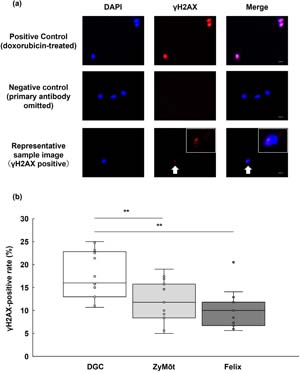
Figure 1. γH2AX immunofluorescence staining and comparison of γH2AX-positive rates among different sperm selection methods(a) Immunofluorescence staining of human sperm nuclei with DAPI and γH2AX. DNA was counterstained with DAPI (blue), and γH2AX immunofluorescence (red) was used to detect nuclear γH2AX signals as a surrogate marker associated with DSB-related DNA damage signaling and chromatin alterations. The positive control (sperm treated with 20 µM doxoru-bicin for 1 h) exhibited clear γH2AX signals, whereas omission of the primary antibody served as a negative control. These controls were used to validate staining performance and were not intended to represent method-specific imag-es. Arrows indicate γH2AX-positive nuclei. Panels from left to right: DAPI, γH2AX, and merged images. Scale bar=5 µm.(b) Comparison of sperm γH2AX-positive rates among different sperm selection methods. Box plots indicate the median, interquartile range (IQR), and individual data points. Overall differences were tested by the Friedman test, followed by Wil-coxon signed-rank tests with Bonferroni correction. Effect sizes are reported as Hodges-Lehmann estimated paired differ-ences (percentage points) with 95% confidence intervals. Asterisks indicate statistically significant differences (**adjusted p<0.01). DAPI, 4',6-diamidino-2-phenylindole.
Additional evaluations
In addition to the γH2AX-positive rate, basic sperm characteristics and processing efficiency were evaluated for each preparation method. The parameters assessed included sperm recovery, total motility, and laboratory workflow processing time.Sperm recovery was determined by measuring the sperm concentration in the 0.5 mL suspension obtained after processing and expressed as the total sperm count (×106 per 0.5 mL). Total motility was calculated as the percentage of motile spermatozoa among the total sperm population observed under a microscope using a Makler chamber. These assessments were performed as part of routine laboratory practice by trained embryologists according to standardized procedures under an internal quality control program. Processing time was defined a priori as the expected duration (min) from the start of sperm preparation to completion of sperm collection based on the standardized protocol for each method, including hands-on steps and device-related waiting periods (e.g., centrifugation, incubation, or electrophoresis). This metric was not subjected to statistical testing because it reflects method-specific workflow rather than participant-level variability.
Statistical analysis
Comparisons among the three sperm preparation methods were performed using the Friedman test, a nonparametric test for related samples. When significant differences were found, pairwise comparisons were conducted using the Wilcoxon signed-rank test with Bonferroni correction for multiple testing. For pairwise comparisons, effect sizes were summarized as Hodges-Lehmann estimated paired differences with 95% confidence intervals. In an exploratory analysis, Spearman’s rank correlation coefficient (ρ) was used to assess the relationship between patient age and the γH2AX-positive rate for each sperm preparation method. Because correlations were examined separately for three methods, results were interpreted cautiously in light of multiplicity. Statistical analyses were performed using EZR (version 1.61; Saitama Medical Center, Jichi Medical University, Japan) (Kanda, 2013), which is a graphical user interface for R, and two-tailed p-values<0.05 were considered statistically significant. All data are presented as medians with an interquartile range (IQR), as required for nonparametric statistical reporting. Processing time was not subjected to statistical testing because it was defined a priori as a method-specific workflow metric rather than measured as a participant-level variable.
RESULTS
Patient characteristics
A total of 15 semen samples were analyzed in this study. The median age of the patients was 33 years (IQR, 31.0-34.0 years). The median time interval from ejaculation to sperm preparation was 130 min (IQR, 30.5-180.0 min). The median semen volume, sperm concentration, and total motility were 3.2 mL (IQR, 2.5-4.6), 68.0 × 10⁶/mL (IQR, 46.5-89.0), and 48.9% (IQR, 45.9-60.7), respectively (Table 1).
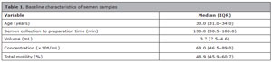
Table 1. Baseline characteristics of semen samplesVariable Median (IQR) Age (years) 33.0 (31.0-34.0) Semen collection to preparation time (min) 130.0 (30.5-180.0) Volume (mL) 3.2 (2.5-4.6) Concentration (×106/mL) 68.0 (46.5-89.0) Total motility (%) 48.9 (45.9-60.7) Semen characteristics of the study samples (n=15). Values are presented as median (IQR). IQR, interquartile range. Ab-stinence duration was not systematically recorded; participants were instructed to maintain 2-7 days of abstinence prior to collection. Motility represents total motility assessed visually using a Makler chamber during routine semen analysis by trained embryologists under routine internal quality control.
γH2AX-positive rate
The proportion of γH2AX-positive spermatozoa differed significantly among the three preparation methods (Friedman χ2=19.61, df=2, p<0.001). The median (IQR) γH2AX-positive rate was 16.0% (14.0-22.7) for DGC, 11.8% (8.4-15.4) for ZyMōt, and 10.0% (7.0-11.0) for Felix. Post hoc Wilcoxon signed-rank tests with Bonferroni correction showed that both ZyMōt and Felix yielded significantly lower γH2AX-positive rates than DGC (Hodges-Lehmann estimated paired difference, DGC minus ZyMōt, 5.88 percentage points; 95% confidence interval, 3.91-8.40; adjusted p=0.0041; and DGC minus Felix, 7.06 percentage points; 95% confidence interval, 5.00-10.66; adjusted p=0.0022), whereas there was no significant difference between ZyMōt and Felix (ZyMōt minus Felix, 2.40 percentage points; 95% confidence interval, -1.20 to 5.34; adjusted p=0.5460; Fig. 1b).
Exploratory correlation between patient age and γH2AX-positive rate
As an exploratory analysis, Spearman’s rank correlation coefficient (ρ) was used to examine the association between patient age and the γH2AX-positive rate for each sperm preparation method. Sperm prepared by DGC showed a moderate positive correlation between age and the proportion of γH2AX-positive spermatozoa (ρ=0.516, p=0.049; Fig. 2a). However, this was an exploratory analysis and multiplicity arising from testing correlations across three methods was not accounted for; therefore, the finding should be interpreted cautiously. In contrast, no significant correlations were observed for ZyMōt (ρ=0.229, p=0.411; Fig. 2b) or Felix (ρ=-0.103, p=0.714; Fig. 2c).
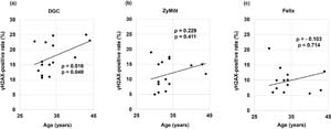
Figure 2. Exploratory correlation between patient age and γH2AX-positive rateScatter plots show the relationship between patient age and the γH2AX-positive rate (%) in sperm prepared by (a) DGC, (b) ZyMōt, and (c) Felix (n=15). Spearman’s rank correlation coefficient (ρ) and the corresponding two-tailed p-value are shown in each panel. This analysis was exploratory; because correlations were assessed across three methods, p-values should be interpreted cautiously. Solid lines indicate linear trend lines for visualization only.
Sperm recovery
A significant overall difference in sperm recovery was observed among the three methods (Friedman χ2=17.39, df=2, p<0.001). The number of recovered spermatozoa (median [IQR], ×106) was 4.5 (3.2-11.5) for DGC, 4.0 (1.9-5.8) for ZyMōt, and 2.25 (0.83-3.65) for Felix. Post-hoc tests with Bonferroni correction showed that DGC yielded significantly higher sperm recovery than ZyMōt (Hodges-Lehmann estimated paired difference, DGC minus ZyMōt, 3.06 × 10⁶; 95% confidence interval, 0.75-6.30; adjusted p=0.020) or Felix (DGC minus Felix, 3.00 × 10⁶; 95% confidence interval, 1.63-6.60; adjusted p<0.001), whereas there was no significant difference between ZyMōt and Felix (ZyMōt minus Felix, 1.13 × 10⁶; 95% confidence interval, -0.52 to 2.72; adjusted p=0.27; Fig. 3a).
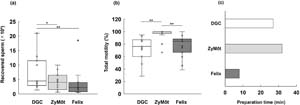
Figure 3. Comparison of outcomes among different sperm selection methods(a) Number of recovered sperm (×106 per 0.5 mL). (b) Sperm total motility (%). (c) Workflow-defined processing time (min). Box plots indicate the median, interquartile range (IQR), and individual data points for (a) and (b). Overall differenc-es were assessed by the Friedman test, followed by Wilcoxon signed-rank tests with Bonferroni correction; effect sizes are reported as Hodges-Lehmann estimated paired differences with 95% confidence intervals. Preparation time (c) represents fixed durations defined a priori by the standardized protocol for each method (hands-on steps plus device-related waiting periods) and therefore was not subjected to statistical testing. Asterisks indicate significant differences after pairwise com-parisons with Bonferroni correction (**adjusted p<0.01, *adjusted p<0.05).
Sperm total motility
Sperm total motility also differed significantly among the three methods (Friedman χ2=19.97, df=2, p<0.001). The median (IQR) total motility was 76.3% (65.7-86.8) for DGC, 84.6% (70.5-87.9) for Felix, and 100% (98.5- 100.0) for ZyMōt. Post-hoc tests with Bonferroni correction indicated that ZyMōt achieved significantly higher total motility than both DGC (Hodges-Lehmann estimated paired difference, ZyMōt minus DGC, 22.67 percentage points; 95% confidence interval, 12.89-31.86; adjusted p<0.001) and Felix (ZyMōt minus Felix, 19.23 percentage points; 95% confidence interval, 10.35-29.55; adjusted p=0.006), whereas no significant difference was observed between DGC and Felix (Felix minus DGC, 4.30 percentage points; 95% confidence interval, -3.50 to 11.84; adjusted p=1.00; Fig. 3b).
Processing time
The workflow-defined processing time (based on the standardized protocol for each method) differed markedly among the three methods: 8 min for Felix, 27 min for DGC, and 32 min for ZyMōt. These values represent typical end-to-end laboratory workflow durations, including hands-on steps and device-related waiting periods, rather than participant-level measured outcomes; therefore, no statistical testing was performed. Accordingly, Felix required a substantially shorter workflow processing time than the other two methods (Fig. 3c).
DISCUSSION
In this study, we compared three sperm selection methods, DGC, microfluidic system (ZyMōt), and electrophoretic system (Felix), focusing on the γH2AX-positive rate as a surrogate marker associated with DSB-related DNA damage signaling, sperm recovery, total motility, and processing time. Both non-centrifugal systems, ZyMōt and Felix, showed significantly lower γH2AX-positive rates than DGC (Fig. 1b). In relative terms, ZyMōt reduced the proportion of γH2AX-positive spermatozoa by approximately 26%, whereas Felix achieved nearly a 38% reduction, suggesting that non-centrifugal systems may be associated with lower levels of DSB-associated DNA damage signaling in this cohort. Furthermore, ZyMōt achieved higher total motility (Fig. 3b), whereas Felix offered a shorter processing time (Fig. 3c), highlighting distinct practical advantages of each system. To our knowledge, few studies have directly compared these three methods under identical conditions, particularly with respect to DSB-associated sperm DNA damage.
Normal embryo development after fertilization requires the genomic integrity of both sperm and oocytes. Considering the highly condensed chromatin, mature spermatozoa have minimal capacity to repair DNA damage; post-fertilization repair therefore depends on maternal factors (Newman et al., 2022). However, the DNA repair capacity of oocytes declines with advancing maternal age. Horta et al. reported that aged mouse oocytes exhibited reduced expression of DNA repair-related genes and diminished repair responses, resulting in a markedly lower blastocyst formation rate when fertilized with sperm populations exhibiting high levels of DNA damage compared with young oocytes (Horta et al., 2020). Setti et al. (2021) demonstrated that advanced maternal age is associated with lower blastocyst formation, pregnancy, and live birth rates, as well as increased miscarriage rates, suggesting that sperm DNA damage may contribute to adverse clinical outcomes. Moreover, because the repair response is substantially reduced after embryonic genome activation, unrepaired damage can manifest as “late paternal effects,” leading to impaired embryo development and adverse clinical outcomes (Tesarik et al., 2004; Pardiñas et al., 2026). Among types of DNA damage, DSBs are generally considered more challenging to repair and have been associated in some studies with adverse ART outcomes (Casanovas et al., 2019; Agarwal et al., 2020a; García-Rodríguez et al., 2018). Thus, DSB-related sperm DNA damage may be clinically relevant in some clinical contexts; however, whether selecting sperm with lower γH2AX positivity improves ART outcomes remains to be determined.
In interpreting our results, it is important to consider prior evidence suggesting that sperm DSBs may represent clinically relevant DNA damage that is not captured by standard semen parameters. Lara-Cerrillo et al. reported that, although SSBs were markedly reduced after Swim-up and ICSI-related sperm selection, DSB levels remained largely unchanged across ejaculated sperm, Swim-up-selected sperm, and sperm used for ICSI (Lara-Cerrillo et al., 2021). Their multimodal assessment, including the neutral Comet assay, further indicated that DSBs were not reliably associated with sperm motility, implying that selecting morphologically normal and progressively motile spermatozoa alone may be insufficient to reduce the DSB burden. Importantly, recurrent miscarriage cases in that study exhibited significantly higher DSB levels despite comparatively normal semen parameters, underscoring the clinical relevance of DSBs as a biomarker of underlying genomic instability rather than conventional semen quality (Lara-Cerrillo et al., 2021). These observations are consistent with reports that DSBs, unlike SSBs, arise predominantly from enzymatic events during meiosis and chromatin remodeling and rely on limited, error-prone repair pathways after fertilization (Agarwal et al., 2020a). Taken together with our γH2AX-based assessment, these data support the concept that DSB-associated sperm DNA damage may represent a relevant subset of sperm DNA damage in ART and that preparation methods capable of lowering the γH2AX-positive rate may be relevant at the laboratory level, although confirmation using prospective clinical outcome data is required.
Among the available methods for detecting DSB-associated sperm DNA damage, γH2AX immunostaining is widely used as a sensitive marker of DNA damage signaling (Heylmann & Kaina, 2016), and its association with clinical outcomes has also been reported (Garolla et al., 2015). However, γH2AX signals in mature sperm should be interpreted cautiously because chromatin compaction and epitope accessibility can influence detection, and γH2AX does not provide a definitive quantification of DSB number or canonical repair-foci dynamics. The use of γH2AX in the present study is therefore appropriate as a laboratory-level surrogate endpoint, but it should be considered complementary to established SDF assays rather than intrinsically superior in clinical relevance in the absence of outcome data. Accordingly, whether lower γH2AX positivity after ZyMōt or Felix translates into improved fertilization, embryo development, or pregnancy outcomes remains to be determined. Integrating γH2AX with established assays such as TUNEL or SCSA may help further clarify its utility as a complementary biomarker.
Our findings are consistent with previous reports, which showed a reduction in the DNA fragmentation index with ZyMōt (Hsu et al., 2023), and decreased DNA damage following Felix treatment, as reported by Shapouri et al. (2023) and Villeneuve et al. (2023), thereby supporting the utility of non-centrifugal sperm selection methods. In DGC, spermatozoa are subjected to centrifugal forces, which can disrupt the delicate structure of the sperm membrane and promote excessive production of ROS within the pellet and surrounding medium (Aitken & Clarkson, 1988). Increased ROS levels can rapidly induce DNA damage, including DSBs, even in spermatozoa that appear morphologically normal and otherwise of good quality. In contrast, microfluidic and electrophoretic approaches avoid centrifugation and therefore may require fewer handling steps and impose less mechanical stress. The ZyMōt system is a microfluidic device that uses parallel microchannels connecting the inlet and outlet chambers; sperm separation relies on the intrinsic motility of higher-quality spermatozoa that actively swim through these channels into the collection chamber, thereby depleting less motile or damaged sperm without exposure to centrifugation or marked shear forces (Gisbert Iranzo et al., 2025). The Felix system is an electrophoretic device that separates spermatozoa in a gentle electric field according to their negative surface charge (zeta potential), a hallmark of mature sperm. By minimizing physical stress and maintaining a near-physiological environment, it is regarded as a low-stress, non-centrifugal alternative (Cariati et al., 2024). The comparable reductions in γH2AX positivity achieved by these two distinct systems are consistent with the broader applicability of these findings, although confirmation in larger studies is required.
In the present cohort, a moderate positive correlation between patient age and the γH2AX-positive rate was observed only in sperm prepared by DGC (Fig. 2a), whereas no significant associations were detected for ZyMōt (Fig. 2b) or Felix (Fig. 2c). While this analysis is exploratory and limited by the small sample size and relatively narrow age range, it raises the possibility that centrifugation-based preparation could be more sensitive to age-related variation in DNA integrity; however, this inference remains speculative and should be interpreted cautiously because multiplicity across three methods was not formally accounted for. Aging is accompanied by increased oxidative stress and a decline in the efficiency of DNA repair pathways in the male germ line and semen (Nguyen-Powanda & Robaire, 2020; Herati et al., 2017; Elías-Llumbet et al., 2025; Nago et al., 2021). In vitro manipulations that involve centrifugation have been suggested to generate additional oxidative stress (Fleming et al., 2025), which could further increase the burden of DSBs in older men. By contrast, non-centrifugal systems such as ZyMōt and Felix may help to minimize exposure to age-related variation in sperm DNA integrity by avoiding high g-forces and reducing the generation of ROS during processing. Larger studies explicitly designed to investigate paternal age across different preparation methods will be required to confirm these hypotheses.
Regarding sperm recovery, the DGC group yielded the highest number of spermatozoa, whereas ZyMōt and Felix resulted in lower recovery (Fig. 3a). This difference is likely attributable to the inherent principles of each method. DGC recovers a broad sperm fraction by centrifugation, achieving high yield efficiently (Fleming et al., 2025). In contrast, ZyMōt employs a microchannel structure that enriches progressively motile sperm capable of migrating through microchannels, and previous studies have also reported lower absolute sperm recovery compared with DGC (Hsu et al., 2023). Similarly, Felix separates spermatozoa based on electrophoretic mobility, enriching mature cells characterized by negative surface charge, intact membranes, and appropriate size (Shapouri et al., 2023; Villeneuve et al., 2023). Regarding sperm total motility, the ZyMōt yielded the highest total motility, whereas Felix showed total motility comparable to DGC (Fig. 3b). This observation is consistent with the underlying principles of each method. ZyMōt selectively isolates sperm capable of actively migrating through microchannels, thereby enriching progressively motile spermatozoa (Hsu et al., 2023). In contrast, Felix separates sperm based on surface charge and membrane integrity rather than motility itself. Consequently, some spermatozoa that retain a negative surface charge but have lost motility may also be recovered, resulting in motility rates similar to those observed with DGC (Shapouri et al., 2023; Villeneuve et al., 2023). Regarding processing time, the Felix group exhibited the shortest duration among the three methods (Fig. 3c). Felix is based on an electrophoretic principle and employs a standardized protocol that enables completion of sperm separation within a few minutes, offering a practical advantage that may improve workflow efficiency, particularly in laboratory settings facing workforce constraints (Shirasawa et al., 2024).
Taken together, these characteristics indicate that the three sperm preparation systems should be regarded as complementary tools that can be selected according to laboratory priorities and practical constraints rather than as direct competitors. From a laboratory standpoint, non-centrifugal approaches may be considered when the goal is to enrich sperm populations with lower γH2AX positivity; however, any clinical benefit remains hypothetical without prospective outcome data. Conversely, in clinical scenarios in which high sperm yield is essential or access to microfluidic and electrophoretic devices is limited, conventional DGC remains a pragmatic option supported by its superior recovery.
A key strength of this study is the within-sample design, in which each ejaculate was processed in parallel by all three methods, thereby controlling for inter-individual variability in baseline semen characteristics and enabling robust paired comparisons. Another strength is the focus on γH2AX immunofluorescence as a surrogate marker associated with DSB-related DNA damage signaling, rather than asserting direct quantification of DSB number or repair foci formation in mature sperm. By combining a DSB-associated surrogate endpoint (γH2AX positivity) with practical laboratory metrics, our study provides information that may support evidence-based selection of sperm preparation methods in ART laboratories.
This study has several limitations. The sample size was small (n=15) and derived from a single center, and we limited our assessment to laboratory outcomes without evaluating fertilization, embryo development, implantation, or clinical pregnancy. In addition, sperm total motility was evaluated visually rather than by computer-assisted analysis. Although γH2AX immunofluorescence was used as a sensitive surrogate marker associated with DSB-related DNA damage signaling, its detection in mature sperm may be influenced by highly compacted chromatin and potential method-related differences in chromatin/ epitope accessibility and antibody binding, which could affect signal intensity and classification. In addition, because sperm concentration and the applied volume for smear preparation were not strictly standardized or recorded prior to staining, method-related differences in smear density and background fluorescence may have influenced γH2AX signal detectability. Scoring was performed by a single blinded evaluator; therefore, inter-observer variability was not applicable, but intra-observer repeatability was not formally assessed. Despite these limitations, our findings provide preliminary evidence that non-centrifugal sperm selection methods may reduce the proportion of γH2AX-positive spermatozoa compared with DGC under our laboratory conditions. Larger, multicenter studies incorporating clinical outcome data are needed to determine whether method-dependent differences in γH2AX positivity are associated with ART outcomes and to optimize protocols for each preparation method.
CONCLUSIONS
This study directly compared three sperm preparation methods under identical conditions and showed that the non-centrifugal systems ZyMōt and Felix yielded lower proportions of γH2AX-positive spermatozoa than DGC while maintaining comparable overall performance. These findings suggest that non-centrifugal sperm preparation may be considered when minimizing DSB-associated DNA damage signaling is a laboratory priority. Larger prospective studies are required to determine whether reductions in γH2AX positivity achieved by these methods translate into improved fertilization, embryo development, pregnancy, and live birth outcomes in ART.
REFERENCES
Agarwal A, Ikemoto I, Loughlin KR. Effect of sperm washing on levels of reactive oxygen species in semen. Arch Androl. 1994;33:157-62. PMID: 7857166 DOI: 10.3109/01485019408987819
Medline
Agarwal A, Mulgund A, Hamada A, Chyatte MR. A unique view on male infertility around the globe. Reprod Biol Endocrinol. 2015;13:37. PMID: 25928197 DOI: 10.1186/ s12958-015-0032-1
Medline
Agarwal A, Barbăroşie C, Ambar R, Finelli R. The impact of singleand double-strand DNA breaks in human spermatozoa on assisted reproduction. Int J Mol Sci. 2020a;21:3882. PMID: 32485940 DOI: 10.3390/ijms21113882
Medline
Agarwal A, Majzoub A, Baskaran S, Panner Selvam MK, Cho CL, Henkel R, Finelli R, Leisegang K, Sengupta P, Barbarosie C, Parekh N, Alves MG, Ko E, Arafa M, Tadros N, Ramasamy R, Kavoussi P, Ambar R, Kuchakulla M, Robert KA, et al. Sperm DNA Fragmentation: A New Guideline for Clinicians. World J Mens Health. 2020b;38:412-71. PMID: 32777871 DOI: 10.5534/wjmh.200128
Medline
Aitken RJ, Clarkson JS. Significance of reactive oxygen species and antioxidants in defining the efficacy of sperm preparation techniques. J Androl. 1988;9:367-76. PMID: 3215823 DOI: 10.1002/j.1939-4640.1988.tb01067.x
Medline
Cariati F, Orsi MG, Bagnulo F, Del Mondo D, Vigilante L, De Rosa M, Sciorio R, Conforti A, Fleming S, Alviggi C. Advanced sperm selection techniques for assisted reproduction. J Pers Med. 2024;14:726. PMID: 39063980 DOI: 10.3390/jpm14070726
Medline
Casanovas A, Ribas-Maynou J, Lara-Cerrillo S, Jimenez-Macedo AR, Hortal O, Benet J, Carrera J, García-Peiró A. Double-stranded sperm DNA damage is a cause of delay in embryo development and can impair implantation rates. Fertil Steril. 2019;111:699-707.e1. PMID: 30826116 DOI: 10.1016/j.fertnstert.2018.11.035
Medline
Coban O, Serdarogullari M, Yarkiner Z, Serakinci N. Investigating the level of DNA double-strand break in human spermatozoa and its relation to semen characteristics and IVF outcome using phospho-histone H2AX antibody as a biomarker. Andrology. 2020;8:421-6. PMID: 31393082 DOI: 10.1111/andr.12689
Medline
Elías-Llumbet A, Lira S, Manterola M. Male aging in germ cells: what are we inheriting? Genet Mol Biol. 2025;47:e20240052. PMID: 39969160 DOI: 10.1590/1678-4685-GMB-2024-0052
Medline
Fleming S, Morroll D, Nijs M. Sperm Separation and Selection Techniques to Mitigate Sperm DNA Damage. Life (Basel). 2025;15:302. PMID: 40003711 DOI: 10.3390/life15020302
Medline
García-Rodríguez A, Gosálvez J, Agarwal A, Roy R, Johnston S. DNA damage and repair in human reproductive cells. Int J Mol Sci. 2018;20:31. PMID: 30577615 DOI: 10.3390/ijms20010031
Medline
Garolla A, Cosci I, Bertoldo A, Sartini B, Boudjema E, Foresta C. DNA double-strand breaks in human spermatozoa can be predictive for assisted reproductive outcome. Reprod Biomed Online. 2015;31:100-7. PMID: 25985994 DOI: 10.1016/j.rbmo.2015.03.009
Medline
Gisbert Iranzo A, Cano-Extremera M, Hervás I, Falquet Guillem M, Gil Juliá M, Navarro-Gomezlechon A, Pacheco-Rendón RM, Garrido N. Sperm selection using microfluidic techniques significantly decreases sperm DNA fragmentation (SDF), enhancing reproductive outcomes: A systematic review and meta-analysis. Biology (Basel). 2025;14:792. PMID: 40723352 DOI: 10.3390/biology14070792
Medline
Halvaei I, Litzky J, Esfandiari N. Advanced paternal age: effects on sperm parameters, assisted reproduction outcomes and offspring health. Reprod Biol Endocrinol. 2020;18:110. PMID: 33183337 DOI: 10.1186/s12958-020-00668-y
Medline
Herati AS, Zhelyazkova BH, Butler PR, Lamb DJ. Age-related alterations in the genetics and genomics of the male germ line. Fertil Steril. 2017;107:319-23. PMID: 28160920 DOI: 10.1016/j.fertnstert.2016.12.021
Medline
Heylmann D, Kaina B. The γH2AX DNA damage assay from a drop of blood. Sci Rep. 2016;6:22682. PMID: 26940638 DOI: 10.1038/srep22682
Medline
Horta F, Catt S, Ramachandran P, Vollenhoven B, Temple-Smith P. Female ageing affects the DNA repair capacity of oocytes in IVF using a controlled model of sperm DNA damage in mice. Hum Reprod. 2020;35:529-44. PMID: 32108237 DOI: 10.1093/humrep/dez308
Medline
Hsu CT, Lee CI, Lin FS, Wang FZ, Chang HC, Wang TE, Huang CC, Tsao HM, Lee MS, Agarwal A. Live motile sperm sorting device for enhanced sperm-fertilization competency: comparative analysis with density-gradient centrifugation and microfluidic sperm sorting. J Assist Reprod Genet. 2023;40:1855-64. PMID: 37300647 DOI: 10.1007/s10815-023-02838-4
Medline
Kaltsas A, Zikopoulos A, Vrachnis D, Skentou C, Symeonidis EN, Dimitriadis F, Stavros S, Chrisofos M, Sofikitis N, Vrachnis N, Zachariou A. Advanced Paternal Age in Focus: Unraveling Its Influence on Assisted Reproductive Technology Outcomes. J Clin Med. 2024;13:2731. PMID: 38792276 DOI: 10.3390/jcm13102731
Medline
Kanda Y. Investigation of the freely available easy-to-use software ‘EZR’ for medical statistics. Bone Marrow Transplant. 2013;48:452-8. PMID: 23208313 DOI: 10.1038/bmt.2012.244
Medline
Lara-Cerrillo S, Ribas-Maynou J, Rosado-Iglesias C, Lacruz-Ruiz T, Benet J, García-Peiró A. Sperm selection during ICSI treatments reduces singlebut not double-strand DNA break values compared to the semen sample. J Assist Reprod Genet. 2021;38:1187-96. PMID: 33660206 DOI: 10.1007/s10815-021-02129-w
Medline
Li N, Wang H, Zou S, Yu X, Li J. Perspective in the mechanisms for repairing sperm DNA damage. Reprod Sci. 2025;32:41-51. PMID: 39333437 DOI: 10.1007/s43032-024-01714-5
Medline
Muratori M, Marchiani S, Tamburrino L, Baldi E. Sperm DNA Fragmentation: Mechanisms of Origin. Adv Exp Med Biol. 2019;1166:75-85. PMID: 31301047 DOI: 10.1007/978-3-030-21664-1_5
Medline
Nago M, Arichi A, Omura N, Iwashita Y, Kawamura T, Yumura Y. Aging increases oxidative stress in semen. Investig Clin Urol. 2021;62:233-8. PMID: 33660452 DOI: 10.4111/icu.20200066
Medline
Newman H, Catt S, Vining B, Vollenhoven B, Horta F. DNA repair and response to sperm DNA damage in oocytes and embryos, and the potential consequences in ART: a systematic review. Mol Hum Reprod. 2022;28:gaab071. PMID: 34954800 DOI: 10.1093/molehr/gaab071
Medline
Nguyen-Powanda P, Robaire B. Oxidative stress and reproductive function in the aging male. Biology (Basel). 2020;9:282. PMID: 32932761 DOI: 10.3390/biology9090282
Medline
Pardiñas ML, de Celis C, Gil J, Ortega-Jaen D, Martin A, Mercader A, de los Santos MJ. Oocyte-mediated repair of sperm DNA fragmentation: a critical determinant of embryo viability. Reprod Biomed Online. 2026;52:105165. PMID: 41198515 DOI: 10.1016/j.rbmo.2025.105165
Medline
Setti AS, Braga DPAF, Provenza RR, Iaconelli A, Borges E. Oocyte ability to repair sperm DNA fragmentation: the impact of maternal age on intracytoplasmic sperm injection outcomes. Fertil Steril. 2021;116:123-9. PMID: 33589137 DOI: 10.1016/j.fertnstert.2020.10.045
Medline
Shapouri F, Mahendran T, Govindarajan M, Xie P, Kocur O, Palermo GD, Bakos HW, Ahlström A, Caisander G, Xu B, Bai S, Lambourne S, Aitken RJ. A comparison between the Felix™ electrophoretic system of sperm isolation and conventional density gradient centrifugation: a multicentre analysis. J Assist Reprod Genet. 2023;40:83-95. PMID: 36515800 DOI: 10.1007/s10815-022-02680-0
Medline
Shirasawa H, Yamada M, Jwa SC, Kuroda K, Harada M, Osuga Y. Assessment of embryologist sufficiency and associated regional disparities in Japanese assisted reproduction facilities using nationwide survey data (IZANAMI project). J Obstet Gynaecol Res. 2024;50:1459-69. PMID: 38961649 DOI: 10.1111/jog.16022
Medline
Simon L, Zini A, Dyachenko A, Ciampi A, Carrell DT. A systematic review and meta-analysis to determine the effect of sperm DNA damage on in vitro fertilization and intracytoplasmic sperm injection outcome. Asian J Androl. 2017;19:80-90. PMID: 27345006. DOI: 10.4103/1008-682X.182822
Medline
Tesarik J, Greco E, Mendoza C. Late, but not early, paternal effect on human embryo development is related to sperm DNA fragmentation. Hum Reprod. 2004;19:611-5. PMID: 14998960 DOI: 10.1093/humrep/deh127
Medline
Vander Borght M, Wyns C. Fertility and infertility: definition and epidemiology. Clin Biochem. 2018;62:2-10. PMID: 29555319 DOI: 10.1016/j.clinbiochem.2018.03.012
Medline
Villeneuve P, Saez F, Hug E, Chorfa A, Guiton R, Schubert B, Force A, Drevet JR. Spermatozoa isolation with Felix™ outperforms conventional density gradient centrifugation preparation in selecting cells with low DNA damage. Andrology. 2023;11:1593-604. PMID: 36629014 DOI: 10.1111/andr.13384
Medline
Zhao J, Zhang Q, Wang Y, Li Y. Whether sperm deoxyribonucleic acid fragmentation has an effect on pregnancy and miscarriage after in vitro fertilization/ intracytoplasmic sperm injection: a systematic review and meta-analysis. Fertil Steril. 2014;102:998-1005.e8. PMID: 25190048 DOI: 10.1016/j.fertnstert.2014.06.033
Medline