JBRA Assist. Reprod. 2026;30(1):132-141
ORIGINAL ARTICLE
doi: 10.5935/1518-0557.20250171
Introducing the Blastocyst Fragmentation Indicator (BFI): A Novel Time-Lapse Metric for Enhanced Aneuploidy Risk Stratification in Non-Invasive Embryo Assessment
1Bios Human Reproduction Center, Clínica Evangelista Torquato, Fortaleza, Ceará, Brazil
CONFLICT OF INTEREST
The authors have no competing interests to declare
ABSTRACT
Objective: To propose an improved blastocyst grading system by integrating dynamic time-lapse parameters with conventional morphological criteria, aiming to enhance embryo selection during assisted reproduction.
Methods: This retrospective cohort study evaluated 1,182 embryos derived from 433 cycles of intracytoplasmic sperm injection. Time-lapse parameters included morula compaction status, blastocyst collapse frequency, and fragmentation of the inner cell mass and trophectoderm. These findings were analyzed in conjunction with standard morphological grading and embryo scoring systems based on implantation data. Embryo outcomes were stratified by chromosomal status determined through preimplantation genetic testing for aneuploidy.
Results: Fully compacted morulae were associated with higher morphological grades; however, the proportion of chromosomally normal embryos did not differ significantly when compared to those derived from partially compacted morulae, suggesting that static morphology alone may not accurately reflect developmental potential. Although most blastocyst collapse events were minor, they correlated with increased fragmentation and reduced embryo quality. Fragmentation of the inner cell mass and/or the trophectoderm was associated with significantly lower rates of chromosomal normalcy. Logistic regression analysis confirmed fragmentation as the strongest predictor of aneuploidy, outperforming both conventional grading systems.
Conclusions: This integrated evaluation model, combining static morphology with dynamic developmental events, offers a more comprehensive and accurate approach to assessing embryo viability. By identifying embryos with higher risk of chromosomal abnormalities, this strategy may enhance embryo selection, improve reproductive outcomes, and contribute to more individualized care in assisted reproduction.
Keywords: Time-lapse monitoring, embryo selection, blastocyst fragmentation, embryo grading, chromosomal abnormalities
INTRODUCTION
The morphological evaluation of embryos, particularly at the blastocyst stage, is pivotal in embryo selection for transfer during in vitro fertilization (IVF) cycles. The Gardner and Schoolcraft grading system (1999) remains widely used for this purpose (Gardner & Schoolcraft, 1999), offering a morphological scoring system based on blastocyst expansion and the quality of both the inner cell mass (ICM) and the trophectoderm (TE). While effective, this approach has limitations due to its static and subjective nature, as it assesses embryos at specific time points without accounting for dynamic events that can influence implantation potential (ALPHA Scientists in Reproductive Medicine & ESHRE Special Interest Group of Embryology, 2011).
In recent years, time-lapse imaging technology has introduced a new dimension to embryo evaluation, enabling continuous observation of morphokinetic parameters in real time. Studies have demonstrated that incorporating dynamic data, such as the timing and synchrony of cell divisions, improves implantation success predictions compared to static morphological evaluation alone (Meseguer et al., 2011).
Time-lapse studies have shown that the timing and completeness of compaction are strong indicators of developmental potential (ESHRE Working group on Time-lapse technology et al., 2020). Fully compacted morulae are generally more likely to progress to high-quality blastocysts with robust chromosomal profiles, while partial or delayed compaction patterns are associated with reduced rates of blastocyst formation (Hur et al., 2023). Nevertheless, recent findings suggest that partially compacted morulae that do reach the blastocyst stage can exhibit euploidy rates comparable to those of fully compacted morulae. Counterintuitively, this indicates that chromosomal integrity can be maintained even in the presence of incomplete compaction (De Martin et al., 2024). These findings highlight morula compaction as a meaningful developmental milestone with predictive value for embryo selection.
Another crucial parameter enabled by time-lapse monitoring is blastocyst collapse, a transient reduction in blastocoel volume followed by re-expansion. Controlled collapse and rapid recovery may be indicative of natural cellular reorganization, while frequent collapses or delayed recovery are often markers of compromised viability (Sciorio & Meseguer, 2021; Cimadomo et al., 2022). These findings underline the value of monitoring collapse events and recovery patterns as indicators of developmental potential.
Finally, ICM and TE fragmentation are essential factors in embryo viability. The ICM, which forms fetal tissues, and the TE, which contributes to placental development, are critical to embryo competence. Excessive fragmentation within these regions may signal reduced developmental potential, with ICM fragmentation affecting fetal development and TE integrity influencing implantation success (Pribenszky et al., 2017; Ozguldez et al., 2020; Ai et al., 2021).
This study prioritizes the evaluation of ploidy status over pregnancy outcomes, in line with the recommendations of Bamford et al. (2023). We propose an integrative blastocyst grading system that complements the conventional Gardner classification by incorporating dynamic and advanced morphological parameters. Specifically, this study introduces inner cell mass (ICM) and trophectoderm (TE) fragmentation as a novel parameter, alongside morula compaction quality and blastocyst collapse frequency. By combining classical morphology assessment with these critical developmental events, our approach seeks to refine embryo selection in IVF and optimize clinical outcomes.
MATERIALS AND METHODS
Study Design
This retrospective cohort study is based on anonymized data from embryos generated by in vitro fertilization (IVF) and cultured in a time-lapse system. All data were collected as part of frozen embryo transfer (FET) IVF cycles performed routinely at a single assisted reproduction center. Only data from cycles involving ovarian stimulation, oocyte retrieval, fertilization via intracytoplasmic sperm injection (ICSI), embryo culture in a time-lapse incubator, and blastocyst biopsy for preimplantation genetic testing for aneuploidy (PGT-A) were included in the study. Written informed consent was obtained from all patients, authorizing the anonymous use of clinical and laboratory data for research purposes.
Data Collection
Data were obtained from 396 patients treated at a single assisted reproduction center. All clinical procedures followed the guidelines of the ASRM, ESHRE, SBRA, and CFM. The database includes 1182 embryos from 433 ICSI cycles performed between September 2022 and December 2024. Inclusion criteria were as follows: (i) embryos individually cultured and monitored from the zygote to blastocyst stage in a time-lapse incubator (EmbryoScope Plus, Vitrolife, Denmark); (ii) blastocysts analyzed by next-generation sequencing (NGS) following a single trophectoderm biopsy. The primary indication for IVF was advanced maternal age (≥ 37 years in 54.8% of cases), with unexplained infertility (11.3%), male factor (23.5%), and other factors (10.4%) accounting for the remainder.
Time-Lapse Monitoring and Assessment
Embryos were cultured in a time-lapse incubator, allowing continuous monitoring without disturbance to the culture environment. Developmental timing parameters were automatically recorded (for descriptive purposes only) and included the time to pronuclear fading (tPNf), 2-cell division (t2), 4-cell division (t4), 5-cell division (t5), 8-cell division (t8), morula stage (tM), onset of cavitation (tSB), full blastocyst formation (tB) and expanded blastocyst formation (tEB).
During monitoring, spontaneous collapse events were recorded and classified as either minor collapses or major collapses, with total counts computed for each type. Additionally, the presence or absence of fragmentation in the inner cell mass (ICM) and trophectoderm (TE) was noted, with N (Normal) indicating no fragmentation and F (Fragmented) indicating the presence of fragmentation. Embryos were then categorized into three groups based on the integrity of these structures: NN: ICM and TE intact (no fragmentation); FN: Fragmented ICM with intact TE; and FF: Fragmentation in both ICM and TE.
Figure 1 illustrates examples of embryos exhibiting ‘minor’ and ‘major’ collapses. A ‘minor collapse’ is defined by a localized or incomplete retraction of the trophectoderm (TE) away from the zona pellucida. A ‘major collapse’, conversely, describes a complete separation or detachment of the trophectoderm (TE) from the entire inner surface of the zona pellucida.
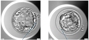
Figure 1. Spontaneous collapses: CL (Minor Collapse); MCL (Major Collapse).
Figure 2 illustrates embryos exhibiting different patterns of ICM/TE fragmentation. Embryos designated as FN are characterized by fragmentation that is either exclusive to, or primarily located in, the inner cell mass (ICMf). In contrast, embryos classified as FF display fragmentation involving both the inner cell mass (ICMf) and the trophectoderm (TEf).
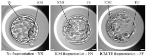
Figure 2. ICM/TE fragmentation.
In addition, blastocyst morphology was classified using the Gardner system and categorized into groups A (comprising AA, AB, BA), B (BB, BC, AC), and C (CA, CB, CC). KIDScore categories (KIDScore D5 v3.1) were defined as A (KIDScore ≥7), B (KIDScore 4-7), and C (KIDScore <4). Morula compaction was assessed and categorized as fully compacted (FCM), partially compacted with excluded cells (Exc-PCM), or partially compacted with extruded cells (Ext-PCM). This classification, which is illustrated in Figure 3, follows the criteria described by Lagalla et al. (2017).
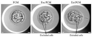
Figure 3. Morula compaction.
Genetic Analysis and Morphological Classification
Blastocysts were assessed for biopsy eligibility on days 5, 6, or 7 of development. Internal diameters were measured using EmbryoViewer software (Vitrolife). Trophectoderm biopsy, restricted to fully expanded blastocysts (diameter >160 μm), involved laser-assisted herniation to facilitate retrieval of 5-7 cells. Retrieved blastomeres underwent preimplantation genetic testing for aneuploidy (PGT-A) via next-generation sequencing to determine ploidy status. Post-biopsy, embryos were vitrified pending PGT-A results.
Statistical Analyses
Categorical variables were summarized as absolute numbers and percentages, and numerical variables as means, standard deviations (SD), and ranges. Trends in categorical variables were analyzed using the chi-square test. A Generalized Linear Model (GLM) with Poisson distribution assessed the association between ICM/TE fragmentation and blastocyst collapse frequency (minor/major). Logistic regression, controlling for maternal age, examined the association between blastocyst classification criteria (Gardner, KIDScore, and ICM/TE fragmentation) and ploidy status, and odds ratios (ORs) with 95% confidence intervals (CIs) were calculated. Statistical analyses were performed with JAMOVI software (version 2.6.13), with p<0.05 considered significant.
RESULTS
This study evaluated 1182 embryos from 433 ICSI cycles (396 patients) reaching the blastocyst stage, subsequently biopsied for PGT-A. Maternal age ranged from 26.0 to 44.9 years (mean 37.6 ± 3.5 years). Analyses were performed on morula compaction patterns, morphology (Gardner’s criteria), morphokinetics (KIDScore), spontaneous collapse events, and ICM/TE fragmentation, stratified by maternal age: <37 years (45.6%) and >37 years (54.4%). Descriptive data are summarized in Figure 4. The overall euploidy rate was 44.5%, with 54.7% and 35.9% observed in patients aged <37 and >37 years, respectively.
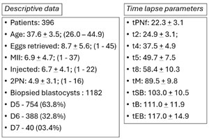
Figure 4. Descriptive data and time-lapse parameters.
Spontaneous Collapses
Minor collapses occurred in 69.0% of blastocysts, while major collapses were identified in 34.3% of embryos. Logistic regression analysis demonstrated a significant association between collapse events and euploidy (p<0.001); however, predictive power was limited, with AUC values of 0.597 for minor collapses and 0.588 for major collapses. Non-fragmented embryos (NN) exhibited the lowest frequencies of minor (0.893) and major collapses (0.243), which increased in partially fragmented (FN, minor = 1.735, major = 0.650) and fully fragmented embryos (FF, minor = 3.015, major = 1.421). GLM analysis (Figure 5) confirmed that ICM/TE fragmentation significantly increased the prevalence of blastocyst collapse. Compared to NN, FN embryos had 1.94- and 2.68-fold higher prevalence of minor (CL) and major (MCL) collapses, while FF embryos showed 3.38- and 5.86-fold increases (Figure 5).
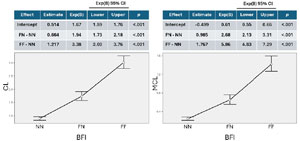
Figure 5. Frequency of Minor and Major Blastocyst Collapses According to Fragmentation Pattern. Generalized linear models (Poisson distribution with Bca bootstrap, 1,000 replications) were used to estimate the frequency of minor (CL, left panel) and major (MCL, right panel) collapse events in blastocysts, based on the distribution of inner cell mass (ICM) and trophectoderm (TE) fragmentation. Embryos were classified into three categories: NN (no fragmentation), FN (fragmentation restricted to the ICM), and FF (fragmentation in both ICM and TE).
Morula Compaction and Euploidy
Fully compacted morulae (FCM) accounted for 73.7% of the embryos analyzed, with 62.7% reaching Gardner grade A. In contrast, partially compacted morulae with excluded cells (Exc PCM) comprised 19.0% of the cohort, and only 20.1% were classified as grade A. Despite these marked differences in morphological quality, the euploidy rates were comparable between FCM (46.8%) and Exc PCM (45.5%), as shown in Figure 6. Notably, the majority of blastocysts exhibiting fragmentation restricted to the inner cell mass (FN) originated from FCM (70.9%), whereas only 21.8% derived from Exc PCM. A similar pattern was observed among blastocysts displaying concurrent fragmentation of both the inner cell mass and trophectoderm (FF), with 50.5% arising from FCM and 24.8% from Exc PCM.
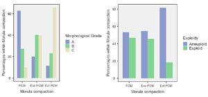
Figure 6. Morphological Scores and Euploidy Rates According to Morula Compaction Patterns. Distribution of morphological grades (A, B, C) (left panel) and euploidy status (euploid vs. aneuploid) (right panel) across three morula compaction categories: fully compacted morulae (FCM), partially compacted morulae with excluded cells (Exc PCM), and partially compacted morulae with extruded cells (Ext PCM).
Comparison of Selection Criteria
Euploidy rates increased alongside improvements in both Gardner grading (A: 56.2%, B: 35.3%, C: 27.9%) and KIDScore (grade A: 61.1%, B: 39.2%, C: 28.0%). However, KIDScore classified fewer embryos as grade A compared to the Gardner system (37.0% vs. 50.8%). In contrast, ICM/TE fragmentation exhibited the most predictive power for euploidy: non-fragmented (NN) embryos showed a 62.5% euploidy rate, whereas this was 20.1% for partially fragmented (FN), and only 6.4% for fully fragmented (FF) embryos. The contrasting patterns, including the much sharper decline in euploidy associated with ICM/TE fragmentation, are clearly presented in Figure 7.
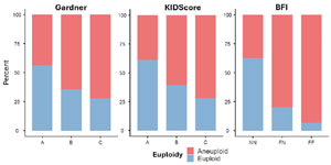
Figure 7. Euploidy Rates According to Gardner Grading, KIDScore, and the Blastocyst Fragmentation Indicator (BFI). Stacked bar plots showing the proportion of euploid (blue) and aneuploid (red) embryos across morphological grades (A, B, C) using the Gardner system (left panel), the KIDScore classification (middle panel), and the BFI categories (right panel): NN (no fragmentation), FN (fragmentation in the inner cell mass), and FF (fragmentation in both the inner cell mass and trophectoderm). The figure illustrates the distribution of chromosomal status within each scoring system.
Logistic regression, adjusted for maternal age, confirmed significant reductions in euploidy likelihood for lower Gardner grades (grade B: OR 0.467, 95% CI 0.353-0.619; grade C: OR 0.335, 95% CI 0.239-0.469) and KIDScore grades (B: OR 0.443, 95% CI 0.336-0.584; C: OR 0.278, 95% CI 0.201-0.386). However, ICM/TE fragmentation showed the most profound effect. Compared to non-fragmented embryos, FN and FF embryos showed an 84% (OR 0.157, 95% CI 0.110-0.225) and 95% (OR 0.047, 95% CI 0.026-0.085) reduction in euploidy likelihood, respectively. AUC values for predicting euploidy were 0.697 for KIDScore, 0.688 for Gardner grading, and 0.791 for ICM/TE fragmentation (Figure 8). Odds ratios for aneuploidy in the lowest categories were 3.593 (KIDScore), 2.986 (Gardner grading), and 21.036 (ICM/TE fragmentation).
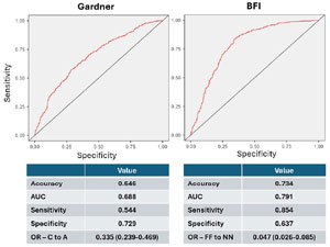
Figure 8. ROC Curves and Performance Metrics Comparing Gardner Grading and ICM/TE Fragmentation (BFI). Receiver operating characteristic (ROC) curves (top) and corresponding diagnostic metrics (bottom) comparing the predictive performance of the Gardner grading system (left panel) and the Blastocyst Fragmentation Indicator (BFI) based on inner cell mass (ICM) and trophectoderm (TE) fragmentation (right panel). Performance measures include accuracy, area under the curve (AUC), sensitivity, specificity, and odds ratios (OR) for the lowest scoring categories (Gardner grade C vs. A; FF vs. NN in BFI).
Association with Specific Types of Aneuploidy
Chi-squared analysis revealed a significant association between ICM/TE fragmentation and the type of aneuploidy (χ2 = 333.252, df = 10, p<0.001), with a graded increase in complex aneuploidies observed with greater fragmentation. Chaotic abnormalities were significantly more frequent in the FF group (31.7%) than the NN group (4.7%). Gardner morphological grading also showed a significant association with aneuploidy types (χ2 = 97.688, df = 10, p<0.001), but less pronounced than with fragmentation. Monosomies were more prevalent than trisomies in the FN (36.8%; 13,7%) and FF (35.1%; 14.4%) groups, whilst more balanced in NN embryos (12.7%; 13.1%). A striking discrepancy in aneuploidy patterns was found when comparing embryos classified by Gardner criteria (C) with those classified by ICM/TE fragmentation (FF), in contrast to the similarity between patterns observed for A and NN embryos (Figure 9).
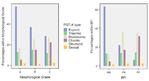
Figure 9. Distribution of PGT-A Aneuploidy Types According to Morphological Grade and BFI Classification. Stacked bar charts showing the relative distribution of preimplantation genetic testing for aneuploidy (PGT-A) results by aneuploidy type across morphological grades (left panel: A, B, C) and Blastocyst Fragmentation Indicator (BFI) categories (right panel: NN = no fragmentation, FN = fragmentation in inner cell mass, FF = fragmentation in both inner cell mass and trophectoderm).
Analysis of euploidy rates within each morphological category (A, B, C) was conducted for embryos ‘without fragmentation’ (NN) versus ‘with fragmentation’ (combining FN and FF). Fragmentation had a significant negative impact on euploidy rates (13.8%) compared with those without fragmentation (62.5%; p<0.001). Moreover, no significant differences in euploidy rate were observed for grade A, B, and C embryos within the fragmented group (12.8%, 12.4%, and 15.5%, respectively; p=0.682). A non-significant trend for euploidy was observed within the non-fragmented group. This confirms that fragmentation, more than morphology alone, serves as a strong negative indicator for euploidy (Figure 10).
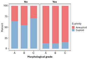
Figure 10. Euploidy Rates Across Morphological Grades According to the Presence or Absence of ICM/TE Fragmentation. Stacked bar plots representing the proportion of euploid (blue) and aneuploid (red) embryos across morphological grades A, B, and C, stratified by the absence (No, left panel) or presence (Yes, right panel) of fragmentation in the inner cell mass (ICM) and/or trophectoderm (TE). Each panel shows the chromosomal status distribution within each morphological category based on fragmentation status.
Novel Classification System
A novel classification system was developed by integrating morula compaction status (FCM = 1, Exc-PCM = 2, Ext-PCM = 3) with ICM/TE integrity (N for normal, F for fragmented). The proposed nomenclature (e.g., 1-NN, 2-FN, 3-FF) enables a dynamic and predictive assessment of embryo quality. No blastocysts with an intact ICM and fragmented TE were observed. Minor and major collapses, whilst recorded by embryologists as supplementary information, are not included in the final classification. This system offers a robust tool for embryo selection, given the independence and predictive strength of ICM/TE fragmentation.
DISCUSSION
This study introduces a simplified blastocyst ranking system based on time-lapse imaging parameters. The system integrates morula compaction, spontaneous blastocyst collapses, and, crucially, fragmentation of the inner cell mass (ICM) and trophectoderm (TE), providing a dynamic and detailed perspective of embryonic development to enhance embryo selection.
The single embryo transfer (SET) policy, widely adopted in assisted reproductive technology (ART) to minimize multiple pregnancy risks (Martin et al., 2017; Practice Committee of the Society for Reproductive Endocrinology and Infertility, 2022), relies on selecting the most viable embryo. While the Gardner scoring system remains prevalent (Gardner & Schoolcraft, 1999), its correlation with ploidy is limited. Morphologically superior blastocysts are more likely to be euploid, but this is not definitive; up to half of morphologically excellent blastocysts may be aneuploid (Alfarawati et al., 2011; Li et al., 2022). This study observed euploidy rates of 56.2% for “A” graded embryos and 27.9% for “C” graded embryos, highlighting the risk of relying solely on morphology. Time-lapse imaging offers a dynamic approach, with parameters such as cleavage timing linked to euploidy (Campbell et al., 2013; Zhan et al., 2020). However, this relationship is not absolute either (Rienzi et al., 2015; Barnes et al., 2023). For instance, optimal morphokinetics do not guarantee euploidy, and vice-versa. In this study, euploidy rates were 61.1% for embryos graded as “A” (KIDScore > 7), and 28.0% for “C” graded embryos (KIDScore<4), demonstrating the limitations of morphokinetic scores alone.
This study introduces ICM/TE fragmentation as a novel parameter. Embryos were classified as NN (no fragmentation), FN (ICM fragmentation), or FF (ICM and TE fragmentation). Euploidy rates were 62.5%, 20.1%, and 6.4% for NN, FN, and FF embryos, respectively, demonstrating the value of this parameter. This study achieved area under the curve (AUC) values for euploidy prediction, adjusted for maternal age, of 0.697, 0.688, and 0.791 for KIDScore, Gardner criteria, and ICM/TE fragmentation, respectively. These values indicate that ICM/TE fragmentation demonstrated better predictive capabilities than existing systems. For comparison, Kato et al. (2023) reported AUCs of 0.666, 0.655 and 0.642 for IDAScore, KIDScore and Gardner criteria (Kato et al., 2023). A recent meta-analysis using artificial intelligence for euploidy prediction yielded a combined AUC of 0.80 (95% CI 0.76 to 0.83) (Xin et al., 2024), consistent with our findings. Furthermore, odds ratios (ORs) for lower-grade classifications were 0.278, 0.335, and 0.047 for KIDScore, Gardner criteria, and ICM/TE fragmentation, indicating reduced euploidy probabilities of 72.2%, 66.5%, and 95.3%, respectively. Conversely, ORs for aneuploidy associated with lower grades were 3.593, 2.986, and 21.036, translating to 2.6-fold, 2.0-fold, and 20.0-fold increased odds of aneuploidy, respectively. These results align with prior studies showing associations between TE grading and euploidy/aneuploidy rates (Minasi et al., 2016; Wang et al., 2018).
Analysis of aneuploidies showed low-grade embryos had significantly higher rates of chaotic aneuploidy, particularly in Gardner grade C, compared with grade A. This was mirrored in ICM/TE fragmentation analysis, with FF embryos exhibiting a markedly increased frequency of chaotic aneuploidy compared with NN embryos. Monosomy predominated over trisomy in FN and FF embryos, whereas NN embryos exhibited a more balanced distribution of monosomies and trisomies, consistent across all Gardner grades. Prior studies demonstrate increased rates of apoptosis in aneuploid TE cells relative to the ICM (Starostik et al., 2020; Martin et al., 2023; Regin et al., 2024; Zhai et al., 2024) and poorer developmental outcomes in monosomic blastomeres (Shahbazi et al., 2020).
Morula compaction was also investigated (Lagalla et al., 2017, 2020). Partially compacted morulae (Exc-PCM) yielded lower morphological grades than fully compacted morulae (FCM), consistent with previous findings (De Martin et al., 2024; Parriego et al., 2024), but euploidy rates were similar. Morphology disparities may be due to excluded blastomeres compressed against the zona pellucida, resulting in a smoother TE appearance. Paradoxically, many FN and FF embryos originated from FCM, suggesting two subtypes within Gardner grade C: FCM-derived embryos with apoptotic events, and Exc-PCM-derived embryos with poorer morphology but potentially better prognosis. The clinical significance of these subtypes is underlined by studies reporting positive outcomes following the transfer of lower-quality embryos, particularly in patients with a poor prognosis (Cimadomo et al., 2019; Hill et al., 2020; Wang et al., 2020; He et al., 2024). These results support the hypothesis that many viable embryos are likely from the Exc-PCM group, emphasizing the importance of incorporating morula compaction as an additional parameter in embryo selection.
Spontaneous blastocyst collapses (Marcos et al., 2015; Sciorio & Meseguer, 2021; Cimadomo et al., 2022), categorized as minor or major, were investigated for predictive value. Minor collapses were more frequent, with limited predictive value for euploidy. However, increased collapse frequency correlated with higher fragmentation levels, especially in FF embryos.
Embryo selection using morphology or morphokinetics faces limitations in identifying meiotic aneuploidies (Fragouli et al., 2014; Reignier et al., 2018; Tartia et al., 2022). While our system has similar limitations, it shows strong correlations between ICM/TE fragmentation and monosomies/complex abnormalities. Euploidy rates remained consistently low in FN and FF embryos, regardless of morphology. Therefore, embryos without fragmentation could be prioritized for transfer or PGT-A.
There are limitations to the present study that warrant further consideration. This retrospective, single-center study used PGT-A results as a ground truth (Bamford et al., 2023; Barnes et al., 2023) and exclusively included embryos with sufficient quality for biopsy. Furthermore, not all embryos underwent PGT-A, particularly in younger women. This selection bias may explain the higher proportion of fully compacted morulae (73%) observed in this study compared with those of 51% (Parriego et al., 2024) and 57% (De Martin et al., 2024), leading to a potential underestimation of ICM/TE fragmentation.
Despite these limitations, this study offers significant strengths, including the standardization of qualitative parameters and the exclusive use of time-lapse imaging, which enables non-invasive embryo assessment. The novel parameters introduced here enhance the precision of embryo selection and hold promise for integration into automated systems, reducing operator variability and supporting broader clinical adoption.
CONCLUSION
This study demonstrates that a dynamic, event-based approach effectively complements traditional blastocyst assessment. The association between blastocyst collapse and subsequent ICM/TE fragmentation emerges as a valuable marker suggestive of monosomies or complex chromosomal abnormalities. In addition, the absence of ICM/TE fragmentation, along with tracking morula compaction patterns, may help identify embryos with developmental potential that are otherwise morphologically underestimated. The Blastocyst Fragmentation Indicator (BFI) provides embryologists with a practical tool to refine embryo selection and gain deeper insight into blastocyst development. Further prospective validation is needed to determine the clinical utility of BFI, particularly in improving the non-invasive identification of embryos with higher implantation potential.
REFERENCES
Ai J, Jin L, Zheng Y, Yang P, Huang B, Dong X. The Morphology of Inner Cell Mass Is the Strongest Predictor of Live Birth After a Frozen-Thawed Single Embryo Transfer. Front Endocrinol (Lausanne). 2021;12:621221. PMID: 33716973 DOI: 10.3389/fendo.2021.621221 Medline
Alfarawati S, Fragouli E, Colls P, Stevens J, Gutiérrez-Mateo C, Schoolcraft WB, Katz-Jaffe MG, Wells D. The relationship between blastocyst morphology, chromosomal abnormality, and embryo gender. Fertil Steril. 2011;95:520-4. PMID: 20537630 DOI: 10.1016/j.fertnstert.2010.04.003 Medline
ALPHA Scientists In Reproductive Medicine; ESHRE Special Interest Group Embryology. Istanbul consensus workshop on embryo assessment: proceedings of an expert meeting. Reprod Biomed Online. 2011;22:632-46. PMID: 39242260 DOI: 10.1016/j.rbmo.2011.02.001 Medline
Bamford T, Easter C, Montgomery S, Smith R, Dhillon-Smith RK, Barrie A, Campbell A, Coomarasamy A. A comparison of 12 machine learning models developed to predict ploidy, using a morphokinetic meta-dataset of 8147 embryos. Hum Reprod. 2023;38:569-81. PMID: 36825452 DOI: 10.1093/humrep/dead034 Medline
Barnes J, Brendel M, Gao VR, Rajendran S, Kim J, Li Q, Malmsten JE, Sierra JT, Zisimopoulos P, Sigaras A, Khosravi P, Meseguer M, Zhan Q, Rosenwaks Z, Elemento O, Zaninovic N, Hajirasouliha I. A non-invasive artificial intelligence approach for the prediction of human blastocyst ploidy: a retrospective model development and validation study. Lancet Digit Health. 2023;5:e28-e40. PMID: 36543475 DOI: 10.1016/S2589-7500(22)00213-8 Medline
Campbell A, Fishel S, Bowman N, Duffy S, Sedler M, Hickman CF. Modelling a risk classification of aneuploidy in human embryos using non-invasive morphokinetics. Reprod Biomed Online. 2013;26:477-85. PMID: 23518033 DOI: 10.1016/j.rbmo.2013.02.006 Medline
Cimadomo D, Soscia D, Vaiarelli A, Maggiulli R, Capalbo A, Ubaldi FM, Rienzi L. Looking past the appearance: a comprehensive description of the clinical contribution of poor-quality blastocysts to increase live birth rates during cycles with aneuploidy testing. Hum Reprod. 2019;34:1206-14. PMID: 31247100 DOI: 10.1093/humrep/dez078 Medline
De Martin H, Bonetti TCS, Nissel CAZ, Gomes AP, Fujii MG, Monteleone PAA. Association of early cleavage, morula compaction and blastocysts ploidy of IVF embryos cultured in a time-lapse system and biopsied for genetic test for aneuploidy. Sci Rep. 2024;14:739. PMID: 38185698 DOI: 10.1038/s41598-023-51087-z Medline
ESHRE Working group on Time-lapse technology; Apter S, Ebner T, Freour T, Guns Y, Kovacic B, Le Clef N, Marques M, Meseguer M, Montjean D, Sfontouris I, Sturmey R, Coticchio G. Good practice recommendations for the use of time-lapse technology. Hum Reprod Open. 2020;2020:hoaa008. PMID: 32206731 DOI: 10.1093/hropen/hoaa008 Medline
Fragouli E, Alfarawati S, Spath K, Wells D. Morphological and cytogenetic assessment of cleavage and blastocyst stage embryos. Mol Hum Reprod. 2014;20:117-26. PMID: 24184690 DOI: 10.1093/molehr/gat073 Medline
Gardner DK, Schoolcraft WB. Culture and transfer of human blastocysts. Curr Opin Obstet Gynecol. 1999;11:307-11. PMID: 10369209 DOI: 10.1097/00001703-199906000-00013 Medline
He T, Xue X, Shi J. Impact of inclusion of a poor-quality embryo with a good-quality embryo on pregnancy outcomes in vitrified-warmed blastocyst transfers. Reprod Biomed Online. 2024;49:104104. PMID: 39032356 DOI: 10.1016/j.rbmo.2024.104104 Medline
Hill MJ, Eubanks AE, Csokmay JM, Christy AY, Jahandideh S, DeCherney AH, Devine K, Levens ED, Connell MT. Is transferring a lower-quality embryo with a good-quality blastocyst detrimental to the likelihood of live birth? Fertil Steril. 2020;114:338-45. PMID: 32624214 DOI: 10.1016/j.fertnstert.2020.03.027 Medline
Hur C, Nanavaty V, Yao M, Desai N. The presence of partial compaction patterns is associated with lower rates of blastocyst formation, sub-optimal morphokinetic parameters and poorer morphologic grade. Reprod Biol Endocrinol. 2023;21:12. PMID: 36709281 DOI: 10.1186/s12958-023-01059-9 Medline
Kato K, Ueno S, Berntsen J, Kragh MF, Okimura T, Kuroda T. Does embryo categorization by existing artificial intelligence, morphokinetic or morphological embryo selection models correlate with blastocyst euploidy rates? Reprod Biomed Online. 2023;46:274-81. PMID: 36470714 DOI: 10.1016/j.rbmo.2022.09.010 Medline
Lagalla C, Coticchio G, Sciajno R, Tarozzi N, Zacà C, Borini A. Alternative patterns of partial embryo compaction: prevalence, morphokinetic history and possible implications. Reprod Biomed Online. 2020;40:347-54. PMID: 32057674 DOI: 10.1016/j.rbmo.2019.11.011 Medline
Lagalla C, Tarozzi N, Sciajno R, Wells D, Di Santo M, Nadalini M, Distratis V, Borini A. Embryos with morphokinetic abnormalities may develop into euploid blastocysts. Reprod Biomed Online. 2017;34:137-46. PMID: 27938863 DOI: 10.1016/j.rbmo.2016.11.008 Medline
Li N, Guan Y, Ren B, Zhang Y, Du Y, Kong H, Lou H. Effect of Blastocyst Morphology and Developmental Rate on Euploidy and Live Birth Rates in Preimplantation Genetic Testing for Aneuploidy Cycles With Single-Embryo Transfer. Front Endocrinol (Lausanne). 2022;13:858042. PMID: 35498424 DOI: 10.3389/fendo.2022.858042 Medline
Marcos J, Pérez-Albalá S, Mifsud A, Molla M, Landeras J, Meseguer M. Collapse of blastocysts is strongly related to lower implantation success: a time-lapse study. Hum Reprod. 2015;30:2501-8. PMID: 26355116 DOI: 10.1093/humrep/dev216 Medline
Martin A, Mercader A, Dominguez F, Quiñonero A, Perez M, Gonzalez-Martin R, Delgado A, Mifsud A, Pellicer A, De Los Santos MJ. Mosaic results after preimplantation genetic testing for aneuploidy may be accompanied by changes in global gene expression. Front Mol Biosci. 2023;10:1180689. PMID: 37122560 DOI: 10.3389/fmolb.2023.1180689 Medline
Martin AS, Chang J, Zhang Y, Kawwass JF, Boulet SL, McKane P, Bernson D, Kissin DM, Jamieson DJ, Collaborative SMART (SMART). Perinatal outcomes among singletons after assisted reproductive technology with single-embryo or double-embryo transfer versus no assisted reproductive technology. Fertil Steril. 2017;107:954-60. PMID: 28292615 DOI: 10.1016/j.fertnstert.2017.01.024 Medline
Meseguer M, Herrero J, Tejera A, Hilligsøe KM, Ramsing NB, Remohí J. The use of morphokinetics as a predictor of embryo implantation. Hum Reprod. 2011;26:2658-71. PMID: 21828117 DOI: 10.1093/humrep/der256 Medline
Minasi MG, Colasante A, Riccio T, Ruberti A, Casciani V, Scarselli F, Spinella F, Fiorentino F, Varricchio MT, Greco E. Correlation between aneuploidy, standard morphology evaluation and morphokinetic development in 1730 biopsied blastocysts: a consecutive case series study. Hum Reprod. 2016;31:2245-54. PMID: 27591227 DOI: 10.1093/humrep/dew183 Medline
Ozguldez HO, Fan R, Bedzhov I. Placental gene editing via trophectoderm-specific Tat-Cre/loxP recombination. Development. 2020;147:dev190371. PMID: 32541013 DOI: 10.1242/dev.190371 Medline
Parriego M, Coll L, Carrasco B, Garcia S, Boada M, Polyzos NP, Vidal F, Veiga A. Blastocysts from partial compaction morulae are not defined by their early mistakes. Reprod Biomed Online. 2024;48:103729. PMID: 38367593 DOI: 10.1016/j.rbmo.2023.103729 Medline
Practice Committee of the Society for Reproductive Endocrinology and Infertility, Quality Assurance Committee of the Society for Assisted Reproductive Technology, and the Practice Committee of the American Society for Reproductive Medicine. Electronic address: asrm@asrm.org Multiple gestation associated with infertility therapy: a committee opinion. Fertil Steril. 2022;117:498-511. PMID: 35115166 DOI: 10.1016/j.fertnstert.2021.12.016 Medline
Pribenszky C, Nilselid AM, Montag M. Time-lapse culture with morphokinetic embryo selection improves pregnancy and live birth chances and reduces early pregnancy loss: a meta-analysis. Reprod Biomed Online. 2017;35:511-20. PMID: 28736152 DOI: 10.1016/j.rbmo.2017.06.022 Medline
Reignier A, Lammers J, Barriere P, Freour T. Can time-lapse parameters predict embryo ploidy? A systematic review. Reprod Biomed Online. 2018;36:380-7. PMID: 29398421 DOI: 10.1016/j.rbmo.2018.01.001 Medline
Rienzi L, Capalbo A, Stoppa M, Romano S, Maggiulli R, Albricci L, Scarica C, Farcomeni A, Vajta G, Ubaldi FM. No evidence of association between blastocyst aneuploidy and morphokinetic assessment in a selected population of poor-prognosis patients: a longitudinal cohort study. Reprod Biomed Online. 2015;30:57-66. PMID: 25458852 DOI: 10.1016/j.rbmo.2014.09.012 Medline
Sciorio R, Meseguer M. Focus on time-lapse analysis: blastocyst collapse and morphometric assessment as new features of embryo viability. Reprod Biomed Online. 2021;43:821-32. PMID: 34593324 DOI: 10.1016/j.rbmo.2021.08.008 Medline
Shahbazi MN, Wang T, Tao X, Weatherbee BAT, Sun L, Zhan Y, Keller L, Smith GD, Pellicer A, Scott RT Jr, Seli E, Zernicka-Goetz M. Developmental potential of aneuploid human embryos cultured beyond implantation. Nat Commun. 2020;11:3987. PMID: 32778678 DOI: 10.1038/s41467-020-17764-7 Medline
Starostik MR, Sosina OA, McCoy RC. Single-cell analysis of human embryos reveals diverse patterns of aneuploidy and mosaicism. Genome Res. 2020;30:814-25. PMID: 32641298 DOI: 10.1101/gr.262774.120 Medline
Tartia AP, Wu CQ, Gale J, Shmorgun D, Léveillé MC. Time-lapse KIDScoreD5 for prediction of embryo pregnancy potential in fresh and vitrified-warmed single-embryo transfers. Reprod Biomed Online. 2022;45:46-53. PMID: 35523712 DOI: 10.1016/j.rbmo.2022.03.019 Medline
Wang A, Kort J, Behr B, Westphal LM. Euploidy in relation to blastocyst sex and morphology. J Assist Reprod Genet. 2018;35:1565-72. PMID: 30030712 DOI: 10.1007/s10815-018-1262-x Medline
Wang W, Cai J, Liu L, Xu Y, Liu Z, Chen J, Jiang X, Sun X, Ren J. Does the transfer of a poor quality embryo with a good quality embryo benefit poor prognosis patients? Reprod Biol Endocrinol. 2020;18:97. PMID: 32998748 DOI: 10.1186/s12958-020-00656-2 Medline
Xin X, Wu S, Xu H, Ma Y, Bao N, Gao M, Han X, Gao S, Zhang S, Zhao X, Qi J, Zhang X, Tan J. Non-invasive prediction of human embryonic ploidy using artificial intelligence: a systematic review and meta-analysis. EClinicalMedicine. 2024;77:102897. PMID: 39513188 DOI: 10.1016/j.eclinm.2024.102897 Medline
Zhai F, Kong S, Song S, Guo Q, Ding L, Zhang J, Wang N, Kuo Y, Guan S, Yuan P, Yan L, Yan Z, Qiao J. Human embryos harbor complex mosaicism with broad presence of aneuploid cells during early development. Cell Discov. 2024;10:98. PMID: 39313513 DOI: 10.1038/s41421-024-00719-3 Medline
Zhan Q, Sierra ET, Malmsten J, Ye Z, Rosenwaks Z, Zaninovic N. Blastocyst score, a blastocyst quality ranking tool, is a predictor of blastocyst ploidy and implantation potential. F S Rep. 2020;1:133-41. PMID: 34223229 DOI: 10.1016/j.xfre.2020.05.004 Medline